. 2017 Jun 1;100(6):969-977.
doi: 10.1016/j.ajhg.2017.05.009.

Mutations in NKX6-2 Cause Progressive Spastic Ataxia and Hypomyelination

Affiliations

Mutations in NKX6-2 Cause Progressive Spastic Ataxia and Hypomyelination

Viorica Chelban et al. Am J Hum Genet. .

Abstract

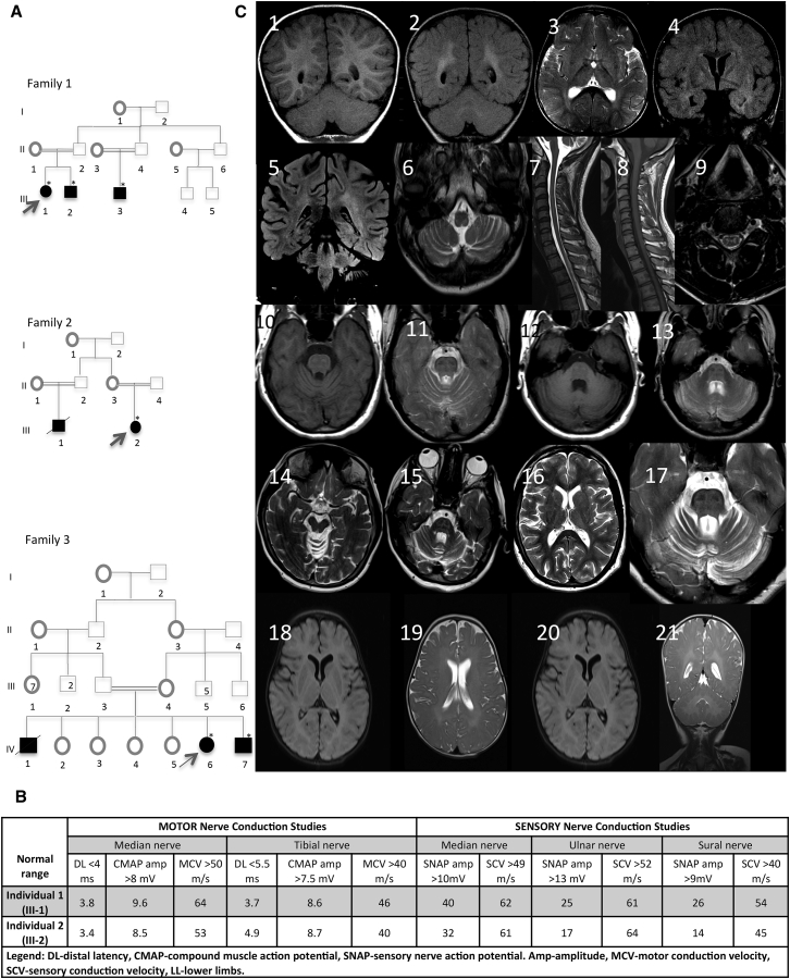
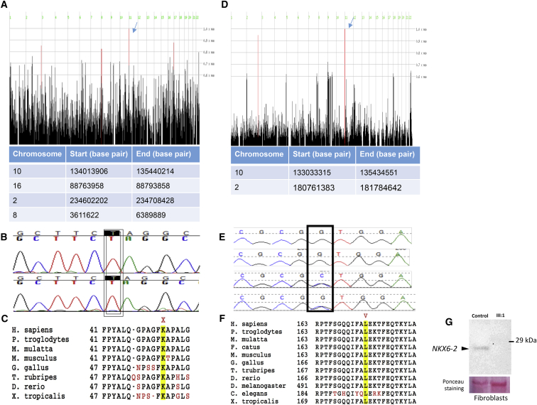
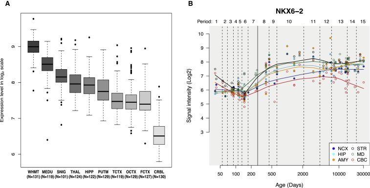
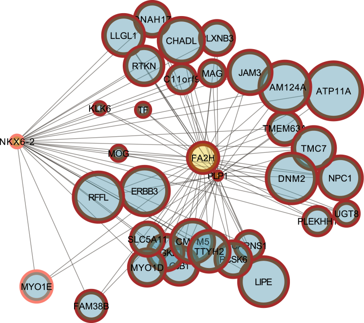
Progressive limb spasticity and cerebellar ataxia are frequently found together in clinical practice and form a heterogeneous group of degenerative disorders that are classified either as pure spastic ataxia or as complex spastic ataxia with additional neurological signs. Inheritance is either autosomal dominant or autosomal recessive. Hypomyelinating features on MRI are sometimes seen with spastic ataxia, but this is usually mild in adults and severe and life limiting in children. We report seven individuals with an early-onset spastic-ataxia phenotype. The individuals come from three families of different ethnic backgrounds. Affected members of two families had childhood onset disease with very slow progression. They are still alive in their 30s and 40s and show predominant ataxia and cerebellar atrophy features on imaging. Affected members of the third family had a similar but earlier-onset presentation associated with brain hypomyelination. Using a combination of homozygozity mapping and exome sequencing, we mapped this phenotype to deleterious nonsense or homeobox domain missense mutations in NKX6-2. NKX6-2 encodes a transcriptional repressor with early high general and late focused CNS expression. Deficiency of its mouse ortholog results in widespread hypomyelination in the brain and optic nerve, as well as in poor motor coordination in a pattern consistent with the observed human phenotype. In-silico analysis of human brain expression and network data provides evidence that NKX6-2 is involved in oligodendrocyte maturation and might act within the same pathways of genes already associated with central hypomyelination. Our results support a non-redundant developmental role of NKX6-2 in humans and imply that NKX6-2 mutations should be considered in the differential diagnosis of spastic ataxia and hypomyelination.

Keywords: NKX6-2; ataxia; genetic; leukodystrophy; recessive; spasticity.


Figures

Figure 1
Figure 1
Identification of Three Families affected by Progressive Spastic Ataxia and Hypomyelination (A) Pedigree of the families with NXK6-2 mutations. (B) Results of nerve-conduction studies showing normal myelination of peripheral nerves in family 1. (C) Magnetic resonance imaging findings in individuals with NKX6-2 mutations. Images 1 and 2 show Coronal T1W and FLAIR sequences demonstrating periventricular FLAIR hyperintensity and corresponding iso- to hyper-intense T1W signal relative to gray matter, suggestive of hypomyelination. Some hyperintensity of the dentate hilus is noted. Images 3 and 4 show axial T2W and coronal FLAIR sequences demonstrating abnormal T2W hyper-intense signal in the periventricular white matter, globi palladi, and external capsules. Images 5 and 6 show coronal FLAIR demonstrating T2W hyperintensity in the periventricular white matter and superior cerebellar peduncles. An axial T2W sequence demonstrating hyperintensity of the inferior cerebellar penduncles is shown. Images 7–9 show sagittal T2W and T1W as well as axial T2W sequences indicating diffuse spinal-cord volume loss and abnormal T2W hyperintensity symmetrically affecting the gray matter of the ventral and dorsal horn cells. Images 10–13 show axial T1W and T2W sequences demonstrating diffuse T2W hyperintensity and T1W iso- to hypo-intensity of the pons along with relative sparing of the cortico-spinal tracts. There is disproportionate volume loss of the cerebellum as well as the superior and middle cerebellar peduncles. Images 14–17 show axial T2W. A subtle T2W hyperintense signal change affects the anterior limbs of the internal capsules, thalami, and left peritrigonal white matter. The image shows a pontine diffuse T2W hyperintense signal sparing the cortico-spinal tracts and volume loss of the cerebellum and superior cerebellar peduncles. Images 18–21 show axial and coronal T2 weighted spin echo and axial FLAIR images indicating diffusely increased T2 signal intensity in cerebral and cerebellar white matter bilaterally and symmetrically, demonstrating delayed myelination in the youngest individuals.
Figure 2
Figure 2
NKX6-2 Mutations (A) Homozygosity mapping in families 1 and 2 identified four homozygous regions that were shared by the three affected individuals from families 1 and 2. The region on chromosome 10 includes NKX6-2 (arrow) shared between the two families. (B) Sanger sequencing confirming c.121A>T in the two families. (C) Conservation of p. Lys41 across species. (D) Homozygosity mapping in family 3 identified two homozygous regions that were shared by the affected individuals. The region on chromosome 10 includes NKX6-2 (arrow). (E) Sanger sequencing confirming c.487C>G and segregation in the family. (F) Conservation of the p.Leu163Val residue within the NKX6-2 homeodomain across species. The mutation is marked in red above the corresponding amino acid. (G) Immunoblot analysis showing a complete absence of NKX6-2 in an affected individual (individual III-1 in family 1 in the pedigree) compared to a control individual.
Figure 3
Figure 3
NKX6-2 Expression in Normal Developing Brain and Adult Brain (A) NKX6-2 expression in different brain areas in adult pathologically normal human brains.NKX6-2 is expressed in all ten brain regions; the highest expression is detected in white matter. Abbreviations are as follows: WHMT, white matter; MEDU, medulla; SNIG, substantia nigra; THAL, thalamus; HIPP, hippocampus; PUTM, putamen; TCTX, temporal cortex; OCTX, occipital cortex; FCTX, frontal cortex; and CRB, cerebellum. (B) NKX6-2 expression in developing brain. Abbreviations are as follows: NCX, neocortex; STR, striatum; HP, hippocampus; AMY, amygdala; MD, midbrain; and CBC, cerebellum.
Figure 4
Figure 4
A bottom-up Plot Showing a Different Connectivity of NKX6-2 Compared to PLP1 and FA2H, Suggesting a Regulatory Role of NKX6-2 Seed genes associated with hereditary spastic paraplegia contain an inner yellow circle, whereas context genes are represented by inner blue circles. The context genes in the plot are enriched for genes that the algorithm for the reconstruction of accurate cellular networks (ARACNe) based on an adaptive partitioning (AP) strategy predicts are found in the regulon of NKX6-2 (23 out of 35, FET p value 2.2 × 10−16).

References

    1. Boespflug-Tanguy O. Inborn errors of brain myelin formation. Handb. Clin. Neurol. 2013;113:1581–1592. - PubMed
    1. Fields R.D. White matter in learning, cognition and psychiatric disorders. Trends Neurosci. 2008;31:361–370. - PMC - PubMed
    1. Paus T., Keshavan M., Giedd J.N. Why do many psychiatric disorders emerge during adolescence? Nat. Rev. Neurosci. 2008;9:947–957. - PMC - PubMed
    1. Vanderver A., Simons C., Helman G., Crawford J., Wolf N.I., Bernard G., Pizzino A., Schmidt J.L., Takanohashi A., Miller D., Leukodystrophy Study Group Whole exome sequencing in patients with white matter abnormalities. Ann. Neurol. 2016;79:1031–1037. - PMC - PubMed
    1. Lek M., Karczewski K.J., Minikel E.V., Samocha K.E., Banks E., Fennell T., O’Donnell-Luria A.H., Ware J.S., Hill A.J., Cummings B.B., Exome Aggregation Consortium Analysis of protein-coding genetic variation in 60,706 humans. Nature. 2016;536:285–291. - PMC - PubMed

MeSH terms

Supplementary concepts