Review
. 2017 Jun 1;169(6):1000-1011.
doi: 10.1016/j.cell.2017.05.015.

Senescence in Health and Disease

Affiliations
Review

Senescence in Health and Disease

Shenghui He et al. Cell. .

Abstract

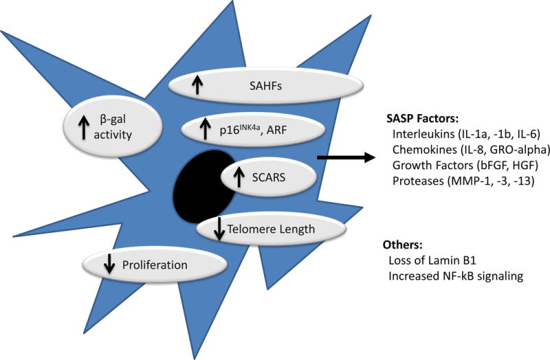
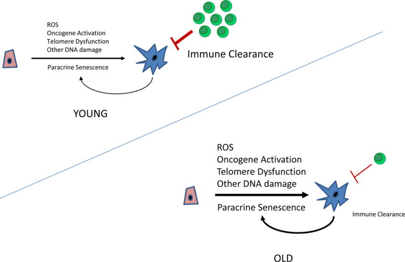
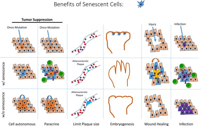
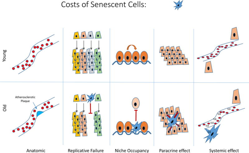
Many cellular stresses activate senescence, a persistent hyporeplicative state characterized in part by expression of the p16INK4a cell-cycle inhibitor. Senescent cell production occurs throughout life and plays beneficial roles in a variety of physiological and pathological processes including embryogenesis, wound healing, host immunity, and tumor suppression. Meanwhile, the steady accumulation of senescent cells with age also has adverse consequences. These non-proliferating cells occupy key cellular niches and elaborate pro-inflammatory cytokines, contributing to aging-related diseases and morbidity. This model suggests that the abundance of senescent cells in vivo predicts "molecular," as opposed to chronologic, age and that senescent cell clearance may mitigate aging-associated pathology.

Keywords: DNA damage; SASP; aging; cancer; cellular senescence; molecular age; p16(INK4a); senolysis; telomere; tumor suppression.


Figures

Figure 1
Figure 1. Characteristics of cellular senescence
Senescent cells exhibit durable growth arrest, increased expression of the products of the CDKN2a locus (p16INK4a and to a lesser extent ARF) and characteristic changes in cellular structures and protein expressions (e.g. elaboration of SASP factors). Senescent cells in vitro exhibit changes in cellular morphology (e.g. increased cell spreading) and increased SA-β-galactosidase activity, but these markers have been less useful for in vivo recognition. Several other markers (e.g. short telomeres; SCARS; activated NF-kB and DNA damage response; SAHFs) are often associated with cellular senescence, but these markers are neither sensitive nor specific for the senescent state. Loss of the Lamin B1 is an interesting, new marker of senescence that is under investigation.
Figure 2
Figure 2. Mechanisms of senescent cell accumulation with aging
The rate at which senescent cells accumulate increases with aging. This may reflect an increased rate of senescent cell production due to changes in DNA repair, telomere dysfunction and/or decreased senescent cell clearance by immune system. It has also been suggested that senescent cells can induce the formation of other senescent cells in a paracrine manner.
Figure 3
Figure 3. Beneficial roles of cellular senescence
Senescence affords tumor suppression in a cell intrinsic manner, and perhaps also by augmenting local anti-tumor immunity. The activation of the CDKN2a locus appears to limit the size of atherosclerotic plaques, thereby reducing anatomic obstruction. Senescence resulting from p21CIP expression during embryogenesis may be required for certain aspects of fetal development. Senescence contributes to wound healing and host immunity.
Figure 4
Figure 4. Detrimental effects of senescent cells
The accumulation of senescent cells can lead to anatomic lesions (e.g. as in an atherosclerotic plaque). The loss of replicative capacity of certain senescent cells (e.g. T cells, pancreatic β-cells) may lead to defects in tissue regeneration. The occupancy of prized physiological niches by senescent cells may impair tissue homeostasis. The paracrine and endocrine elaboration of pro-inflammatory hormones and enzymes promotes tissue dysfunction locally and at the organismal level.

References

    1. Acosta JC, Banito A, Wuestefeld T, Georgilis A, Janich P, Morton JP, Athineos D, Kang TW, Lasitschka F, Andrulis M, et al. A complex secretory program orchestrated by the inflammasome controls paracrine senescence. Nature cell biology. 2013;15:978–990. - PMC - PubMed
    1. Acosta JC, O’Loghlen A, Banito A, Guijarro MV, Augert A, Raguz S, Fumagalli M, Da Costa M, Brown C, Popov N, et al. Chemokine signaling via the CXCR2 receptor reinforces senescence. Cell. 2008;133:1006–1018. - PubMed
    1. Akbar AN, Henson SM. Are senescence and exhaustion intertwined or unrelated processes that compromise immunity? Nat Rev Immunol. 2011;11:289–295. - PubMed
    1. Armanios M, Blackburn EH. The telomere syndromes. Nature reviews Genetics. 2012;13:693–704. - PMC - PubMed
    1. Baker DJ, Childs BG, Durik M, Wijers ME, Sieben CJ, Zhong J, R AS, Jeganathan KB, Verzosa GC, Pezeshki A, et al. Naturally occurring P16-positive cells shorten healthy lifespan. Nature 2016 - PMC - PubMed