. 2017 Jun 2;14(1):112.
doi: 10.1186/s12974-017-0890-x.

B cells do not have a major pathophysiologic role in acute ischemic stroke in mice

Affiliations

B cells do not have a major pathophysiologic role in acute ischemic stroke in mice

Michael K Schuhmann et al. J Neuroinflammation. .

Abstract

Background: Lymphocytes have been shown to play an important role in the pathophysiology of acute ischemic stroke, but the properties of B cells remain controversial. The aim of this study was to unravel the role of B cells during acute cerebral ischemia using pharmacologic B cell depletion, B cell transgenic mice, and adoptive B cell transfer experiments.

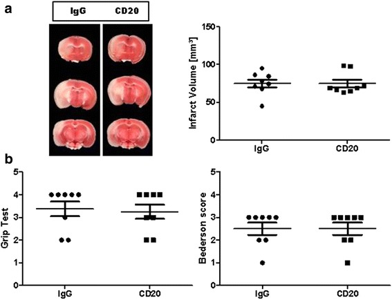
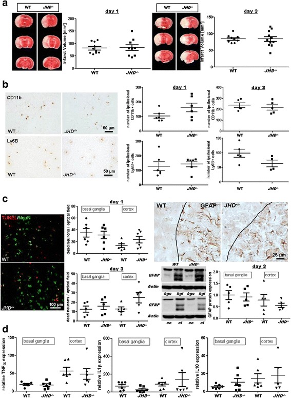
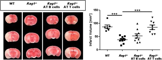
Methods: Transient middle cerebral artery occlusion (60 min) was induced in wild-type mice treated with an anti-CD20 antibody 24 h before stroke onset, JHD -/- mice and Rag1 -/- mice 24 h after adoptive B cell transfer. Stroke outcome was assessed at days 1 and 3. Infarct volumes were calculated from 2,3,5-triphenyltetrazolium chloride (TTC)-stained brain sections, and neurological scores were evaluated. The local inflammatory response was determined by real-time PCR and immunohistochemistry. Apoptosis was analyzed by TUNEL staining, and astrocyte activation was revealed using immunohistochemistry and Western blot.

Results: Pharmacologic depletion of B cells did not influence infarct volumes and functional outcome at day 1 after stroke. Additionally, lack of circulating B cells in JHD -/- mice also failed to influence stroke outcome at days 1 and 3. Furthermore, reconstitution of Rag1 -/- mice with B cells had no influence on infarct volumes.

Conclusion: Targeting B cells in experimental stroke did not influence lesion volume and functional outcome during the acute phase. Our findings argue against a major pathophysiologic role of B cells during acute ischemic stroke.

Keywords: B cells; Ischemic stroke; Transient middle cerebral artery occlusion.


Figures

Fig. 1
Fig. 1
Pharmacologic prestroke B cell depletion does not alter stroke outcome in a transient middle cerebral artery occlusion (tMCAO) model. a Left, representative 2,3,5-triphenyltetrazolium chloride stains of three corresponding brain sections of an IgG-treated C57BL/6 mouse (IgG) and a C57BL/6 mouse at day 1 after stroke, treated with a specific antibody against CD20 1 day before 60-min tMCAO. a Right, infarct volumes are similar (n = 8/group) between the two treatment groups (unpaired, two-tailed Student’s t test). b B cell depletion does not improve functional outcome on day 1 after tMCAO (n = 8/group) as assessed by the grip test (left) and Bederson score (right), Mann–Whitney test
Fig. 2
Fig. 2
Lack of B cells does not impact stroke outcome in a transient middle cerebral artery occlusion (tMCAO) model. a Representative 2,3,5-triphenyltetrazolium chloride stains of three corresponding brain sections and infarct volumes of control (WT) and B cell-deficient (JHD −/−) mice at days 1 (left) and 3 (right) after 60-min tMCAO (n = 9–11/group). Infarct volumes are similar between the two groups for both time points (unpaired, two-tailed Student’s t test). b Left, representative immunocytologic staining of CD11b+ and Ly6B+ cells within the ischemic brains of WT and JHD −/− mice. b Right, quantification revealed comparable numbers of CD11b+ monocytes and Ly6B+ neutrophils in the ipsilesional hemispheres of WT and JHD −/− mice at days 1 (n = 6/group) and 3 (n = 4–5/group) after tMCAO (unpaired, two-tailed Student’s t test). c Left, representative brain sections from a WT and a JHD −/− mouse stained for the neuronal marker NeuN and subjected to TUNEL assay to visualize apoptosis. Quantification of dead neurons per optical field in basal ganglial as well as cortical regions at days 1 (n = 6/group) and 3 (n = 5/group) was comparable between WT and JHD −/− mice (unpaired, two-tailed Student’s t test). c Right, representative immunocytologic staining of glial fibrillary acidic protein (GFAP) immunoreactivity in the penumbra (black lines) of the ischemic cortex of a WT and a JHD −/− mouse at day 3 after tMCAO (top). Representative anti-GFAP Western blot analysis (cc cortex contralesional, ci cortex ipsilesional, bgc basal ganglia contralesional, bgi basal ganglia ipsilesional) and densitometric quantification of ipsilesional GFAP protein expression in the basal ganglial as well as cortical regions at day 3 (n = 5/group) after tMCAO (bottom) was comparable between WT and JHD −/− mice (unpaired, two-tailed Student’s t test). d Relative gene expression of tumor necrosis factor α (TNFα), interleukin (IL)-1β, and IL-10 in the ischemic basal ganglia and cortex are similar at day 1 after tMCAO of WT and JHD −/− mice (n = 5–6/group) when normalized to sham operation (unpaired, two-tailed Student’s t test)
Fig. 3
Fig. 3
Prestroke B cell adoptive transfer (AT) into Rag1 −/− mice does not worsen stroke outcome in a transient middle cerebral artery occlusion (tMCAO) model. Left, representative 2,3,5-­triphenyltetrazolium chloride stains of three corresponding brain sections of a C57BL/6 mouse (WT), a Rag1 −/− mouse without adoptive cell transfer (Rag1 -/-) and with adoptively transferred B cells (Rag1 −/−> AT B cells) or T cells (Rag1 −/− AT B cells) on day 1 after 60-min tMCAO. Right, infarct volumes are reduced in Rag1 −/− mice (n = 10) compared with those in WT mice (n = 6). Only AT of T cells (n = 8), but not B cells (n = 9), increased infarct volumes (one-way analysis of variance with post hoc Bonferroni adjustment for P values). ***P < 0.001

References

    1. Stoll G, Kleinschnitz C, Nieswandt B. Combating innate inflammation: a new paradigm for acute treatment of stroke? Ann N Y Acad Sci. 2010;1207:149–54. doi: 10.1111/j.1749-6632.2010.05730.x. - DOI - PubMed
    1. Gelderblom M, Leypoldt F, Steinbach K, Behrens D, Choe CU, Siler DA, Arumugam TV, Orthey E, Gerloff C, Tolosa E, Magnus T. Temporal and spatial dynamics of cerebral immune cell accumulation in stroke. Stroke. 2009;40:1849–57. doi: 10.1161/STROKEAHA.108.534503. - DOI - PubMed
    1. Yilmaz G, Arumugam TV, Stokes KY, Granger DN. Role of T lymphocytes and interferon-gamma in ischemic stroke. Circulation. 2006;113:2105–12. doi: 10.1161/CIRCULATIONAHA.105.593046. - DOI - PubMed
    1. Ren X, Akiyoshi K, Dziennis S, Vandenbark AA, Herson PS, Hurn PD, Offner H. Regulatory B cells limit CNS inflammation and neurologic deficits in murine experimental stroke. J Neurosci. 2011;31:8556–63. doi: 10.1523/JNEUROSCI.1623-11.2011. - DOI - PMC - PubMed
    1. Chen Y, Bodhankar S, Murphy SJ, Vandenbark AA, Alkayed NJ, Offner H. Intrastriatal B-cell administration limits infarct size after stroke in B-cell deficient mice. Metab Brain Dis. 2012;27:487–93. doi: 10.1007/s11011-012-9317-7. - DOI - PMC - PubMed

Publication types

MeSH terms