Review
. 2017 Jul;12(3):269-288.
doi: 10.1016/j.cpet.2017.02.001.

Human Epidermal Growth Factor Receptor 2-Targeted PET/Single- Photon Emission Computed Tomography Imaging of Breast Cancer: Noninvasive Measurement of a Biomarker Integral to Tumor Treatment and Prognosis

Affiliations
Review

Human Epidermal Growth Factor Receptor 2-Targeted PET/Single- Photon Emission Computed Tomography Imaging of Breast Cancer: Noninvasive Measurement of a Biomarker Integral to Tumor Treatment and Prognosis

Kelly E Henry et al. PET Clin. 2017 Jul.

Abstract

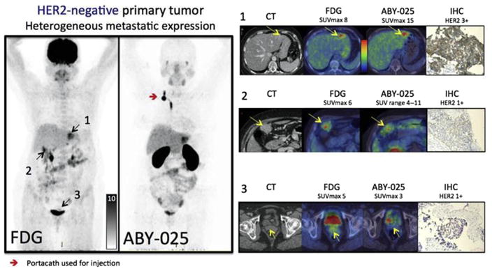
Increased human epidermal growth factor receptor 2 (HER2) expression is a hallmark of aggressive breast cancer. Imaging modalities have the potential to diagnose HER2-positive breast cancer and detect distant metastases. The heterogeneity of HER2 expression between primary and metastatic disease sites limits the value of tumor biopsies. Molecular imaging is a noninvasive tool to assess HER2-positive primary lesions and metastases. Radiolabeled antibodies, antibody fragments, and affibody molecules devise a reliable and quantitative method for detecting HER2-positive cancer using PET. HER2-targeted PET imaging is a valuable clinical tool with respect to both the care and maintenance of patients with breast cancer.

Keywords: Breast cancer; CT; HER2; Imaging; Metastasis; PET; Radiotracers; SPECT.


Figures

Fig. 1
Fig. 1
89Zr-trastuzumab biodistribution in time. (AC) Three 89Zr-trastuzumab scans of a patient already on tras-tuzumab treatment (cohort 3) show the increase over time in the tumor/nontumor ratio with regard to uptake of the tracer. Arrow indicates 89Zr-trastuzumab uptake in the only lesion. p.i., postinjection; 89Zr, zirconium-89. (From Dijkers EC, Oude Munnink TH, Kosterink JG, et al. Biodistribution of 89Zr-trastuzumab and PET imaging of HER2-positive lesions in patients with metastatic breast cancer. Clin Pharmacol Ther 2010;87(5):588; with permission.)
Fig. 2
Fig. 2
Examples of fusion images from human epidermal growth factor receptor 2 (HER2) PET and MR imaging scans. (A) In a vertebral metastasis seen on MR imaging but unapproachable for biopsy, HER2 status was revealed by 89Zr-trastuzumab uptake on PET imaging. (B) Example of HER2-positive brain lesion undetected by conventional scans, revealed by 89Zr-trastuzumab PET imaging, and subsequently confirmed by MR imaging. Arrows indicate lesions. HER2, human epidermal growth factor receptor 2; 89Zr, zirconium-89. (From Dijkers EC, Oude Munnink TH, Kosterink JG, et al. Biodistribution of 89Zr-trastuzumab and PET imaging of HER2-positive lesions in patients with metastatic breast cancer. Clin Pharmacol Ther 2010;87(5):589; with permission.)
Fig. 3
Fig. 3
A 41-year-old woman with primary ER-positive, human epidermal growth factor receptor 2 (HER2)-negative invasive ductal breast carcinoma and recurrence in the thoracic nodes. (A) Immunohistochemistry score of primary breast malignancy was 11 (at 20 × magnification), consistent with HER2-negative malignancy. (B) Axial computed tomography (CT) and 89Zr-trastuzumab PET/CT demonstrated 89Zr-trastuzumab avidity in enlarged right supraclavicular nodes (arrows, maximum standardized uptake value of 4.6) and left internal mammary nodes (not shown). (C) Biopsy of right supraclavicular node demonstrated metastatic breast carcinoma with immunohistochemistry score of 31 (at 20 × magnification), consistent with HER2-positive disease. The patient began systemic treatment including trastuzumab and pertuzumab. (D) Follow-up axial CT after 2 months of treatment demonstrated resolution of nodes on CT examination (arrow). (From Ulaner GA, Hyman DM, Ross DS, et al. Detection of HER2-positive metastases in patients with HER2-negative primary breast cancer using 89Zr-trastuzumab PET/CT. J Nuc Med 2016;57(10):1526; with permission.)
Fig. 4
Fig. 4
An 83-year-old woman with primary ER-positive/human epidermal growth factor receptor 2 (HER2)-negative invasive ductal breast carcinoma. (A) Immunohistochemistry score of primary breast malignancy was 11 (at 400 × magnification), consistent with HER2-negative malignancy. (B) 89Zr-trastuzumab maximum intensity projection demonstrates several foci of 89Zr-trastuzumab avidity that localize to osseous structures (arrows). Avidity in liver and bowel is considered physiologic. (C) Axial computed tomography (CT) and 89Zr-trastuzumab PET/CT demonstrate 89Zr-trastuzumab avidity in proximal left femur (arrow; maximum standardized uptake value of 7.7). (D) Biopsy of proximal left femur demonstrated metastatic breast carcinoma with immunohistochemistry score of 11 (at 400 × magnification), consistent with HER2-negative disease. (From Ulaner GA, Hyman DM, Ross DS, et al. Detection of HER2-positive metastases in patients with HER2-negative primary breast cancer using 89Zr-trastuzumab PET/CT. J Nuc Med 2016;57(10):1527; with permission.)
Fig. 5
Fig. 5
Whole-body copper-64-DOTA-trastuzumab PET images at 1, 24, and 48 hours after injection (patient 4). (From Tamura K, Kurihara H, Yonemori K, et al. 64Cu-DOTA-trastuzumab PET imaging in patients with HER2-positive breast cancer. J Nucl Med 2013;54(11):1871; with permission.)
Fig. 6
Fig. 6
Copper-64 (64Cu)-DOTA-trastuzumab PET images of human epidermal growth factor receptor 2 (HER2)-positive metastatic brain lesions (arrows). (A) Brain metastases were clearly visualized by 64Cu-DOTA-trastuzumab PET in patient 1. Significant uptake values were found in areas corresponding to brain metastatic lesions that were detected by MR imaging. Some brain metastases could not be detected on conventional CT. (B) In patient 4, 64Cu-DOTA-trastuzumab PET imaging could detect solitary brain metastasis that had also been identified in similar location by MR imaging. Gd, gadolinium. (From Tamura K, Kurihara H, Yonemori K, et al. 64Cu-DOTA-trastuzumab PET imaging in patients with HER2-positive breast cancer. J Nucl Med 2013;54(11):1874; with permission.)
Fig. 7
Fig. 7
Copper-64 (64Cu)-DOTA-trastuzumab PET images of metastatic brain tumors in patients with human epidermal growth factor receptor 2 (HER2)-positive primary breast tumors. The white arrows show the metastatic brain tumors. Upper panels: 64Cu-DOTA-trastuzumab PET images. Lower panels: gadolinium (Gd)-DTPA-enhanced T1-weighted MR imaging images. White arrows indicate metastatic brain lesions detectable by both MR imaging and 64Cu-DOTA-trastuzumab PET, and the red arrow indicates a lesion detectable by MR imaging but not by 64Cu-DOTA-trastuzumab PET. In the PET image from patient 2, nonspecific high uptake in the blood was noted. (From Kurihara H, Hamada A, Yoshida M, et al. 64Cu-DOTA-trastuzumab PET imaging and HER2 specificity of brain metastases in HER2-positive breast cancer patients. EJNMMI Res 2015;5(8):4; with permission.)
Fig. 8
Fig. 8
Serial MIP images (patient 6) displayed at the same relative intensity. Although a slight decrease in blood pool is present, blood pool activity dominates the distribution. At 58, 109, and 189 minutes, the blood pool has a maximum standardized uptake value of 16.9, 13.6, and 9.8, respectively. There is slight increased uptake over time in the liver and kidneys. The patient had human epidermal growth factor 2 (HER2)-positive disease and was receiving lapatinib. Tumors in the left adrenal, hilar nodes, and bones were not visualized on Ga-PET. HER2, human epidermal growth factor receptor 2; MIP, maximum intensity projection; p.i, postinjection. (From Beyergil V, Morris PG, Smith-Jones PM, et al. Pilot study of 68Ga-DOTA-F(ab′)2-trastuzumab in patients with breast cancer. Nucl Med Commun 2013;34(12):1162; with permission.)
Fig. 9
Fig. 9
Lytic lesion in the left calvarium with gallium-68 (68Ga)-DOTA-F(ab′)2-trastuzumab uptake (patient 1). 68Ga-DOTA-F(ab′)2-trastuzumab, 68Ga-1,4,7,10-tetraazacyclododecane- N,N′,N′,N‴-tetraacetic acid (DOTA)-F(ab′) 2-trastuzumab. (From Beyergil V, Morris PG, Smith-Jones PM, et al. Pilot study of 68Ga-DOTA-F(ab′)2-trastuzumab in patients with breast cancer. Nucl Med Commun 2013;34(12):1163; with permission.)
Fig. 10
Fig. 10
In patient 2, fused images obtained at B1 hours show mild 68Ga-DOTAF(ab′)2-trastuzumab uptake in a lung metastasis (arrows). 68Ga-DOTA-F(ab′)2- trastuzumab, 68Ga-1,4,7,10-tetraazacyclododecane-N,N′,N′,N‴-tetraacetic acid (DOTA)-F(ab′′)2-trastuzumab. (From Beyergil V, Morris PG, Smith-Jones PM, et al. Pilot study of 68Ga-DOTA-F(ab′)2-trastuzumab in patients with breast cancer. Nucl Med Commun 2013; 34(12):1163; with permission.)
Fig. 11
Fig. 11
Representative whole-body images at 10, 30, 60, 120, and 240 minutes after injection of a low and high peptide dose in the same patient. (From Sandström M, Lindskog K, Velikyan I, et al. Bio-distribution and radiation dosimetry of the anti-HER2 affibody molecule 68Ga-ABY-025 in breast cancer patients. J Nuc Med 2016; 57(6):868; with permission.)
Fig. 12
Fig. 12
Uptake images of gallium-68 (68Ga)-BY-025 with low (LD) and high administered peptide dose (HD) at 1, 2 and 4 hours after injection in patient 2. All images are normalized to the same scale of standardized uptake value 10. A rainbow color scale is used: red color, standardized uptake value of greater than 10. Normal soft tissue uptake was higher with LD. Note the gradually higher contrast of metastases in liver and bone (arrows), owing to both the disappearance of normal liver uptake and tumor uptake accumulation. (From Sörensen J, Velikyan I, Sandberg D, et al. Measuring HER2-receptor expression in metastatic breast cancer using [68Ga]ABY-025 affibody PET/CT. Theranostics 2016;6(2):266; with permission.)
Fig. 13
Fig. 13
Based on the results from gallium-68-ABY-025 PET/computed tomography (CT), mixed expression of human epidermal growth factor receptor 2 (HER2) in metastatic breast cancer was seen in several patients (arrows) and confirmed by biopsies in 2. Patient 9 had HER2-negative primary tumor and was enrolled as negative control. Fluorodeoxyglucose (FDG)-PET/CT showed metastases in left liver lobe, peritoneal lymph nodes and cervix of uterus (arrows). ABY-025 uptake was high in the liver metastasis, low in peritoneal metastases and absent in the cervical region (not shown). According to immunohistochemistry (IHC), the liver finding was true positive and both other sites were true negative. SUVmax, maximum standardized uptake volume. (From Sörensen J, Velikyan I, Sandberg D, et al. Measuring HER2-receptor expression in metastatic breast cancer using [68Ga] ABY-025 affibody PET/CT. Theranostics 2016;6(2):268; with permission.)
Fig. 14
Fig. 14
Representative maximum intensity projection images at 10, 60, and 90 minutes after injection of gallium-68 (68Ga)-human epidermal growth factor receptor 2 (HER2)-nanobody for different mass subgroups. (A) Patient 4, injected with 0.01 mg of 68Ga-HER2-nanobody. (B) Patient 12, injected with 0.1 mg. (C) Patient 17, injected with 1.0 mg. (From Keyaerts M, Xavier C, Heemskerk J, et al. Phase I study of 68Ga-HER2-nanobody for PET/CT assessment of HER2 expression in breast carcinoma. J Nuc Med 2016;57(1):30; with permission.)
Fig. 15
Fig. 15
Uptake of gallium-68 (68Ga)-human epidermal growth factor receptor 2-nanobody in primary breast carcinoma lesions (arrows) on PET/computed tomography (CT) images (top) and PET images (bottom). (A) Patient 14 showed highest tracer uptake (mean standardized uptake value, 11.8). (B) Patient 15 showed moderate tracer uptake, which was easily discernable from background (mean standardized uptake value, 4.9). (C) Patient 6 showed no uptake (mean standardized uptake value, 0.9), with CT showing marker clip at tumor region. (From Keyaerts M, Xavier C, Heemskerk J, et al. Phase I study of 68Ga-HER2-nanobody for PET/CT assessment of HER2 expression in breast carcinoma. J Nuc Med 2016;57(1):31; with permission.)
Fig. 16
Fig. 16
Uptake of gallium-68 (68Ga)-human epidermal growth factor receptor 2-nanobody in metastatic lesions on PET/computed tomography images (top) and PET images (bottom). (A) Patient 18, with invaded lymph nodes in mediastinum and left hilar region. (B) Patient 20, with bone metastasis in pelvis. (From Keyaerts M, Xavier C, Heemskerk J, et al. Phase I study of 68Ga-HER2-nanobody for PET/CT assessment of HER2 expression in breast carcinoma. J Nuc Med 2016;57(1):32; with permission.)

References

    1. Weigelt B, Peterse JL, van’t Veer LJ. Breast cancer metastasis: markers and models. Nat Rev Cancer. 2005;5(8):591–602. - PubMed
    1. Dunnwald LK, Rossing MA, Li CI. Hormone receptor status, tumor characteristics, and prognosis: a prospective cohort of breast cancer patients. Breast Cancer Res. 2007;9(1):1–10. - PMC - PubMed
    1. van de Vijver M, Bilous M, Hanna W, et al. Chromogenic in situ hybridisation for the assessment of HER2 status in breast cancer: an international validation ring study. Breast Cancer Res. 2007;9(5):R68–77. - PMC - PubMed
    1. Arteaga CL, Sliwkowski MX, Osborne CK, et al. Treatment of HER2-positive breast cancer: current status and future perspectives. Nat Rev Clin Oncol. 2012;9(1):16–32. - PubMed
    1. Capala J, Bouchelouche K. Molecular imaging of HER2-positive breast cancer - a step toward an individualized “Image and Treat” strategy. Curr Opin Oncol. 2010;22(6):559–66. - PMC - PubMed

MeSH terms