. 2017 Jul 11:976:63-73.
doi: 10.1016/j.aca.2017.04.014. Epub 2017 Apr 10.

Chloroformate derivatization for tracing the fate of Amino acids in cells and tissues by multiple stable isotope resolved metabolomics (mSIRM)

Affiliations

Chloroformate derivatization for tracing the fate of Amino acids in cells and tissues by multiple stable isotope resolved metabolomics (mSIRM)

Ye Yang et al. Anal Chim Acta. .

Abstract

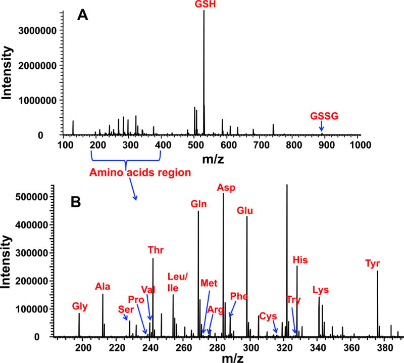
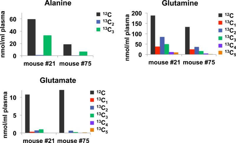
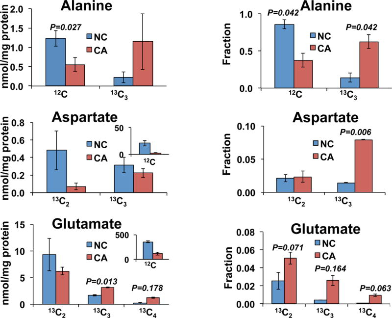
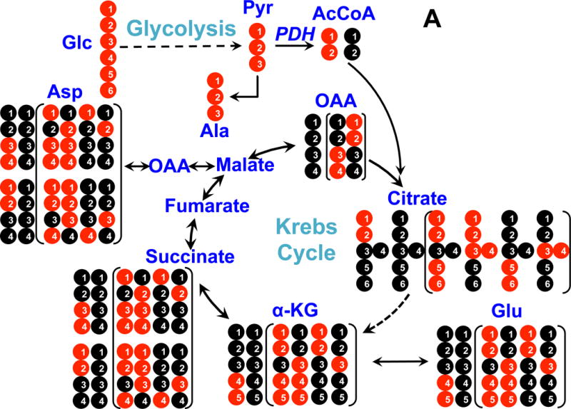
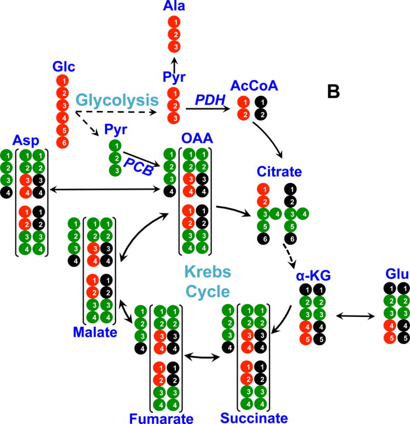
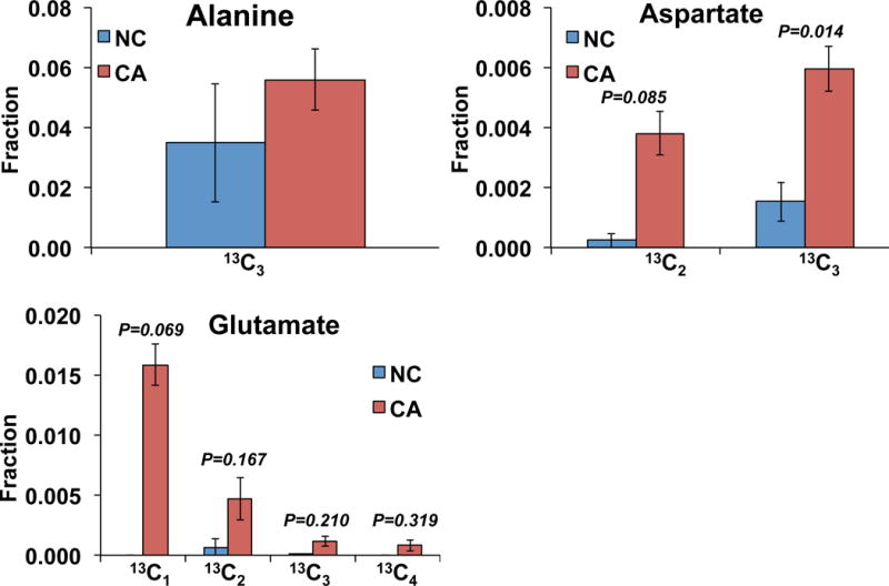
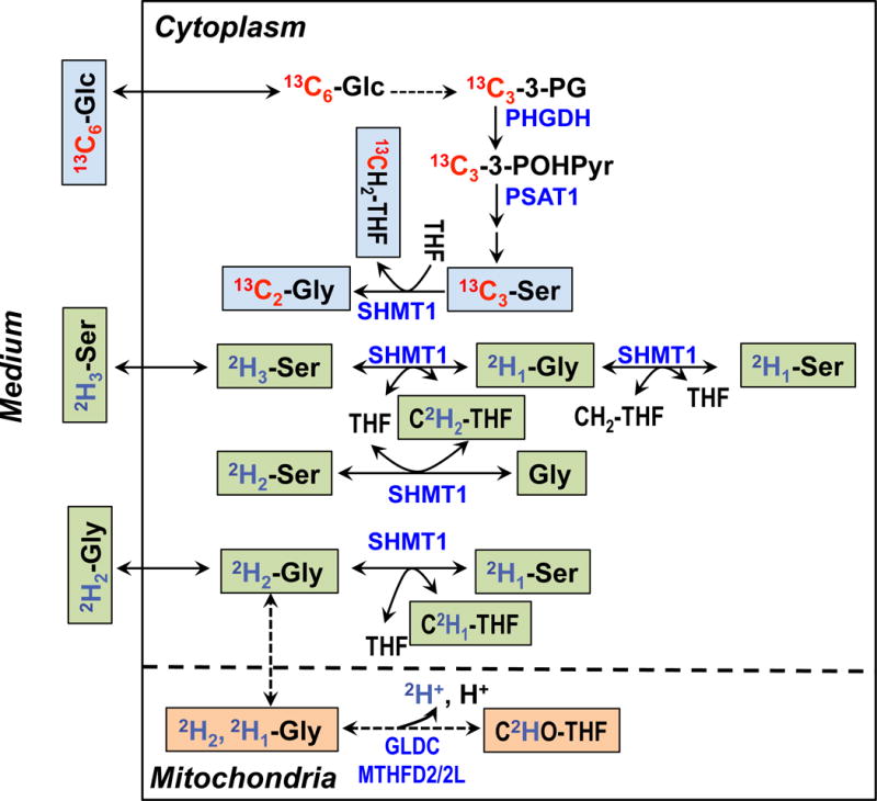
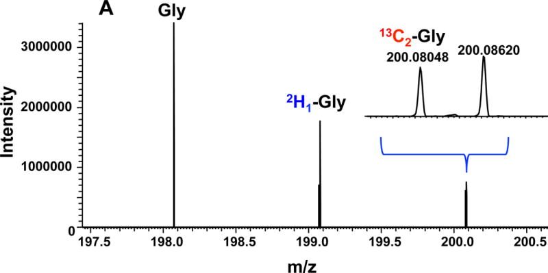
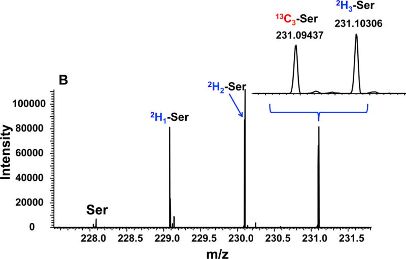
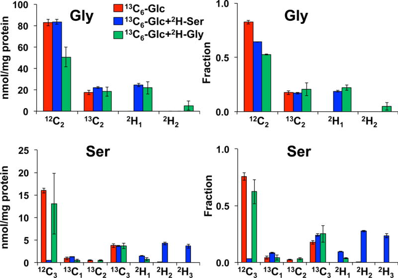
Amino acids have crucial roles in central metabolism, both anabolic and catabolic. To elucidate these roles, steady-state concentrations of amino acids alone are insufficient, as each amino acid participates in multiple pathways and functions in a complex network, which can also be compartmentalized. Stable Isotope-Resolved Metabolomics (SIRM) is an approach that uses atom-resolved tracking of metabolites through biochemical transformations in cells, tissues, or whole organisms. Using different elemental stable isotopes to label multiple metabolite precursors makes it possible to resolve simultaneously the utilization of these precursors in a single experiment. Conversely, a single precursor labeled with two (or more) different elemental isotopes can trace the allocation of e.g. C and N atoms through the network. Such dual-label experiments however challenge the resolution of conventional mass spectrometers, which must distinguish the neutron mass differences among different elemental isotopes. This requires ultrahigh resolution Fourier transform mass spectrometry (UHR-FTMS). When combined with direct infusion nano-electrospray ion source (nano-ESI), UHR-FTMS can provide rapid, global, and quantitative analysis of all possible mass isotopologues of metabolites. Unfortunately, very low mass polar metabolites such as amino acids can be difficult to analyze by current models of UHR-FTMS, plus the high salt content present in typical cell or tissue polar extracts may cause unacceptable ion suppression for sources such as nano-ESI. Here we describe a modified method of ethyl chloroformate (ECF) derivatization of amino acids to enable rapid quantitative analysis of stable isotope labeled amino acids using nano-ESI UHR-FTMS. This method showed excellent linearity with quantifiable limits in the low nanomolar range represented in microgram quantities of biological specimens, which results in extracts with total analyte abundances in the low to sub-femtomole range. We have applied this method to profile amino acids and their labeling patterns in 13C and 2H doubly labeled PC9 cell extracts, cancerous and non-cancerous tissue extracts from a lung cancer patient and their protein hydrolysates as well as plasma extracts from mice fed with a liquid diet containing 13C6-glucose (Glc). The multi-element isotopologue distributions provided key insights into amino acid metabolism and intracellular pools in human lung cancer tissues in high detail. The 13C labeling of Asp and Glu revealed de novo synthesis of these amino acids from 13C6-Glc via the Krebs cycle, specifically the elevated level of 13C3-labeled Asp and Glu in cancerous versus non-cancerous lung tissues was consistent with enhanced pyruvate carboxylation. In addition, tracking the fate of double tracers, (13C6-Glc + 2H2-Gly or 13C6-Glc + 2H3-Ser) in PC9 cells clearly resolved pools of Ser and Gly synthesized de novo from 13C6-Glc (13C3-Ser and 13C2-Gly) versus Ser and Gly derived from external sources (2H3-Ser, 2H2-Gly). Moreover the complex 2H labeling patterns of the latter were results of Ser and Gly exchange through active Ser-Gly one-carbon metabolic pathway in PC9 cells.

Keywords: Amino acids; Direct infusion nano-electrospray; Stable isotope resolved metabolomics; Ultrahigh resolution fourier transform mass spectrometry.


Figures

Figure 1
Figure 1. Typical positive ion mode spectrum of an unlabeled polar extract of PC9 cells after derivatization with ECF
Panel A shows the full m/z range profile spectrum, which included the Na adducts of GSH and GSSG derivatives along with amino acid derivatives. Panel B shows the zoom-in profile spectrum in the amino acid range. The m/z peaks were assigned using “PREMISE” [13]. Most of the amino acids shown represented Na adduct species except for Arg and His, which were protonated species.
Figure 2
Figure 2. 13C labeled amino acids in blood plasma of mice fed with a 13C6-glucose diet
Blood plasma from mice fed with the 13C6-glucose diet was obtained and treated as described in the methods. Isotopologues of the amino acids were determined via the ECF derivatives by UHR-FTMS. High 13C enrichment was observed in Ala (ca. 33%) and Gln (ca. 50%) but not in Glu (<10–15%).
Figure 3
Figure 3. 13C labeling in human lung tissues
The polar extract of lung tissues slices incubated for 24 h in the presence of 13C6-glucose were processed as described in the methods. The amounts of different isotopologues of three amino acids were normalized to the tissue protein weight (left) and the fractional enrichments were calculated (right). Non-cancerous (NC, blue) and cancerous (CA, red) tissue slices procured from a lung cancer patient (UK018). N=2; error bars represent standard error of mean (SEM).
Figure 4
Figure 4. 13C atom-resolved tracing of 13C6-Glc oxidation through the interconnecting cytoplasmic glycolysis and mitochondrial Krebs cycle
Panel A shows 13C incorporation from 13C6-Glc into various metabolites via the PDH (pyruvate dehydrogenase) pathway; panel B shows 13C incorporation from 13C6-Glc into metabolites via the PCB (pyruvate carboxylase) pathway. ●: 12C; ●: 13C; ●,● indicates 13C derived from PDH or PCB mediated Krebs cycle reactions, respectively; In panel A, the labeled patterns of the first and second (in brackets) turns of the PDH-initiated Krebs cycle are shown. In panel B, 13C3-pyruvate (Pyr) is carboxylated to form 13C3-oxaloacetate (OAA), leading to the formation of 13C3-Asp, -malate, -fumarate, and -succinate (patterns outside brackets); the labeled structures in brackets are derived from the reaction of 13C3-OAA with 13C2- or unlabeled acetyl CoA (AcCoA) to form other labeled Krebs cycle intermediates after one cycle turn. α-KG: α-ketoglutarate; not all expected labeled patterns of metabolites are shown [10].
Figure 5
Figure 5. Fractional enrichment of isotopologues in different amino acids hydrolyzed from non-cancerous or cancerous tissue proteins
N=2; error bars represent SEM.
Figure 6
Figure 6. De novo synthesis of Ser and Gly from glucose via glycolysis and the 3-phosphoglycerate (3-PG) pathway and exchanges of exogenously derived Ser and Gly via one-carbon metabolism
Selected Ser and Gly exchange reactions are depicted for the cytoplasmic and mitochondrial compartments to illustrate the production of different 2H isotopologues of Gly and Ser in Figs. 7 and 8. For example, the serine hydroxymethyl transferase (SHMT) reaction was shown for the cytoplasm (SHMT1) but omitted for the mitochondria (SHMT2) [10, 46]. Blue text blocks depict the de novo synthesis carbon pathway from 13C6-glucose while green and beige text blocks respectively depict the hydrogen exchange pathway of Gly and Ser via 1-carbon metabolism catalyzed by SHMT1, glycine decarboxylase complex (GLDC), and methylenetetrahydrofolate dehydrogenase (MTHFD). PHGDH: phosphoglycerate dehydrogenase; PSAT1: phosphoserine amino transferase 1; MTHFD2: mitochondrial NAD+-dependent methylene tetrahydrofolate dehydrogenase/methylene tetrahydrofolate cyclohydrolase; MTHFD2L: mitochondrial NADP+-dependent methylene tetrahydrofolate dehydrogenase.
Figure 7
Figure 7. UHR-FTMS of an extract of PC9 cells
Panel A shows the UHR-FTMS profile spectrum of the Gly-ECF region of a polar extract of PC9 cells grown in a medium containing 13C6-Glc+2H2-Gly. Panel B shows the UHR-FTMS profile spectrum of the Ser-ECF region of a polar extract of 13C6-Glc+2H3-Ser labeled PC9 cells.
Figure 8
Figure 8
13C and 2H Isotopologue distribution of Ser and Gly in dually labeled PC9 cell extracts indicates extensive exchanges between the two amino acids. N=3; error bars represent SEM.

Similar articles

Cited by

References

    1. Lehninger AL, Nelson DL, Cox MM. Lehninger principles of biochemistry. New York: Worth Publishers; 2000.
    1. Hosios AM, Hecht VC, Danai LV, Johnson MO, Rathmell JC, Steinhauser ML, Manalis SR, Vander Heiden MG. Amino Acids Rather than Glucose Account for the Majority of Cell Mass in Proliferating Mammalian Cells. Dev Cell. 2016;36(5):540–9. - PMC - PubMed
    1. Lane AN, Fan TWM. Regulation of mammalian nucleotide metabolism and biosynthesis. Nucleic Acids Res. 2015;43:2466–2485. - PMC - PubMed
    1. Vogler AP, Trentmann S, Lengeler JW. Alternative route for biosynthesis of amino sugars in Escherichia coli K-12 mutants by means of a catabolic isomerase. J Bacteriol. 1989;171(12):6586–92. - PMC - PubMed
    1. Ha HC, Sirisoma NS, Kuppusamy P, Zweier JL, Woster PM, Casero RA., Jr The natural polyamine spermine functions directly as a free radical scavenger. Proc Natl Acad Sci U S A. 1998;95(19):11140–5. - PMC - PubMed