. 2018 Feb;153(2):339-348.
doi: 10.1016/j.chest.2017.05.018. Epub 2017 May 30.

CA-125 in Disease Progression and Treatment of Lymphangioleiomyomatosis

Affiliations

CA-125 in Disease Progression and Treatment of Lymphangioleiomyomatosis

Connie G Glasgow et al. Chest. 2018 Feb.

Abstract

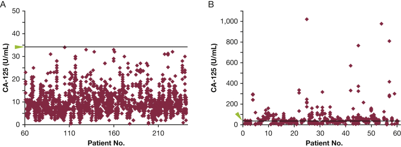
Background: Lymphangioleiomyomatosis (LAM) is a destructive lung disease of women caused by proliferation of neoplastic-like LAM cells, with mutations in the TSC1/2 tumor suppressor genes. Based on case reports, levels of cancer antigen 125 (CA-125), an ovarian cancer biomarker, can be elevated in patients with LAM. We hypothesized that elevated serum CA-125 levels seen in some patients with LAM were due to LAM, not other malignancies, and might respond to sirolimus treatment.

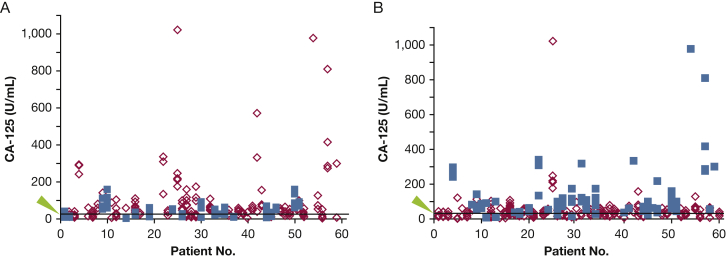
Methods: Serum CA-125 levels were measured for 241 patients at each visit. Medical records were reviewed for co-morbidities, disease progression, and response to sirolimus treatment. CA-125 expression in LAM cells was determined by using immunohistochemical analysis.

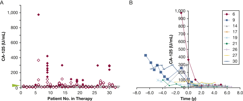
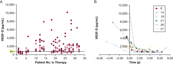
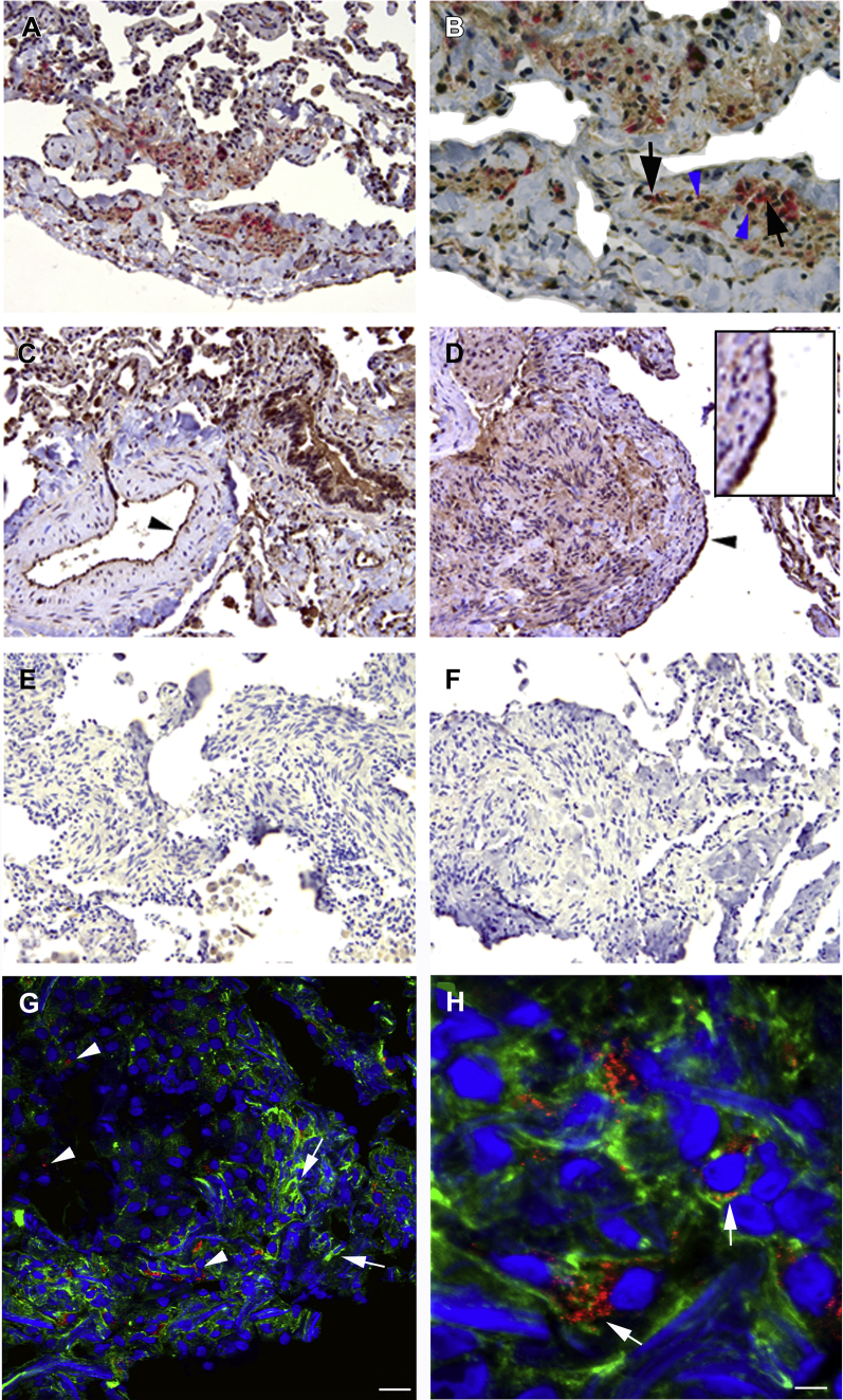
Results: Almost 25% of patients with LAM had at least one elevated serum CA-125 measurement. Higher serum CA-125 levels correlated with lower FEV1, premenopausal status, and pleural effusion in a multivariate model (each P < .001). Serum CA-125 levels decreased following sirolimus treatment (P = .002). CA-125 and α-smooth muscle actin were co-expressed in LAM lung nodules.

Conclusions: Higher serum CA-125 levels were associated with pleural effusions and reduced pulmonary function and were decreased with sirolimus therapy. LAM cells express CA-125. Some elevated serum CA-125 levels may reflect serosal membrane involvement.

Keywords: CA-125; lymphangioleiomyomatosis; mTOR; pleural effusion; tuberous sclerosis.


Figures

Figure 1
Figure 1
A, B, Distribution of serum CA-125 levels for (A) patients (numbers 61-241) who never had an above-normal level of CA-125 (> 34 U/mL) or (B) for patients (numbers 1-60) who had at least one above-normal level of CA-125. Each diamond represents the level of serum CA-125 at a different visit. The green arrowhead and line mark the threshold level of 34 U/mL. CA-125 = cancer antigen 125.
Figure 2
Figure 2
A, B, Serum CA-125 levels are associated with (A) menopausal status (P < .001) and (B) pleural effusions (P < .001). Each diamond or square represents the level of serum CA-125 at a visit. A, Blue squares are levels in postmenopausal patients, and red diamonds are levels in premenopausal patients. B, Blue squares are levels in patients with pleural effusions, and red diamonds are levels in patients without pleural effusions. The arrowhead and line mark the threshold level of 34 U/mL. See Figure 1 legend for expansion of abbreviation.
Figure 3
Figure 3
A, B, Levels of serum CA-125 in patients taking sirolimus. A, Solid red diamonds represent levels of CA-125 prior to sirolimus treatment, and empty red diamonds represent levels following sirolimus treatment. Although a decrease in serum CA-125 levels is associated with sirolimus treatment (P = .002), of the 32 patients taking sirolimus, 17 had normal serum CA-125 levels both prior to and following treatment. Nine patients had many visits with serum CA-125 levels > 65 U/mL prior to and/or following treatment. The arrowhead and line mark the threshold level of 34 U/mL. B, Serum CA-125 levels in these nine patients over time prior to and following the start of sirolimus treatment. See Figure 1 legend for expansion of abbreviation.
Figure 4
Figure 4
A, B, Levels of serum VEGF-D in patients taking sirolimus. A, Solid red diamonds represent levels of VEGF-D prior to sirolimus treatment, and empty red diamonds represent levels following sirolimus treatment. The arrowhead and line mark the threshold level of 800 pg/mL. B, Serum VEGF-D levels in patients over time prior to and following the start of sirolimus treatment. VEGF-D = vascular endothelial growth factor D.
Figure 5
Figure 5
A-H, Immunoreactivity of anti-CA-125 antibodies in lymphangioleiomyomatosis (LAM) lung tissue sections. A, Anti-CA-125 antibodies (brown) and HMB45 (red) reactivity in cells within a LAM lung nodule. B, Higher magnification of graphic A showed cells within the LAM nodule reactive to anti-CA-125 antibodies (blue arrowheads) adjacent to cells reactive to HMB45 (black arrows). C, Bronchial cells reacted with anti-CA-125 antibodies. Cells lining an artery reacted positively to anti-CA-125 antibodies in normal areas of LAM lung (black arrowhead). D, Type II pneumocytes, lining the surface of the LAM lung nodule, were reactive to anti-CA-125 antibodies (arrowhead and insert). Negative controls (omitting primary antibody) were consistently negative. E, Rabbit, hydrogen peroxidase/diaminobenzidine; F, mouse, alkaline phosphatase/vector red. Magnification: A, C, and D: 20x; B: 40x; E and F: 10x. G, Indirect fluorescent immunochemistry illustrated abundant and diffuse expression of alpha-smooth muscle actin (green, illustrated with arrows), and moderate reactivity with anti-CA-125 antibodies (red, arrowheads) in cells located within a LAM lung nodule. H, In a higher magnification of graphic G, cells react to both anti-CA-125 and anti-a-smooth muscle actin antibodies (merge yellow-orange; arrows). 20-µm sections. See Figure 1 legend for expansion of abbreviation.

Comment in

References

    1. Taveira-DaSilva A.M., Moss J. Clinical features, epidemiology, and therapy of lymphangioleiomyomatosis. Clin Epidemiol. 2015;7:249–257. - PMC - PubMed
    1. Glasgow C.G., Steagall W.K., Taveira-Dasilva A. Lymphangioleiomyomatosis (LAM): molecular insights lead to targeted therapies. Respir Med. 2010;104(suppl 1):S45–S58. - PMC - PubMed
    1. Laplante M., Sabatini D.M. mTOR signaling in growth control and disease. Cell. 2012;149(2):274–293. - PMC - PubMed
    1. Goncharova E.A., Goncharov D.A., Spaits M. Abnormal growth of smooth muscle-like cells in lymphangioleiomyomatosis: role for tumor suppressor TSC2. Am J Respir Cell Mol Biol. 2006;34(5):561–572. - PMC - PubMed
    1. Benjamin D., Colombi M., Moroni C., Hall M.N. Rapamycin passes the torch: a new generation of mTOR inhibitors. Nat Rev Drug Discov. 2011;10(11):868–880. - PubMed

Publication types

MeSH terms