. 2017 Sep:105:84-98.
doi: 10.1016/j.nbd.2017.05.014. Epub 2017 May 30.

α-Synuclein fibril-induced inclusion spread in rats and mice correlates with dopaminergic Neurodegeneration

Affiliations

α-Synuclein fibril-induced inclusion spread in rats and mice correlates with dopaminergic Neurodegeneration

Hisham Abdelmotilib et al. Neurobiol Dis. 2017 Sep.

Erratum in

Abstract

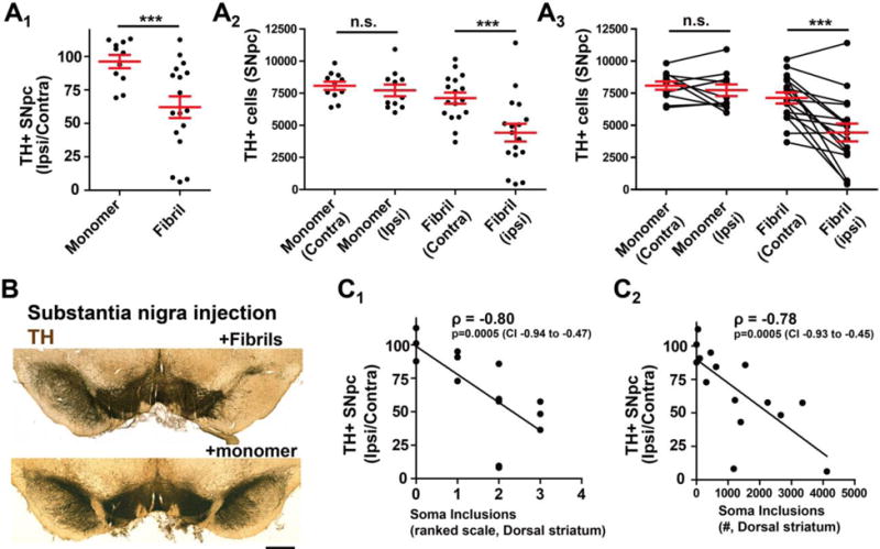
Proteinaceous inclusions in neurons, composed primarily of α-synuclein, define the pathology in several neurodegenerative disorders. Neurons can internalize α-synuclein fibrils that can seed new inclusions from endogenously expressed α-synuclein. The factors contributing to the spread of pathology and subsequent neurodegeneration are not fully understood, and different compositions and concentrations of fibrils have been used in different hosts. Here, we systematically vary the concentration and length of well-characterized α-synuclein fibrils and determine their relative ability to induce inclusions and neurodegeneration in different hosts (primary neurons, C57BL/6J and C3H/HeJ mice, and Sprague Dawley rats). Using dynamic-light scattering profiles and other measurements to determine fibril length and concentration, we find that femptomolar concentrations of fibrils are sufficient to induce robust inclusions in primary neurons. However, a narrow and non-linear dynamic range characterizes fibril-mediated inclusion induction in axons and the soma. In mice, the C3H/HeJ strain is more sensitive to fibril exposures than C57BL/6J counterparts, with more inclusions and dopaminergic neurodegeneration. In rats, injection of fibrils into the substantia nigra pars compacta (SNpc) results in similar inclusion spread and dopaminergic neurodegeneration as injection of the fibrils into the dorsal striatum, with prominent inclusion spread to the amygdala and several other brain areas. Inclusion spread, particularly from the SNpc to the striatum, positively correlates with dopaminergic neurodegeneration. These results define biophysical characteristics of α-synuclein fibrils that induce inclusions and neurodegeneration both in vitro and in vivo, and suggest that inclusion spread in the brain may be promoted by a loss of neurons.

Keywords: Aggregation; NACP; Parkinson disease; Prion; SNCA.


Figures

Figure 1
Figure 1. Electron microscopy characterization of α-syn fibrils after extensive sonication
A) Denaturing gel electrophoresis and Coomassie stain for volumetric sedimentation analysis of a pellet fraction (P100) or supernatant fraction (S100) of recombinant mouse α-syn fibrils, post-ultracentrifugation (100,000 × g for 1 hour). B) Non-denaturing blue-native PAGE, with the indicated sonication treatment time above each lane. Monomer α-syn migrated at ~ 56 kDa. C) Representative negative-stain transmission electron micrographs of unsonicated mouse α-syn fibrils, monomer, or fibrils sonicated for 30 sec, and up to 240 sec, as indicated. Scale bars are 50 nm. D–G) Histogram representation of more than 1,000 particles measured from each condition from randomly captured electron microscopy images. Selected sonication times, as indicated, are shown from representative preparations. Red bars represent group median, with mean and corresponding group standard deviation indicated in bold font. H) Summary graph of the effect of sonication on fibril length. Data points represent group means.
Figure 2
Figure 2. Prediction of α-syn fibril length and concentration by dynamic light scattering
A) Thioflavin binding assay of unsonicated (195 nm) and sonicated mouse α-syn fibrils, with equal α-syn mass (i.e., 5 μg in 100 μl) included in each condition. B) Fibril outgrowth assay based on recruitment of free monomer protein (mouse α-syn) with and without 29 nm seeds (mouse α-syn). Seeds were included at a ratio of 1:20 fibril to monomer (weight). Heavily sonicated fibrils retained the ability to seed new fibril growth as evidenced by thioflavin binding. C) Representative circular dichroism profiles of sonicated α-syn fibril preparations, with fibril profiles indistinguishable between each other, but readily distinguished from monomeric protein. D) Histograms depict dynamic light scattering profiles obtained for each sonicated α-syn fibril preparation, plotted relative to the intensity-distribution predicted molecular weight (Dyna V6.3.4 software). E) Relationship between the average in-solution molecular weight and the calculated molarity (light scattering profiles) of sonicated fibril preparations in a typical solution strength (i.e., 0.2 μg mL−1) used to study α-syn fibrils in cell culture experiments (e.g., Figure 3). F) Thioflavin binding over time, with monomer (mouse α-syn) included in reactions at 5 mg mL−1, with or without the addition of 1 mg mL−1 of the indicated fibrils (mouse α-syn). All results are representative of at least three independent experiments.
Figure 3
Figure 3. Femptomolar concentrations of α-syn fibrils template inclusions in the soma and axon in primary neurons
A) Denaturing PAGE and silver stain of rodent and human α-syn fibril preparations (49 nm fibrils, 30 sec sonication), with fibrils solubilized and boiled in SDS buffer prior to gel loading. The indicated amount of fibrils loaded to each gel is indicated. B–F) Representative wide-field fluorescence images gathered by automated microscopy from neurons in culture. Immunofluorescence depicts tau signal (red) in primary hippocampal neurons (DIV 21), treated with the indicated concentration of 49 nm human α-syn fibrils at DIV 7 (i.e., 14-day incubation). Green depicts signal from pS129-α-syn antibody. The concentration of endogenous α-syn in these cultures is given in Supplemental Figure 1, and results in α-syn knockout neurons (to control for pS129-α-syn staining unrelated to newly generated α-syn fibrils) is given in Supplemental Figure 2. Scale bar shows 40 μm.
Figure 4
Figure 4. α-Syn fibril concentration and length on templating new inclusions in primary neurons
A) Unbiased quantification of the percent occupancy of α-syn inclusions in tau-positive processes, caused by exposure to the indicated concentration of 49 nm mouse or human fibrils (as indicated). Control images from monomer α-syn treatments were used as a threshold for pS129-α-syn inclusion signal (background). B) Unbiased quantification of the percentage of NeuN bodies in each image that overlap with an pS129-α-syn inclusion. C,D) The average tau-area and number of NeuN bodies analyzed per data point for each group are given as column graphs. E) Unbiased quantification of the percent occupancy of pS129-α-syn inclusions in tau-positive processes, and F) soma inclusions, caused by the indicated (mouse) fibril preparation. G,H) The average tau-area and number of NeuN bodies analyzed per data point for each group in panels E and F are given as column graphs. Over one-hundred images were analyzed in at least three independent experiments for each group. * n.s. is not significant, * is p<0.05, ** p<0.01, *** p<0.001, as determined by one-way ANOVA with a Tukey’s post-hoc test. Supplemental Table 1 shows results of post-hoc tests of all groups.
Figure 5
Figure 5. Short α-syn fibrils induce inclusions in C57BL/6J spiny projection neurons
Adult (~P90) mice were unilaterally injected with the indicated mouse α-syn fibril preparation, or monomeric α-syn. Six months later, mice were sacrificed and the dorsal striatum was analyzed by immunohistochemistry for pS129-α-syn inclusions by DAB immunohistochemistry and by confocal microscopy. A–E) Representative lower magnification images in coronal dorsal striatum sections adjacent to the needle track of α-syn (mouse) fibril injection. Scale bars are 200 μm and 100 μm (Panel E). F–H) Higher magnification of individual NeuN-positive neurons or fibers in the striatum, with NeuN (Panel F) or tyrosine-hydroxylase (red, Panels G–J) staining. Scale bars are 40 (Panel F) and 20 μm. K,L) pS129-α-syn inclusions (green) and acetyl cholinesterase (red) in the dorsal striatum, scale bars are 100 and 20 μm, respectively. Results are representative of at least three mice analyzed per group.
Figure 6
Figure 6. Short α-syn fibrils induce pS129-α-syn inclusions in brain nuclei that project to the dorsal striatum
Adult (~P90) C57BL/6J mice were injected unilaterally in the dorsal striatum with 10 μg of the indicated mouse α-syn fibril preparation (195 nm or 29 nm), or 10 μg of monomer α-syn, and analyzed 6 months later by immunohistochemistry for pS129-α-syn and by confocal microscopy in brain nuclei that project to the dorsal striatum (i.e., amygdala, cortex [piriform], SNpc). A1–3) Representative immunohistochemistry (DAB, brown coloration for pS129-α-syn) in the ipsilateral amygdala, scale bars are 100 μm. A4) Representative triple-label confocal microscopy showing pS129-α-syn (green), DAPI (blue) and NeuN (red), in the amygdala. pS129-α-Syn inclusions could be detected both adjacent to NeuN-positive neuronal nuclei, as well as NeuN bodies. Scale bar is 50 μm. B1–3) Representative immunohistochemistry (DAB, brown coloration) in ipsilateral SNpc (highlighted by dashed lines). Scale bars 0.2 mm. B4–6) Quadruple-label confocal microscopy showing several tyrosine-hydroxylase (TH) positive neurons in the ipsilateral substantial nigra pars compacta, with pS129-α-syn (green), TH (red), NeuN (Blue) and DAPI (white). Panel B4 shows a NeuN-negative TH-positive neuron with a pS129-α-syn inclusion, B5 shows another NeuN negative neuron with an inclusion and a pyknotic nucleus at higher magnification, and B6 shows a TH-positive/NeuN positive neuron with much smaller pS129-α-syn inclusions. C1–3) Ipsilateral somatosensory cortex with pS129-α-syn immunohistochemistry (DAB, brown coloration), scale bars 100 μm. C4) Confocal microscopy with pS129-α-syn (green), and C5) NeuN (red), and DAPI (Blue). C4 shows a cross section (coronal) of the ipsilateral somatosensory cortex (scale bar is 100 μm). C5 shows two pyramidal neurons in the piriform cortex, one positive with an inclusion next to one without (scale bar is 25 μm). Results are representative of at least three mice analyzed per group.
Figure 7
Figure 7. Both long and short α-syn fibrils induce inclusions in C3H/HeJ mice
Adult (~P90) C3H/HeJ mice were injected unilaterally in the dorsal striatum with 10 μg of the indicated mouse α-syn fibril preparation (195 nm, 49 nm, or 29 nm), or 10 μg of monomer α-syn, and analyzed 6 months later by immunohistochemistry for pS129-α-syn in the A) dorsal striatum (scale bar 0.1 mm), B) amygdala (scale bar 0.1 mm), C) piriform cortex (scale bar 0.1 mm), and D) SNpc (highlighted by dashed lines, scale bar 0.2 mm). Representative immunohistochemistry (DAB, brown coloration for pS129-α-syn) in the ipsilateral nucleus is shown. Results are representative of at least three mice analyzed per group.
Figure 8
Figure 8. Short α-syn fibrils induce inclusions in Sprague Dawley rats
Adult (~P90) rats were unilaterally injected with 20 μg of the indicated rodent α-syn fibril preparation, or 20 μg of monomer α-syn. Six months later, rats were sacrificed and coronal sections were analyzed by immunohistochemistry for pS129-α-syn (DAB, brown coloration). A1–4) Representative images in sections adjacent to the needle track of the fibril injection. Scale bars are 200 μm. B1–4) Representative images in the ipsilateral somatosensory cortex (scale bars 100 μm), C1–4) SNpc (highlighted by dashed lines, scale bar is 200 μm), or D1–D4) amygdala (scale bar is 200 μm). Images from each brain nuclei are representative of at least three rats analyzed per group.
Figure 9
Figure 9. α-Syn inclusion burden correlates to the loss of dopaminergic neurons in the SNpc
Adult (~P90) mice (C57BL/6J and C3H/HeJ, as indicated) and Sprague Dawley rats were unilaterally injected in the dorsal striatum with 10 μg (mice) or 20 μg (rats) of fibrils (mouse) of the indicated composition of fibrils (195-29 nm, monomer controls included). Six months later, all rodents were sacrificed and coronal sections through the brain were analyzed by immunohistochemistry. A) Unbiased stereological counts of the number of soma pS129-α-syn inclusions through the SNpc. B) Observer-ranked pS129-α-syn inclusion burden. “Pathology score” in each brain region (piriform cortex, amygdala, thalamus) is assigned a rank of 0–5 (see Methods). C) Representative immunohistochemistry of the SNpc and VTA for TH reactivity. A light Nissl stain is in blue to identify the SNpc, scale bar is 0.5 mm. D) Comparison of stereological counts of inclusions throughout the SNpc (Panel A) to the amount of dopaminergic neurodegeneration measured in the same animal using stereological counts (see Methods). E) Ranked inclusion burden in the cortex, amygdala and thalamus, (Panel B) plotted against the amount of dopaminergic neurodegeneration. Linear trend line fits are presented along with Spearman rank correlation values in a 95% confidence interval. Error bars are S.E.M. Results are from at least three rodents analyzed per group.
Figure 10
Figure 10. α-Syn fibril injections in the rat SNpc induce robust inclusions
A) Adult (~P90) rats were unilaterally injected in the SNpc or dorsal striatum with 20 μg of 49 nm fibrils, and 6 months later, rats were sacrificed and coronal sections were analyzed by immunohistochemistry for pS129-α-syn (DAB, brown coloration). B1–5) Representative bright field images of brains injected in the striatum. Prominent pS129-α-syn pathology localizes near the injection site in the striatum (shown), in the amygdala, some inclusions in the SNpc, and widespread inclusions throughout the cortex. C) Injection directly in the SNpc produces fewer inclusions in the striatum (C1), a comparable inclusion load in the amygdala and SNpc (C2 and C3), and very few or no inclusions through much of the cortex (C4 and C5). Scale bars are 100 μm. Results are representative from over 20 rats analyzed.
Figure 11
Figure 11. Inclusion spread from the SNpc to striatal projection neurons correlates with dopaminergic neurodegeneration
Adult (~P90) rats were unilaterally injected in the SNpc with 20 μg of 49 nm fibrils, and 6 months later, rats were sacrificed and A) coronal sections were analyzed by immunohistochemistry for pS129-α-syn (DAB, brown coloration) and tyrosine-hydroxylase (TH, with Nissl, blue coloration). Unbiased stereological counts for Nissl+ neurons, defined by either TH expression or more rarely (e.g., <5% of total cells) Nissl-alone reactivity in large (>~40 μm) Nissl-positive bodies, as presented in column graphs that show mean and S.E.M. with red bars. Each data point represents a rat. A one-way ANOVA was performed (monomer and fibril rats, ipsi versus contra counts) and was significant (p<0.001), and a post-hoc analysis (Tukey’s) revealed the fibril treated ipsilateral SNpc is different from the other groups. B) Representative immunohistochemistry for the fibril-injected rat group (TH stain, brown, with a light Nissl stain, blue), scale bar is 0.5 mm. C) Scatterplot for a ranked-scale of striatum pathology (see Methods section) and stereological counts for soma inclusions scored through the rat striatum, compared to the amount of neurodegeneration (presented as ipsilateral counts over contralateral counts), with a linear trend line fit, and a Spearman rank correlation value shown in bold font with a corresponding 95% confidence interval.

References

    1. Ayers JI, et al. Robust Central Nervous System Pathology in Transgenic Mice following Peripheral Injection of alpha-Synuclein Fibrils. J Virol. 2017;91 - PMC - PubMed
    1. Bousset L, et al. An Efficient Procedure for Removal and Inactivation of Alpha-Synuclein Assemblies from Laboratory Materials. J Parkinsons Dis. 2016;6:143–51. - PMC - PubMed
    1. Brahic M, et al. Axonal transport and secretion of fibrillar forms of alpha-synuclein, Abeta42 peptide and HTTExon 1. Acta Neuropathol. 2016;131:539–48. - PMC - PubMed
    1. Fellner L, et al. Toll-like receptor 4 is required for alpha-synuclein dependent activation of microglia and astroglia. Glia. 2013;61:349–60. - PMC - PubMed
    1. Fitzpatrick AW, et al. Nanomechanics and intermolecular forces of amyloid revealed by four-dimensional electron microscopy. Proc Natl Acad Sci U S A. 2015;112:3380–5. - PMC - PubMed

MeSH terms