

Oral Vitamin D Rapidly Attenuates Inflammation from Sunburn: An Interventional Study
- PMID: 28576736
- PMCID: PMC5610950
- DOI: 10.1016/j.jid.2017.04.040
Oral Vitamin D Rapidly Attenuates Inflammation from Sunburn: An Interventional Study
Abstract
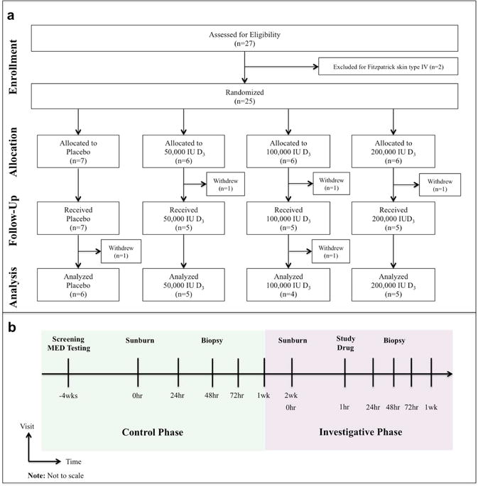
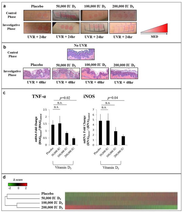
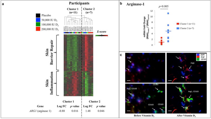
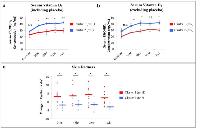
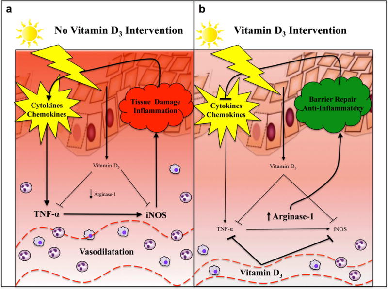
The diverse immunomodulatory effects of vitamin D are increasingly being recognized. However, the ability of oral vitamin D to modulate acute inflammation in vivo has not been established in humans. In a double-blinded, placebo-controlled interventional trial, 20 healthy adults were randomized to receive either placebo or a high dose of vitamin D3 (cholecalciferol) one hour after experimental sunburn induced by an erythemogenic dose of UVR. Compared with placebo, participants receiving vitamin D3 (200,000 international units) demonstrated reduced expression of proinflammatory mediators tumor necrosis factor-α (P = 0.04) and inducible nitric oxide synthase (P = 0.02) in skin biopsy specimens 48 hours after experimental sunburn. A blinded, unsupervised hierarchical clustering of participants based on global gene expression profiles revealed that participants with significantly higher serum vitamin D3 levels after treatment (P = 0.007) demonstrated increased skin expression of the anti-inflammatory mediator arginase-1 (P = 0.005), and a sustained reduction in skin redness (P = 0.02), correlating with significant expression of genes related to skin barrier repair. In contrast, participants with lower serum vitamin D3 levels had significant expression of proinflammatory genes. Together the data may have broad implications for the immunotherapeutic properties of vitamin D in skin homeostasis, and implicate arginase-1 upregulation as a previously unreported mechanism by which vitamin D exerts anti-inflammatory effects in humans.
Copyright © 2017 The Authors. Published by Elsevier Inc. All rights reserved.
Conflict of interest statement
Figures
Comment in
-
A vitamin to D-crease sunburn.Sci Transl Med. 2017 Jun 14;9(394):eaan5233. doi: 10.1126/scitranslmed.aan5233. Sci Transl Med. 2017. PMID: 28615360
-
Vitamin D Prevents Sunburn: Tips for the Summer?J Invest Dermatol. 2017 Oct;137(10):2045-2047. doi: 10.1016/j.jid.2017.07.840. J Invest Dermatol. 2017. PMID: 28941472 Free PMC article.
Similar articles
-
Vitamin D supplementation and systemic inflammation in relapsing-remitting multiple sclerosis.J Neurol. 2015 Dec;262(12):2713-21. doi: 10.1007/s00415-015-7902-5. Epub 2015 Oct 1. J Neurol. 2015. PMID: 26429571 Clinical Trial.
-
Effect of weekly high-dose vitamin D3 supplementation on serum cholecalciferol concentrations in pregnant women.J Steroid Biochem Mol Biol. 2016 Apr;158:76-81. doi: 10.1016/j.jsbmb.2016.01.007. Epub 2016 Jan 16. J Steroid Biochem Mol Biol. 2016. PMID: 26784272 Clinical Trial.
-
A randomized controlled trial of green tea catechins in protection against ultraviolet radiation-induced cutaneous inflammation.Am J Clin Nutr. 2015 Sep;102(3):608-15. doi: 10.3945/ajcn.115.107995. Epub 2015 Jul 15. Am J Clin Nutr. 2015. PMID: 26178731 Free PMC article. Clinical Trial.
-
Response of Vitamin D Concentration to Vitamin D3 Administration in Older Adults without Sun Exposure: A Randomized Double-Blind Trial.J Am Geriatr Soc. 2016 Jan;64(1):65-72. doi: 10.1111/jgs.13774. J Am Geriatr Soc. 2016. PMID: 26782853 Free PMC article. Clinical Trial.
-
Transdermal vitamin D supplementation-A potential vitamin D deficiency treatment.J Cosmet Dermatol. 2020 Jan;19(1):28-32. doi: 10.1111/jocd.13085. Epub 2019 Jul 25. J Cosmet Dermatol. 2020. PMID: 31343822 Review.
Cited by
-
Reply to Jakovac and to Rocha et al.: Can vitamin D prevent or manage COVID-19 illness?Am J Physiol Endocrinol Metab. 2020 Aug 1;319(2):E455-E457. doi: 10.1152/ajpendo.00348.2020. Am J Physiol Endocrinol Metab. 2020. PMID: 32787704 Free PMC article. No abstract available.
-
Nongenomic Activities of Vitamin D.Nutrients. 2022 Dec 1;14(23):5104. doi: 10.3390/nu14235104. Nutrients. 2022. PMID: 36501134 Free PMC article. Review.
-
Cholecalciferol-load films for the treatment of nasal burns caused by cauterization of the hypertrophied inferior turbinate: formulation, in vivo study, and clinical assessment.Drug Deliv Transl Res. 2023 Apr;13(4):1102-1115. doi: 10.1007/s13346-022-01275-7. Epub 2022 Dec 12. Drug Deliv Transl Res. 2023. PMID: 36509965 Clinical Trial.
-
Genetic and non-genetic effects of increased sun and vitamin D exposure: role in the observed healthy changes in cardiometabolic risk factors in Iranian children.Public Health Nutr. 2018 Dec;21(17):3125-3128. doi: 10.1017/S1368980018001180. Epub 2018 Jun 6. Public Health Nutr. 2018. PMID: 29871711 Free PMC article. No abstract available.
-
Characterization of the effects of vitamin D synthesis and sunburn in the population due to solar radiation exposure using PROBIT methodology.Heliyon. 2024 May 11;10(10):e30864. doi: 10.1016/j.heliyon.2024.e30864. eCollection 2024 May 30. Heliyon. 2024. PMID: 38784536 Free PMC article.
References
-
- Abd-El-Aleem SA, Ferguson MW, Appleton I, Kairsingh S, Jude EB, Jones K, et al. Expression of nitric oxide synthase isoforms and arginase in normal human skin and chronic venous leg ulcers. The Journal of pathology. 2000;191(4):434–42. - PubMed
-
- Baeke F, Takiishi T, Korf H, Gysemans C, Mathieu C. Vitamin D: modulator of the immune system. Current opinion in pharmacology. 2010;10(4):482–96. - PubMed
-
- Benjamini Y, Hochberg Y. Controlling the False Discovery Rate: A Practical and Powerful Approach to Multiple Testing. Journal of the Royal Statistical Society Series B (Methodological) 1995;57(1):289–300.
Publication types
MeSH terms
Substances
Grants and funding
LinkOut - more resources
Full Text Sources
Other Literature Sources
Medical
Molecular Biology Databases
Research Materials
