. 2017 Jul 1;144(13):2490-2503.
doi: 10.1242/dev.151951. Epub 2017 Jun 2.

Incompatibility between mitochondrial and nuclear genomes during oogenesis results in ovarian failure and embryonic lethality

Affiliations

Incompatibility between mitochondrial and nuclear genomes during oogenesis results in ovarian failure and embryonic lethality

Chunyang Zhang et al. Development. .

Abstract

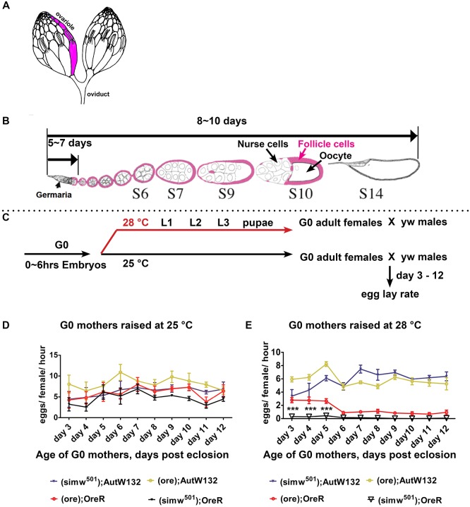
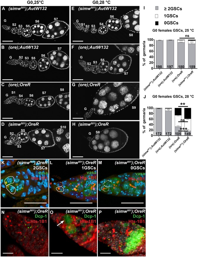
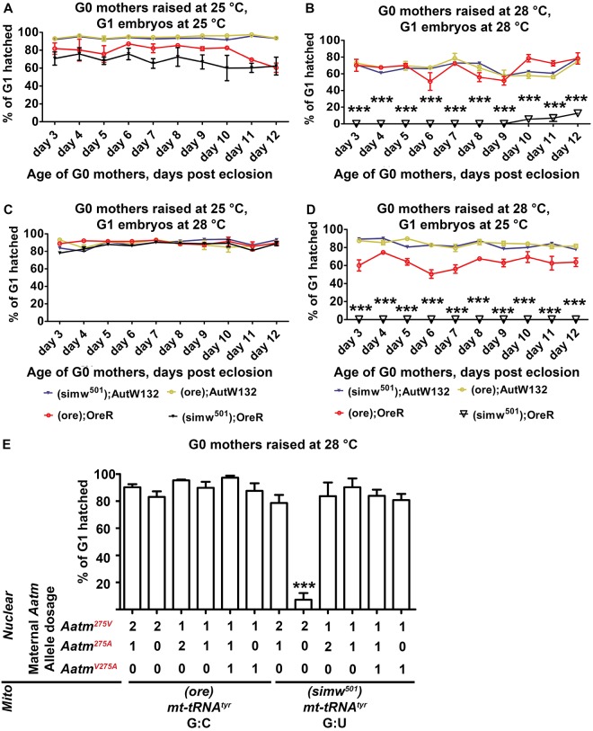
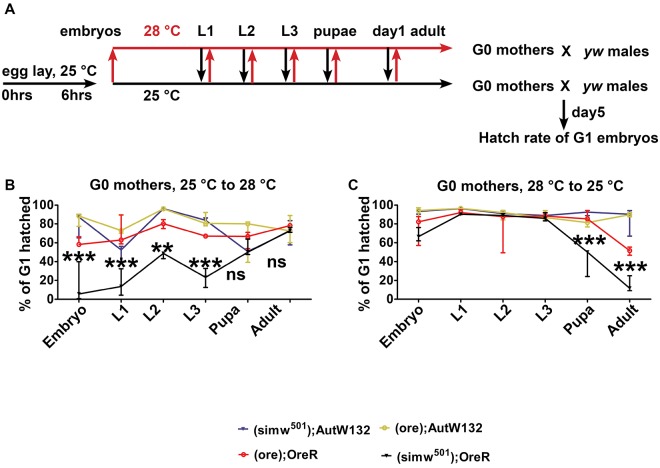
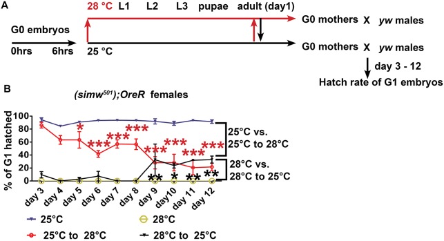
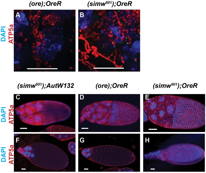
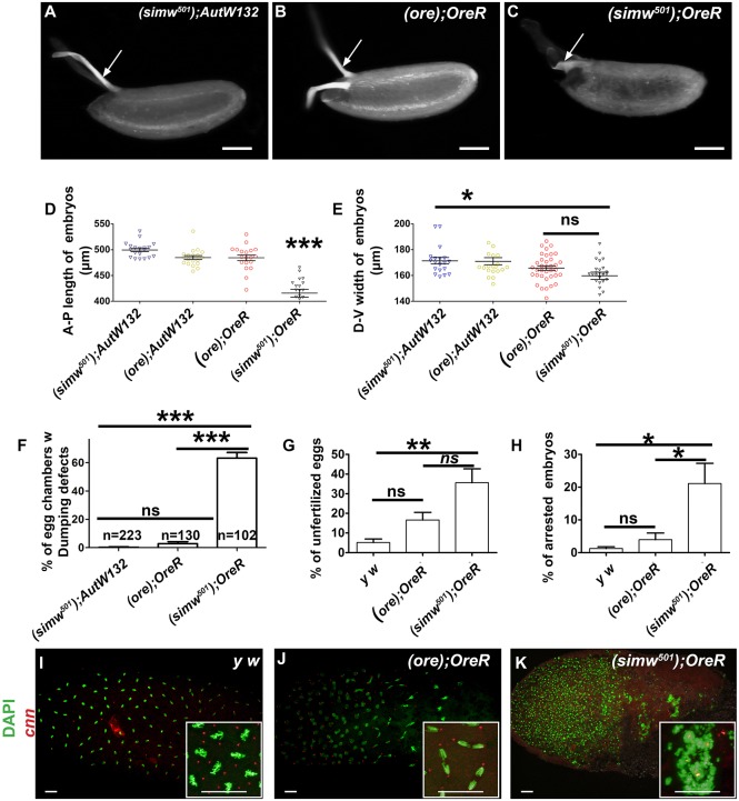
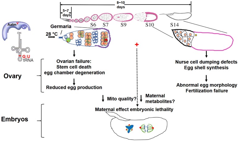
Mitochondrial dysfunction can cause female infertility. An important unresolved issue is the extent to which incompatibility between mitochondrial and nuclear genomes contributes to female infertility. It has previously been shown that a mitochondrial haplotype from D. simulans (simw501 ) is incompatible with a nuclear genome from the D. melanogaster strain Oregon-R (OreR), resulting in impaired development, which was enhanced at higher temperature. This mito-nuclear incompatibility is between alleles of the nuclear-encoded mitochondrial tyrosyl-tRNA synthetase (Aatm) and the mitochondrial-encoded tyrosyl-tRNA that it aminoacylates. Here, we show that this mito-nuclear incompatibility causes a severe temperature-sensitive female infertility. The OreR nuclear genome contributed to death of ovarian germline stem cells and reduced egg production, which was further enhanced by the incompatibility with simw501 mitochondria. Mito-nuclear incompatibility also resulted in aberrant egg morphology and a maternal-effect on embryonic chromosome segregation and survival, which was completely dependent on the temperature and mito-nuclear genotype of the mother. Our findings show that maternal mito-nuclear incompatibility during Drosophila oogenesis has severe consequences for egg production and embryonic survival, with important broader relevance to human female infertility and mitochondrial replacement therapy.

Keywords: Drosophila; Embryogenesis; Mitochondria; Mitochondrial-nuclear incompatibility; Oogenesis; Stem cell.


Conflict of interest statement

Competing interestsThe authors declare no competing or financial interests.

Figures

Fig. 1.
Fig. 1.
The (ore); OreR and (simw501); OreR females have a lower oviposition rate at a higher temperature. (A) An illustration of a pair of Drosophila ovaries with one ovariole indicated in pink. (B) A single ovariole with the developmental timeline of Drosophila oogenesis. Somatic follicle cells (pink) surround the germline nurse cells and oocyte to form an egg chamber. (C) Experimental scheme for the temperature-shift and female egg lay rate assay. (D,E) Oviposition rate of the indicated mito-nuclear females raised at 25°C (D) or 28°C (E) measured over 1 h. Fifty females per genotype, n=six biological replicates; data are mean±s.e.m. ***P<0.001 comparing (simw501); OreR with (ore); OreR using two-way ANOVA with Bonferroni correction. A and B are adapted, with the permission of the Genetics Society of America, from Ables (2015).
Fig. 2.
Fig. 2.
The OreR nuclear genotype contributes to a temperature-sensitive ovarian failure that is enhanced by simw501 mitochondria. Ovarioles labeled with DAPI from different mito-nuclear females at day 3 of adulthood raised at either 25°C (A-D) or 28°C (E-H). The germarium (G) and stages (S) of egg chamber maturation are indicated. Scale bars: 50 µm. (I,J) Quantification of the percentage of germaria with different numbers of germline stem cells (GSCs) from the indicated mito-nuclear females raised at either 25°C (I) or 28°C (J). The numbers on the bars represent the total number of germaria analyzed. Comparison of (ore); OreR and (simw501); OreR at 28°C, ***P<0.001 for two GSC, **P<0.01 for zero GSC, ns, non-significant for one GSC. Data are mean±s.e.m. (K-M) Germaria from (simw501); OreR females raised at 28°C with two (K), one (L) or zero (M) GSCs, labeled with antibodies against Hts (red) and Vasa (green) proteins, and DAPI (blue). Solid outlines: GSCs with Hts-labeled spherical spectrosomes. Dotted outlines: niche positions without a GSC. Scale bars: 20 µm. (N-P) Germline cell death in germaria from (simw501); OreR females raised at 28°C, labeled with antibodies against Hts (red) and cleaved caspase Dcp-1 (green). Images show germaria with no Dcp-1 labeling (N), a Dcp-1 labeled GSC (arrow) (O) or a Dcp-1 labeled 16 cell cyst (P). Scale bars: 10 µm.
Fig. 3.
Fig. 3.
A temperature-sensitive mito-nuclear incompatibility in the mother severely reduces embryonic hatch rate. (A-D) Embryonic hatch rate depends on the temperature of the (simw501); OreR mother. G0 mothers were raised at 25°C or 28°C and their G1 embryos allowed to develop at the same or reciprocal temperature. Embryonic hatch rates were measured for mothers of different ages post eclosion (x axis). Data are mean percentage of G1 hatched eggs±s.e.m. for three biological replicates. (A) Mothers at 25°C and embryos at 25°C. (B) Mothers at 28°C and embryos 28°C. (C) Mothers at 25°C and embryos at 28°C. (D) Mothers at 28°C and embryos 25°C. ***P<0.001 comparing (ore); OreR and (simw501); OreR. n=3. (E) The compatible AatmA allele in the mother rescues embryonic hatch rate. Hatch rates of embryos were measured from mothers with different mitochondria and doses of nuclear Aatm alleles, as indicated below the x axis. Mothers were at 28°C and embryos at 25°C. n=3 biological replicates; data are mean±s.e.m. (***P<0.001). See Fig. S5 for cross scheme.
Fig. 4.
Fig. 4.
The temperature-sensitive period in the mito-nuclear incompatible mothers begins at the L3-to-pupa transition. (A) Experimental scheme for the reciprocal temperature-shift experiments. The G0 females were shifted from 25°C to 28°C (red arrows) or from 28°C to 25°C (black arrows) during different larval stages (L), white pre-pupae (pupae) or day 1 of adulthood. The resulting G0 adult females were crossed to y w males, and on day five post-eclosion G1 embryos were collected, allowed to develop at 25°C, and hatch rate counted after 36 h of embryogenesis. (B) Hatch rate of G1 embryos when their G0 mothers were shifted from a permissive (25°C) up to a restrictive (28°C) temperature at the indicated developmental times. (C) Hatch rate of G1 embryos when their G0 mothers were shifted from a restrictive (28°C) to a permissive (25°C) temperature at the indicated developmental times. n=3 biological replicates; data are mean±s.e.m. **P<0.01 and ***P<0.001 for comparison between (ore); OreR and (simw501); OreR.
Fig. 5.
Fig. 5.
Adult temperature shifts have a delayed impact on the maternal effect. (A) (simw501); OreR females were raised at 25°C or 28°C, kept at the same temperature, or shifted to the reciprocal temperature on day 1 of adulthood, and then crossed to y w males. Embryos from these females were collected on days 3-12 of adulthood, allowed to develop at 25°C and hatch rates were measured at 36 h. (B) Hatch rates of embryos from (simw501); OreR mothers who were treated as described in A. n=3 biological replicates; data are mean±s.e.m.. Red asterisks represent P values for comparison of 25-28°C shift (red line) versus constant 25°C (blue line). Black asterisks represent P values for comparison of 28-25°C shift (black line) versus constant 28°C (gold line). *P<0.05; **P<0.01; ***P<0.001. n=3.
Fig. 6.
Fig. 6.
Mitochondrial abundance and morphology in the ovary. (A,B) Super resolution image of germaria labeled with anti-ATP5a and DAPI from (ore); OreR (A) and (simw501); OreR (B) females raised at 28°C. Scale bars: 5 µm. (C-H) Confocal images of a stage 10 (C-E) and stage 12-13 (F-H) egg chambers labeled with anti-ATP5a and DAPI from (simw501); AutW132 (C,F), (ore); OreR (D,G) and (simw501); OreR (E,H) females that were raised at 28°C. Scale bars: 50 µm.
Fig. 7.
Fig. 7.
The (simw501); OreR mito-nuclear incompatibility compromises egg morphology, fertilization and embryonic cell divisions. (A-C) Bright-field images of eggs laid by (simw501); AutW132 (A) (ore); OreR (B) and (simw501); OreR (C) females at 28°C. Eggs from (simw501); OreR females are shorter, with soft eggshells, and unusual dorsal appendage morphology (arrow). Scale bars: 100 µm. (D,E) Measurement of anterior-posterior length (D) and dorsal-ventral width (E) of ∼0-2 h embryos from mothers of the indicated genotypes at 28°C (*P<0.05, ***P<0.001. n=19 per genotype). (F) Percentage of stage 11-13 egg chambers with reduced transfer of nurse cell cytoplasm into oocyte. (G) Percentage of unfertilized embryos from mothers of the indicated genotypes at 28°C determined by anti-Cnn labeling (n=4, **P<0.01). (H) Percentage of fertilized embryos that were arrested by 8 h after egg lay (AEL). (n=4, *P<0.05). (I-K) Confocal images of embryos during nuclear cleavage cycles, 0-2 h AEL. Embryos are from mothers raised at 28°C and of the indicated genotype. Centrosomes are labeled with anti-Cnn antibody (red) and nuclear DNA is labeled with DAPI (green). The insets show higher magnifications of mitotic chromosome segregation. Scale bars for panels and insets: 20 µm.
Fig. 8.
Fig. 8.
Temperature-sensitive mito-nuclear incompatibility has pleiotropic effects on female fertility. At a higher temperature (28°C), the OreR nuclear genotype causes loss of GSCs, reduced egg chamber production and degeneration of egg chambers through apoptosis and autophagy, all of which are enhanced by incompatibility with simw501 mitochondria. These ovarian phenotypes together contribute to greatly decreased egg production. Temperature-sensitive mito-nuclear incompatibility in (simw501); OreR females caused unique nurse cell dumping and eggshell synthesis defects during later oogenesis. Below: temperature-sensitive mito-nuclear incompatibility in (simw501); OreR mothers resulted in mitotic errors in their embryos, which may be caused by insufficient maternal metabolites or maternal inheritance of sub-functional mitochondria.

References

    1. Ables E. T. (2015). Drosophila oocytes as a model for understanding meiosis: an educational primer to accompany “corolla is a novel protein that contributes to the architecture of the synaptonemal complex of Drosophila”. Genetics 199, 17-23. 10.1534/genetics.114.167940 - DOI - PMC - PubMed
    1. Andersen D. and Horne-Badovinac S. (2016). Influence of ovarian muscle contraction and oocyte growth on egg chamber elongation in Drosophila. Development 143, 1375-1387. 10.1242/dev.131276 - DOI - PMC - PubMed
    1. Belles X. and Piulachs M.-D. (2015). Ecdysone signalling and ovarian development in insects: from stem cells to ovarian follicle formation. Biochim. Biophys. Acta 1849, 181-186. 10.1016/j.bbagrm.2014.05.025 - DOI - PubMed
    1. Benkhalifa M., Ferreira Y. J., Chahine H., Louanjli N., Miron P., Merviel P. and Copin H. (2014). Mitochondria: participation to infertility as source of energy and cause of senescence. Int. J. Biochem. Cell Biol. 55, 60-64. 10.1016/j.biocel.2014.08.011 - DOI - PubMed
    1. Bentov Y., Yavorska T., Esfandiari N., Jurisicova A. and Casper R. F. (2011). The contribution of mitochondrial function to reproductive aging. J. Assist. Reprod. Genet. 28, 773-783. 10.1007/s10815-011-9588-7 - DOI - PMC - PubMed

Publication types

MeSH terms

LinkOut - more resources