. 2017 Aug 15;220(Pt 16):2932-2938.
doi: 10.1242/jeb.154484. Epub 2017 Jun 2.

miR-210 expression is associated with methionine-induced differentiation of trout satellite cells

Affiliations

miR-210 expression is associated with methionine-induced differentiation of trout satellite cells

Mary Latimer et al. J Exp Biol. .

Abstract

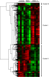
In fish, data on microRNAs (miRNAs) involved in myogenesis are scarce. In order to identify miRNAs involved in satellite cell differentiation, we used a methionine depletion/replenishment protocol to synchronize myogenic cell differentiation. Our results validated that methionine removal (72 h) from the medium strongly decreased myoD1 and myogenin expression, indicating differentiation arrest. In contrast, methionine replenishment rescued expression of myoD1 and myogenin, showing a resumption of differentiation. We performed a miRNA array analysis of myogenic cells under three conditions: presence of methionine for 72 h (control), absence of methionine for 72 h (Meth-) and absence of methionine for 48 h followed by 24 h of methionine replenishment (Meth-/+). A clustering analysis identified three clusters: cluster I corresponds to miRNA upregulated only in Meth-/+ conditions; cluster II corresponds to miRNA downregulated only in Meth-/+ conditions; cluster III corresponds to miRNAs with high expression in control, low expression in Meth- conditions and intermediate expression after methionine replenishment (Meth-/+). Cluster III was very interesting because it fitted with the data obtained for myoD1 and myogenin (supporting an involvement in differentiation) and contained seven miRNAs with muscle-related function (e.g. miR-133a) and one (miR-210) with unknown function. Based on our previously published miRNA repertoire ( Juanchich et al., 2016), we confirmed miR-133a was expressed only in white muscle and showed that miR-210 had strong expression in white muscle. We also showed that miR-210 expression was upregulated during differentiation of satellite cells, suggesting that miR-210 was potentially involved in the differentiation of satellite cells.

Keywords: Methionine depletion; Myo-miR; Myoblast; Myogenesis; miRNA.


Conflict of interest statement

Competing interestsThe authors declare no competing or financial interests.

Figures
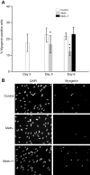
Fig. 1.
Fig. 1.
Immunocytochemical staining of cells for myogenin. Cells were assigned to one of three treatments: presence of methionine for 72 h (control), absence of methionine for 72 h (Meth−) and absence of methionine for 48 h followed by methionine replenishment for 24 h (Meth−/+). (A) Percentage of mygenin-positive cells. (B) Immunofluorescence analysis of myogenin expression. *Significant difference (one-way ANOVA, P<0.05; N=4).
Fig. 2.
Fig. 2.
mRNA expression of myogenin and myoD following methionine restriction in vitro. Data (means±s.d.) are for cells grown under control, Meth− and Meth−/+ conditions (see Fig. 1). mRNA levels were estimated using qPCR and normalized to levels of 18S mRNA. (A) Myogenin expression. (B) MyoD expression. *Significant difference (one-way ANOVA, P<0.05; N=6).
Fig. 3.
Fig. 3.
Hierarchical clustering of differentially expressed microRNA (miRNA) following methionine restriction. Each row represents the expression pattern of miRNA and each column corresponds to a single sample from cells grown under control, Meth− and Meth−/+ conditions (see Fig. 1). The expression levels are represented by different colors, with red indicating the highest levels of expression and green the lowest.
Fig. 4.
Fig. 4.
Expression of miR-133a and miR-210 following methionine restriction and during satellite cell differentiation. For expression studies (A,B,D,E), cells were grown under control, Meth− and Meth−/+ conditions (see Fig. 1). For kinetics of differentiation (C,F), cells were grown in proliferation medium (PM) and then in differentiation medium for 3 days (DM). Data (means+s.d.) were extracted from the microarray (A,D) or obtained by qPCR (B,C,E,F). Different letters indicate a significant difference between treatments (one-way ANOVA and Tukey's multiple comparisons test, P<0.05; N=6).

References

    1. Abdullah A. I., Zhang H., Nie Y., Tang W. and Sun T. (2016). CDK7 and miR-210 co-regulate cell-cycle progression of neural progenitors in the developing neocortex. Stem Cell Rep. 7, 69-79. 10.1016/j.stemcr.2016.06.005 - DOI - PMC - PubMed
    1. Bower N. I. and Johnston I. A. (2009). Selection of reference genes for expression studies with fish myogenic cell cultures. BMC Mol. Biol. 10, 80 10.1186/1471-2199-10-80 - DOI - PMC - PubMed
    1. Cardinali B., Castellani L., Fasanaro P., Basso A., Alemà S., Martelli F. and Falcone G. (2009). Microrna-221 and microrna-222 modulate differentiation and maturation of skeletal muscle cells. PLoS ONE 4, e7607 10.1371/journal.pone.0007607 - DOI - PMC - PubMed
    1. Chan Y. C., Banerjee J., Choi S. Y. and Sen C. K. (2012). miR-210: the master hypoxamir. Microcirculation 19, 215-223. 10.1111/j.1549-8719.2011.00154.x - DOI - PMC - PubMed
    1. Chargé S. B. and Rudnicki M. A. (2004). Cellular and molecular regulation of muscle regeneration. Physiol. Rev. 84, 209-238. 10.1152/physrev.00019.2003 - DOI - PubMed

Publication types