. 2017 Aug;234(2):217-232.
doi: 10.1530/JOE-17-0250. Epub 2017 Jun 2.

Origin of a rapidly evolving homeostatic control system programming testis function

Affiliations

Origin of a rapidly evolving homeostatic control system programming testis function

Pengli Bu et al. J Endocrinol. 2017 Aug.

Abstract

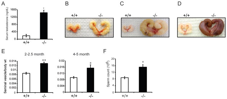
Mammals share common strategies for regulating reproduction, including a conserved hypothalamic-pituitary-gonadal axis; yet, individual species exhibit differences in reproductive performance. In this report, we describe the discovery of a species-restricted homeostatic control system programming testis growth and function. Prl3c1 is a member of the prolactin gene family and its protein product (PLP-J) was discovered as a uterine cytokine contributing to the establishment of pregnancy. We utilized mouse mutagenesis of Prl3c1 and revealed its involvement in the regulation of the male reproductive axis. The Prl3c1-null male reproductive phenotype was characterized by testiculomegaly and hyperandrogenism. The larger testes in the Prl3c1-null mice were associated with an expansion of the Leydig cell compartment. Prl3c1 locus is a template for two transcripts (Prl3c1-v1 and Prl3c1-v2) expressed in a tissue-specific pattern. Prl3c1-v1 is expressed in uterine decidua, while Prl3c1-v2 is expressed in Leydig cells of the testis. 5'RACE, chromatin immunoprecipitation and DNA methylation analyses were used to define cell-specific promoter usage and alternative transcript expression. We examined the Prl3c1 locus in five murid rodents and showed that the testicular transcript and encoded protein are the result of a recent retrotransposition event at the Mus musculus Prl3c1 locus. Prl3c1-v1 encodes PLP-J V1 and Prl3c1-v2 encodes PLP-J V2. Each protein exhibits distinct intracellular targeting and actions. PLP-J V2 possesses Leydig cell-static actions consistent with the Prl3c1-null testicular phenotype. Analysis of the biology of the Prl3c1 gene has provided insight into a previously unappreciated homeostatic setpoint control system programming testicular growth and function.

Keywords: Leydig cells; prolactin family; testis; transposable elements.


Conflict of interest statement

Declaration of interest: There is no conflict of interest that could be perceived as prejudicing the impartiality of the research reported.

Figures

Fig. 1
Fig. 1. Prl3c1 mutant mice exhibit an abnormal male reproductive tract phenotype
A, Schematic of the mouse Prl3c1 locus and the targeting construct. B, PCR analysis of genomic Prl3c1 locus from wild type (+/+), heterozygous (+/−) and null (−/−) animals. C, Gross appearance of the scrotal area on postnatal day 21 of wild type (+/+) and Prl3c1 null (−/−) mice. D, Gross appearance of adult testis of wild type and Prl3c1 null mice. E, Testis-to-body weight ratio of wild type (2–2.5 months, n=14; 4–5 months, n=8) and Prl3c1 null (2–2.5 months, n=20; 4–5 months, n=15) mice. F, CYP11A1 immunofluorescence staining of testis cross-sections from wild type and Prl3c1 null mice. G, Quantification of CYP11A1 positive staining area/total cross-section area of wild type and Prl3c1 null testes, n=5 testes per group. H–M, MKI67 immunofluorescence staining of testis cross-sections from wild type and Prl3c1 null mice. Scale bar: 50 μm. *P<0.05; **P<0.001.
Fig. 2
Fig. 2. Assessment of Leydig cell developmental states in wild type and Prl3c1 null mice
A, qRT-PCR analysis of transcripts associated with the progenitor Leydig cell state in wild type postnatal day 15 (PND 15), adult (2.5–4 months) wild type and Prl3c1 null testes. PND15 represents a transition point from progenitor to adult Leydig cells. qRT-PCR analysis of transcripts for Prl3c1 (B) and transcripts encoding proteins involved in steroid hormone biosynthesis (B, Hsd3b, Srd5a2, and Srd5a3; C, Star, Cyp11a1, Cyp17a1, and Hsd17b3) in adult wild type and Prl3c1 null testes (n=6; *P<0.05, **P<0.001).
Fig. 3
Fig. 3. Measures of testicular function in wild type and Prl3c1 null mice
A, Serum testosterone levels from wild type (n=8) and Prl3c1 null (n=10) male mice (3.5–4.5 months). B, Gross appearance of seminal vesicles from 4–5 month wild type and Prl3c1 null mice. C and D, Gross appearance of seminal vesicles from 11–12 month wild type and Prl3c1 null mice. E, Seminal vesicle-to-body weight ratio of wild type (2–2.5 months, n=14; 4–5 months, n=8) and Prl3c1 null (2–2.5 months, n=20; 4–5 months, n=15) mice. F, Total sperm count of wild type and Prl3c1 null mice (n=5 per genotype). Scale bar: 50 μm. *P<0.05; **P<0.001.
Fig. 4
Fig. 4. Prl3c1 transcript and the PLP-J protein are expressed in Leydig cells of the testis
A, Tissue survey (RT-PCR analysis) for Prl3c1 transcripts in various tissues from male C57BL/6 mice. B, RT-PCR analysis for Prl3c1 transcripts in testicular interstitium and seminiferous tubule compartments. Insl3 and Dmrt1 were included as controls for the interstitium and seminiferous tubule compartments, respectively. C, Immunohistochemical (IHC) staining for PLP-J on tissue sections from wild type testis. D, The Prl3c1 locus is transcriptionally active in Leydig cells of mice with β-galactosidase (LacZ) knocked into the Prl3c1 locus. Left panel: LacZ reporter activity; middle panel: immunofluorescence staining for CYP11A1; right panel: merged image of LacZ and CYP11A1 staining. LacZ reporter activity was localized to CYP11A1 positive Leydig cells in mice. Scale bar: 250 μm.
Fig. 5
Fig. 5. Ontogeny of Prl3c1 and steroidogenic enzyme transcripts in testes of wild type mice
A, RT-PCR analysis of Prl3c1 transcript expression in testes from postnatal days 5 to 45 (n=3 per day). Positive control (P.C.) is adult testis. Insl3 and Gapdh transcript analyses were used as internal controls. B and C, qRT-PCR measurements of transcripts for Prl3c1 (B) and transcripts encoding proteins involved in steroid hormone biosynthesis (B, Hsd3b and Srd5a2; C, Hsd17b3, Hsd3b1, Srd5a3) in testes at postnatal days 15 (D15), 21 (D21), and 45 (D45) (n=6, *P<0.05, ** P<0.001, compared to D15).
Fig. 6
Fig. 6. The testicular homeostatic setpoint control system includes the pituitary
A, Serum LH levels in wild type (n=8) and Prl3c1 null (n=10) adult male mice. *P<0.05. B, Lhb transcript levels in pituitaries of wild type (n=5) and Prl3c1 null (n=7) adult male mice analyzed by qRT-PCR. **P<0.001. C, Serum FSH levels in wild type (n=18) and Prl3c1 null (n=18) adult male mice. D, Fshb transcript levels in pituitaries of wild type (n=5) and Prl3c1 null (n=7) adult male mice analyzed by qRT-PCR. **P<0.001.
Fig. 7
Fig. 7. The Prl3c1 gene is a template for two transcripts with distinct tissue expression patterns
A, Prl3c1 transcript-specific RT-PCR analysis revealed tissue-restricted expression patterns for Prl3c1-v1 (decidua) and Prl3c1-v2 (testis). B, ChIP-qPCR analysis for RNA POL II at regions proximal to Exons 1a of Prl3c1-v1 (V1) and 1b of Prl3c1-v2 (V2) in decidua and Leydig cells. Locations of primer sets used in ChIP-qPCR are depicted as arrowheads shown in panel C (n=4, *P<0.05 compared to IgG control). C, Schematic representation of transcription start sites for Prl3c1-v1 (decidua) and Prl3c1-v2 (Leydig cells) as determined by 5′RACE (see also Supplementary Fig. 1A,B).
Fig. 8
Fig. 8. Origin and species specificity of the testicular homeostatic setpoint control system
A, RT-PCR analysis for Prl3c1 transcripts with primers targeting various exons of the rat Prl3c1 gene in testes of Fischer 344 (F344), Holtzman Sprague Dawley (HSD) and Brown Norway (BN) rats. Decidua tissue (HSD, gestation day 7.5) was included as a positive control. B, PCR amplification of the first intron of the Prl3c1 gene from the mouse (C57BL/6) and rat (BN). Schematic diagram of exon-intron structure indicates the locations of the primers used for amplification. C, Schematic comparison of the mouse and rat Prl3c1 genes reveals a longer first intron in the mouse due to the insertion of a composite transposable element (TE), which contains Exon 1b of Prl3c1-v2. D, PCR amplification of the first introns of the Prl3c1 genes from M. musculus, M. spretus, M. caroli, M. pahari, and R. norvegicus. E, Schematic representations of the first introns of the Prl3c1 genes from M. musculus, M. spretus, M. caroli, M. pahari, and R. norvegicus. Sequence identity comparisons are with the M. musculus sequence. F, Phylogenetic tree based on the nucleotide sequences for the first introns of Prl3c1 genes from M. musculus, M. spretus, M. caroli, M. pahari, and R. norvegicus. G, Primer validation for M. caroli and M. pahari. Schematic diagram indicates the locations of the two sequence-specific primer sets used for detection of Prl3c1 transcripts in testes from M. caroli and M. pahari. Primer sequences were based on partially sequenced coding regions of M. caroli (GenBank Accession No., KJ125427) and M. pahari (GenBank Accession No., KJ125428) Prl3c1 genes and validated by PCR amplification of corresponding Prl3c1 genomic regions with genomic DNA from these species. H, RT-PCR analysis for Prl3c1 transcripts in testes of M. musculus, M. spretus, M. caroli, and M. pahari. Two sets of sequence specific primers were used for M. caroli and M. pahari. I, Annotated schematic of the first intron of Prl3c1 from M. musculus and M. caroli. Note the prominent differences in the organization of RMER17D in M. musculus versus M. caroli.
Fig. 9
Fig. 9. Intracellular distribution and distinct functions of PLP-J V1 versus PLP-J V2
FLAG-tagged PLP-J V1 and FLAG-tagged PLP-J V2 were ectopically expressed in MLTC-1 Leydig cells or COS7 cells (1 μg/ml). Cells transfected with an empty vector were used as a control (1 μg/ml). A, Subcellular fractionation followed by western blotting for FLAG (Cyto: cytoplasm; Nu: nucleus). Western blotting for GAPDH and Histone H3 were used to monitor the integrity of the cytoplasmic and nuclear preparations, respectively. B, Effects of ectopic expression of Prl3c1-v1 (left panel) or Prl3c1-v2 (right panel) on MLTC-1 Leydig cell numbers following 72 h of culture. Cells were cultured in 96 well plates with 0.1 ml of medium/well (n=6, **P<0.001 compared to empty vector control). C, Representative images of MLTC-1 cells transfected with empty vector, Prl3c1-v1, or Prl3c1-v2 (1 μg/ml) following 72 h of culture.

Similar articles

Cited by

References

    1. Achermann JC, Jameson JL. Fertility and infertility: genetic contributions from the hypothalamic-pituitary-gonadal axis. Molecular Endocrinology. 1999;13:812–818. - PubMed
    1. Ain R, Dai G, Dunmore JH, Godwin AR, Soares MJ. A prolactin family paralog regulates reproductive adaptations to a physiological stressor. Proceedings of the National Academy of Sciences USA. 2004;101:16543–16548. - PMC - PubMed
    1. Alam SMK, Konno T, Dai G, Lu L, Wang D, Dunmore JH, Godwin AR, Soares MJ. A uterine decidual cell cytokine ensures pregnancy-dependent adaptations to a physiological stressor. Development. 2007;134:407–415. - PubMed
    1. Alam SM, Konno T, Sahgal N, Lu L, Soares MJ. Decidual cells produce a heparin-binding prolactin family cytokine with putative intrauterine regulatory actions. Journal of Biological Chemistry. 2008;283:18957–18968. - PMC - PubMed
    1. Amory JD, Bremner W. Endocrine regulation of testicular function in men: implications for contraceptive development. Molecular and Cellular Endocrinology. 2001;182:175–179. - PubMed

LinkOut - more resources