. 2017 Aug 3;130(5):666-676.
doi: 10.1182/blood-2017-02-765206. Epub 2017 Jun 2.

Genome editing of factor X in zebrafish reveals unexpected tolerance of severe defects in the common pathway

Affiliations

Genome editing of factor X in zebrafish reveals unexpected tolerance of severe defects in the common pathway

Zhilian Hu et al. Blood. .

Abstract

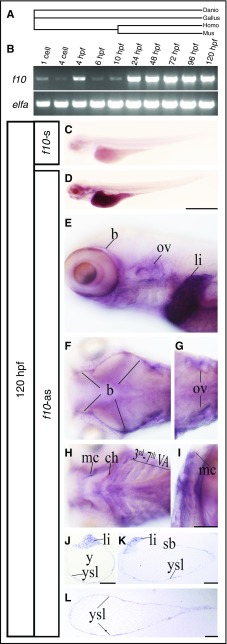
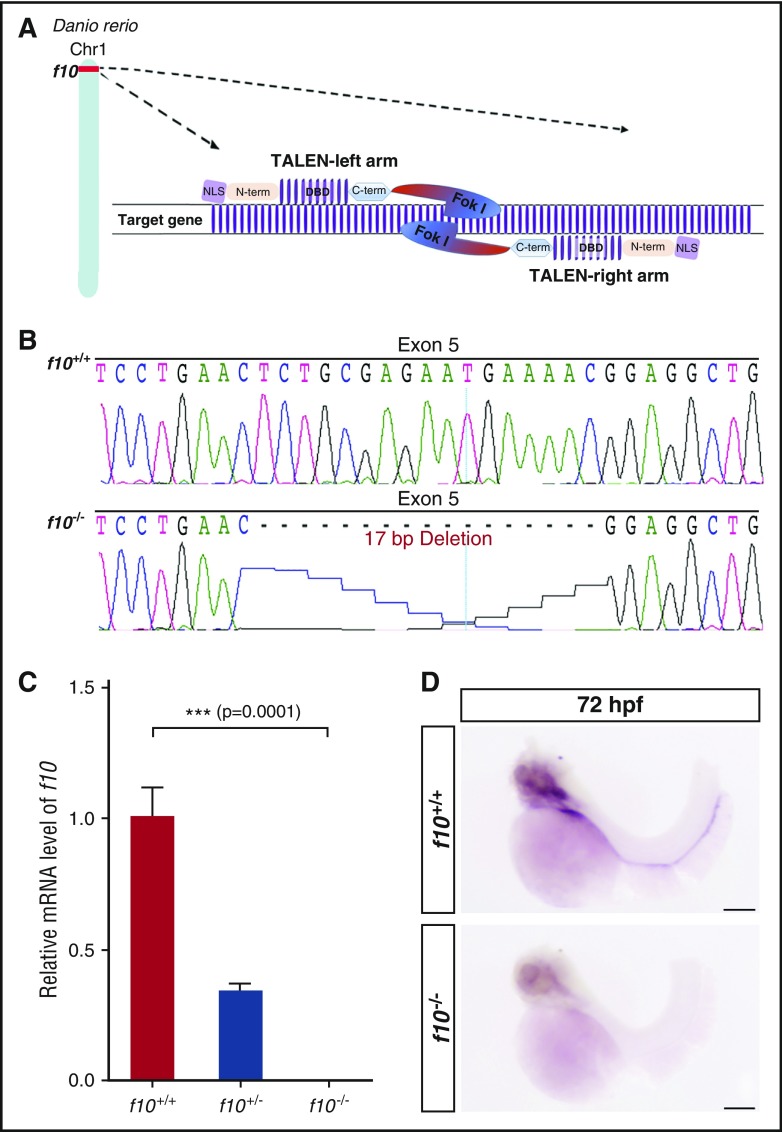
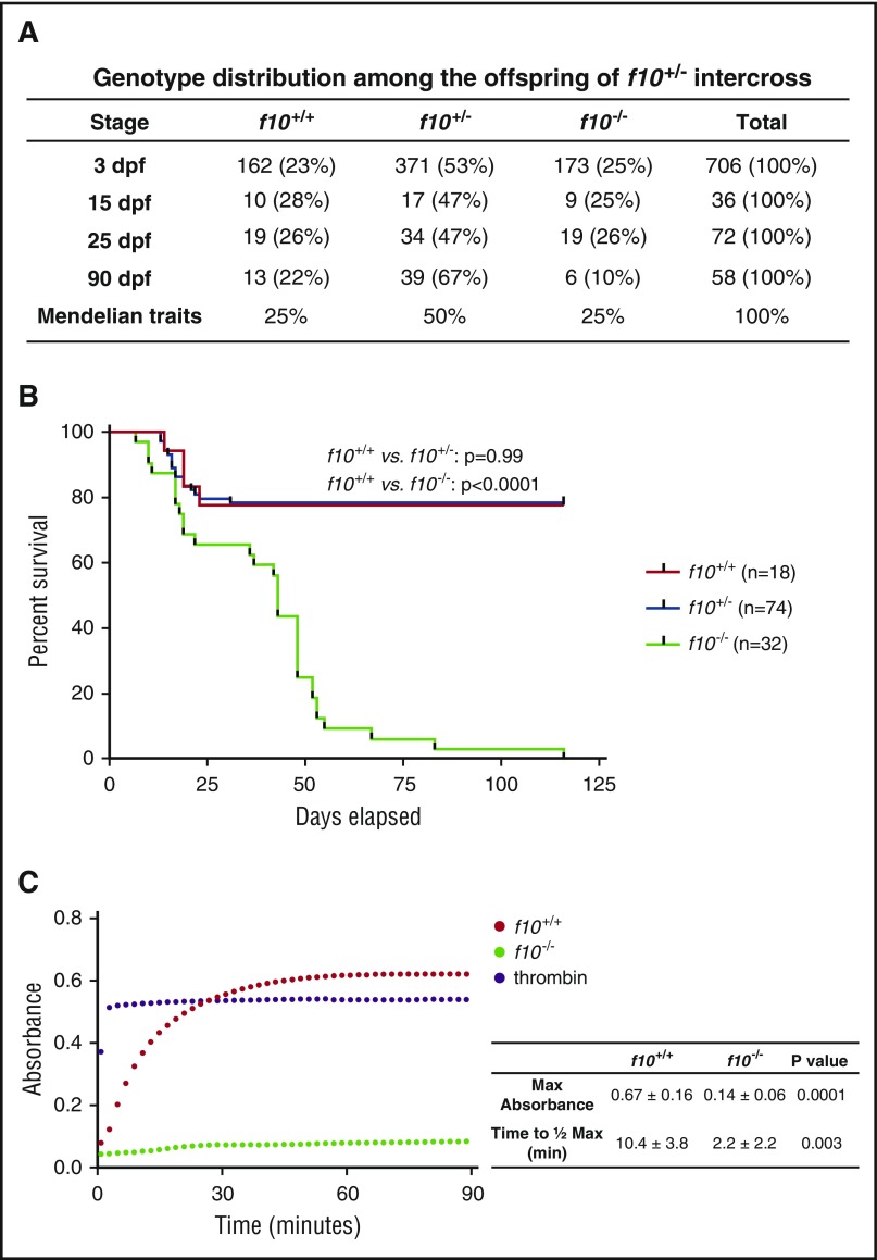
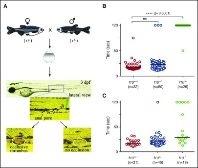
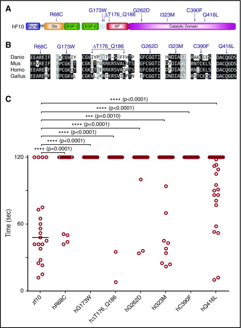
Deficiency of factor X (F10) in humans is a rare bleeding disorder with a heterogeneous phenotype and limited therapeutic options. Targeted disruption of F10 and other common pathway factors in mice results in embryonic/neonatal lethality with rapid resorption of homozygous mutants, hampering additional studies. Several of these mutants also display yolk sac vascular defects, suggesting a role for thrombin signaling in vessel development. The zebrafish is a vertebrate model that demonstrates conservation of the mammalian hemostatic and vascular systems. We have leveraged these advantages for in-depth study of the role of the coagulation cascade in the developmental regulation of hemostasis and vasculogenesis. In this article, we show that ablation of zebrafish f10 by using genome editing with transcription activator-like effector nucleases results in a major embryonic hemostatic defect. However, widespread hemorrhage and subsequent lethality does not occur until later stages, with absence of any detectable defect in vascular development. We also use f10-/- zebrafish to confirm 5 novel human F10 variants as causative mutations in affected patients, providing a rapid and reliable in vivo model for testing the severity of F10 variants. These findings as well as the prolonged survival of f10-/- mutants will enable us to expand our understanding of the molecular mechanisms of hemostasis, including a platform for screening variants of uncertain significance in patients with F10 deficiency and other coagulation disorders. Further study as to how fish tolerate what is an early lethal mutation in mammals could facilitate improvement of diagnostics and therapeutics for affected patients with bleeding disorders.


Conflict of interest statement

Conflict-of-interest disclosure: J.A.S. has been a consultant for Bayer, Shire, CSL Behring, Grifols, and Octopharma. F.P. has received honoraria for participating as a speaker at satellite symposia and educational meetings organized by Bayer, Biotest, CSL Behring, Grifols, Novo Nordisk, and Sobi; is the recipient of research grant funding from Alexion, Biotest, Kedrion Biopharma, and Novo Nordisk paid to Fondazione Luigi Villa; and has received consulting fees from Kedrion Biopharma, LFB, and Octapharma. She is member of the Ablynx scientific advisory board. J.K.J. has financial interests in Beacon Genomics, Beam Therapeutics, Editas Medicine, Poseida Therapeutics, and Transposagen Biopharmaceuticals. J.K.J.’s interests were reviewed and are managed by Massachusetts General Hospital and Partners HealthCare in accordance with their conflict-of-interest policies. The remaining authors declare no competing financial interests.

Figures

Figure 1.
Figure 1.
Spatiotemporal expression of f10 in the developing zebrafish. (A) Phylogenetic tree of F10 from zebrafish (Danio), mouse (Mus), chicken (Gallus), and human (Homo). (B) RT-PCR from whole embryos demonstrates that zebrafish f10 mRNA is expressed during early embryonic development beginning at the 1-cell stage. Expression is relatively stable from 24 through 120 hpf. The expression of elongation factor 1-α (elfa) was used as an internal control. (C-L) WISH analysis of 120-hpf larvae shows that f10 expression is strong in the liver (D), but relatively weak in the yolk syncytial layer and brain (D-F), otic vesicles (E, G), and arches (H-I) by using an antisense probe (f10-as). (C) A sense control (f10-s) did not show any expression. (J-L) Plastic sections (5 µm) of stained larvae in transverse (J), sagittal (K), and coronal (L) planes are shown. Anterior is toward the left in panels C-H and toward the top in panel I. Scale bars (C-L), 100 µm. b, brain; ch, ceratohyal [2nd visceral pharyngeal arch (VA)]; li, liver; mc, mandibular cartilage (1st VA); ov, otic vesicle; sb, swim bladder; y, yolk; ysl, yolk syncytial layer.
Figure 2.
Figure 2.
Genome editing of f10 using TALENs results in a frameshift and null allele. (A) Schematic diagram of TALENs used for targeted mutagenesis of f10. (B) Targeting of f10 exon 5 using a TALEN resulted in frameshift mutations. The 17-bp deletion mutant used for subsequent studies is shown. (C) Expression of f10 mRNA is reduced in heterozygous and undetectable in homozygous mutants, as evaluated by RT-PCR (each genotype was evaluated in triplicate). (D) Whole-mount in situ hybridization with an antisense probe demonstrates absence of expression in f10−/− mutants. Anterior is toward the left, and dorsal is toward the top. Scale bar, 100 µm. Chr1, chromosome 1; C-term, C-terminal domain; DBD, DNA-binding domain; Fok I, Fok I nuclease; NLS, nuclear localization signal; N-term, N-terminal domain.
Figure 3.
Figure 3.
Complete loss of F10 results in progressive adult lethality. (A) Genotype distributions of offspring from separate f10+/− incrosses evaluated at various stages demonstrate loss of homozygous mutants after 25 dpf. (B) Survival curves of closely monitored clutches of zebrafish offspring from f10+/− incrosses demonstrate progressive loss of 75% of homozygotes by 50 dpf and 100% by 115 dpf. There was no significant loss of heterozygotes (P > .05 by log-rank testing). Larvae were genotyped at 3 dpf and selected individuals were observed daily. There was ∼20% background loss of individual fish across all genotypes up to 20 dpf, which is typical during wild-type fish development. (C) Citrated plasmas from 1-month-old fish were recalcified and incubated with human fibrinogen for 90 minutes and absorbance (405 nm) was measured every 2 minutes. Data shown are the average of 2 experiments (n = 5 total pairs of fish for each genotype). The average time to half-maximal absorbance and maximum absorbance were calculated for each genotype and data were analyzed by using the Student t test. Bovine thrombin was used as a positive control. Max, maximum; min, minutes.
Figure 4.
Figure 4.
Loss of F10 results in late-onset hemorrhage at multiple sites. (A) Grossly visible hemorrhaging occurred in f10−/− mutant fish as early as 27 dpf, but not in wild-type or heterozygous siblings. Massive hemorrhages were observed in the brain, muscle, gill filaments, and abdomen as shown in viable f10−/− mutants. (B) Hematoxylin and eosin stained histologic sections of f10−/− mutants confirmed substantial intracranial, abdominal, and intramuscular hemorrhage at 27 dpf. Arrows indicate sites of hemorrhage. Locations of magnified insets (×4) are indicated by the smaller boxes in the same panel. Anterior is toward the left, and dorsal is toward the top. Scale bar, 100 µm. ad, abdomen; fb, forebrain; gf, gill filament; hb, hindbrain; mb, midbrain.
Figure 5.
Figure 5.
Absence of hemostasis in f10 mutant larvae. (A) Schematic diagram of laser-induced endothelial injury of the PCV at the 5th somite (s5) caudal to the anal pore in larvae at 3 dpf. Hemostasis was evaluated by documenting the time to occlusion ≤120 seconds (sec) after laser-induced endothelial injury. (B) The time to occlusion was significantly prolonged in f10−/− larvae in comparison with f10+/+and f10+/− siblings (P < .0001, Mann-Whitney U test). (C) Injection of wild-type zebrafish f10 complementary DNA (cDNA) under control of the ubi promoter into 1-cell–stage embryos resulted in significant rescue of the hemostatic defect in 72% of f10−/− larvae at 3 dpf when compared with uninjected mutants (P < .05). Horizontal bars represent the median time to occlusion. ns, not significant.
Figure 6.
Figure 6.
Treatment with ε-aminocaproic acid does not reverse the hemostatic defect in f10 mutants. Offspring from f10+/− incrosses were treated with ε-aminocaproic acid at 24 hpf and tested for the ability to form a clot in the PCV in response to laser-mediated endothelial injury at 3 dpf, after which genotyping was performed. There was a slight increase in the percentage of occlusion in treated f10−/− larvae (12.8% vs 6.7% in controls). However, this increase was not statistically significant and likely represents background thrombus formation occasionally observed in homozygous mutants. Horizontal bars represent the median time to occlusion. ns, not significant; sec, second.
Figure 7.
Figure 7.
In vivo functional evaluation identifies causative variants for human F10 deficiency. (A) Schematic diagram of the human F10 domain structure and position of known and suspected F10 variants associated with bleeding. (B) Multiple sequence alignment of peptides containing human F10 variants shows conservation across vertebrate species. Protein sequences are from human (Homo; NP_000495.1), mouse (Mus; NP_001229297.1), chicken (Gallus; NP_990353.1), and zebrafish (Danio; NP_958870.2). The altered residues are marked by arrows. (C) Human F10 variants were engineered into the orthologous positions of the zebrafish f10 cDNA and placed under the control of the ubi promoter. The expression vectors were injected into the cytoplasm of 1-cell–stage offspring from f10+/− incrosses. The endothelium of the PCV was injured by laser ablation at 3 dpf, and the time to complete occlusion recorded, after which genotyping was performed. Numbering represents the human amino acid positions. The first 2 variants tested were G262D and C390F, both of which are known to cause clinically significant human F10 deficiency. The subsequent variants, R68C, G173W, ∆T176_Q186, I323M, Q416L, were identified in patients with F10 deficiency and clinically significant bleeding, but are not yet proven to be causative. Although I323M and Q416L showed a trend toward occlusion, none of the variants examined could significantly rescue the hemostatic defect of mutants (P < .001 by Mann-Whitney U test). Horizontal bars represent the median time to occlusion. n ≥ 18 for each variant tested. AP, activation peptide; EGF-1/2, epidermal growth factor-like domains 1/2; Gla, Gla domain; PP, propeptide.

Comment in

Similar articles

Cited by

References

    1. Furie B, Furie BC. The molecular basis of blood coagulation. Cell. 1988;53(4):505-518. - PubMed
    1. Menegatti M, Peyvandi F. Factor X deficiency. Semin Thromb Hemost. 2009;35(4):407-415. - PubMed
    1. Di Scipio RG, Hermodson MA, Yates SG, Davie EW. A comparison of human prothrombin, factor IX (Christmas factor), factor X (Stuart factor), and protein S. Biochemistry. 1977;16(4):698-706. - PubMed
    1. van Dieijen G, Tans G, Rosing J, Hemker HC. The role of phospholipid and factor VIIIa in the activation of bovine factor X. J Biol Chem. 1981;256(7):3433-3442. - PubMed
    1. Rao LV, Rapaport SI. Activation of factor VII bound to tissue factor: a key early step in the tissue factor pathway of blood coagulation. Proc Natl Acad Sci USA. 1988;85(18):6687-6691. - PMC - PubMed

Publication types