. 2017 Aug;58(8):1579-1590.
doi: 10.1194/jlr.M075333. Epub 2017 Jun 2.

Cholesteryl esters of ω-(O-acyl)-hydroxy fatty acids in vernix caseosa

Affiliations

Cholesteryl esters of ω-(O-acyl)-hydroxy fatty acids in vernix caseosa

Aneta Kalužíková et al. J Lipid Res. 2017 Aug.

Abstract

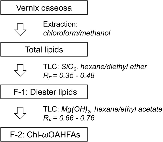
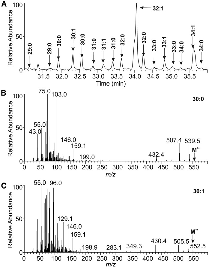
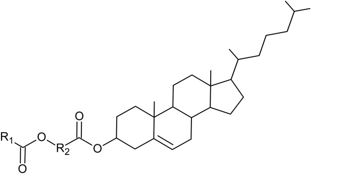
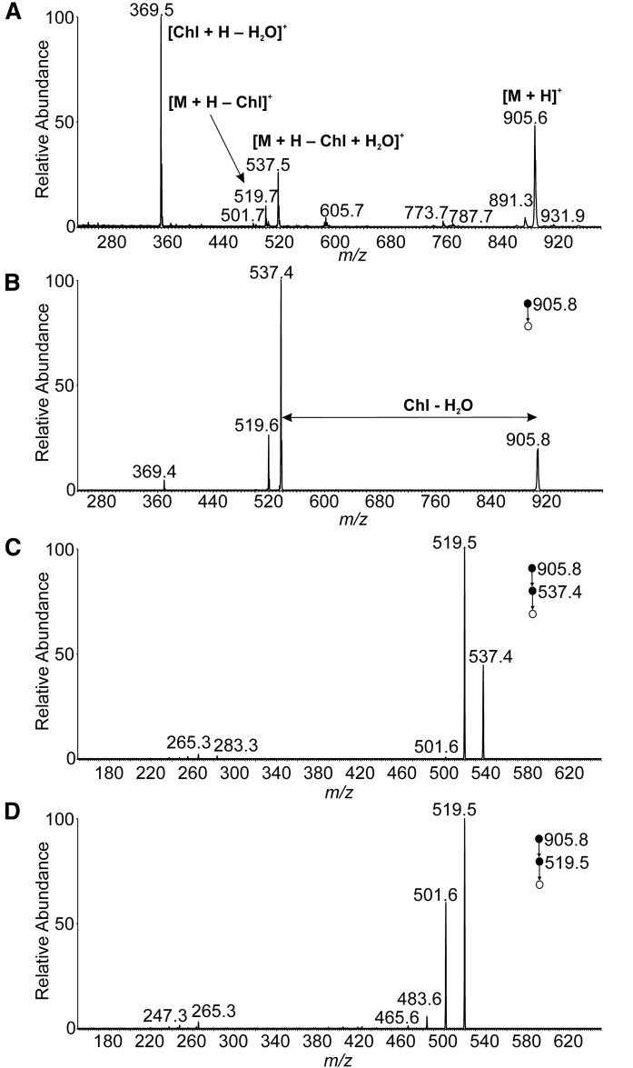
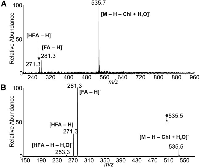
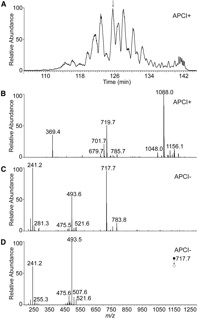
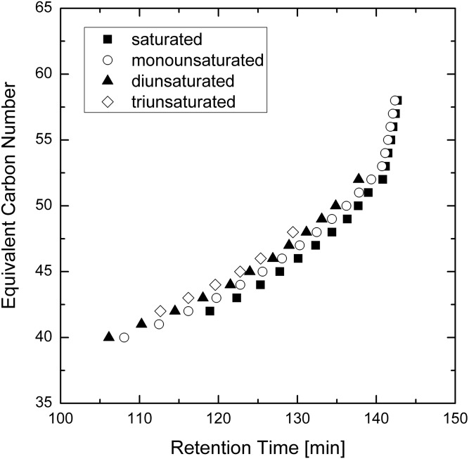
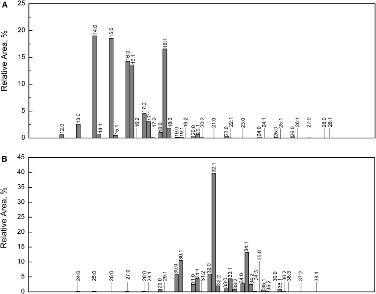
Cholesteryl esters of ω-(O-acyl)-hydroxy FAs (Chl-ωOAHFAs) were identified for the first time in vernix caseosa and characterized using chromatography and MS. Chl-ωOAHFAs were isolated using adsorption chromatography on silica gel and magnesium hydroxide. Their general structure was established using high-resolution and tandem MS of intact lipids, and products of their transesterification and derivatizations. Individual molecular species were characterized using nonaqueous reversed-phase HPLC coupled to atmospheric pressure chemical ionization. The analytes were detected as protonated molecules, and their structures were elucidated in the negative ion mode using controlled thermal decomposition and data-dependent fragmentation. About three hundred molecular species of Chl-ωOAHFAs were identified in this way. The most abundant Chl-ωOAHFAs contained 32:1 ω-hydroxy FA (ω-HFA) and 14:0, 15:0, 16:0, 16:1, and 18:1 FAs. The double bond in the 32:1 ω-HFA was in the n-7 and n-9 positions. Chl-ωOAHFAs are estimated to account for approximately 1-2% of vernix caseosa lipids.

Keywords: cholesterol; lipidomics; mass spectrometry; neutral lipids; skin lipids.


Figures

Fig. 1.
Fig. 1.
Scheme of the isolation and fractionation procedure.
Fig. 2.
Fig. 2.
A section of GC/MS chromatogram reconstructed for m/z 75 showing TMS derivatives of HFAMEs from Chl-ωOAHFAs (A). The EI-MS (70 eV) spectrum of TMS derivative of HFAME 30:0 (tR = 31.9 min) (B). The EI-MS (70 eV) spectrum of TMS derivative of HFAME 30:1 (tR = 32.3 min) (C).
Fig. 3.
Fig. 3.
The general structure of Chl-ωOAHFAs (R1, aliphatic chain of FA; R2, aliphatic chain of HFA).
Fig. 4.
Fig. 4.
APCI mass spectra of 18:1(n-9)/16:0-Chl in the positive ion mode. Full scan spectrum (A). CID MS2 spectrum of the protonated molecule (m/z 905.8; normalized collision energy 15%) (B). CID MS3 spectrum of [M + H − Chl + H2O]+ (m/z 537.4; normalized collision energy 16%) (C). CID MS3 spectrum of [M + H − Chl]+ (m/z 519.5; normalized collision energy 16%) (D). The chloroform solution (1 mg/ml) delivered by a syringe pump (5 μl/min) was mixed with acetonitrile:ethyl acetate (45:55, by volume) flowing at 150 μl/min. The mixture was directly infused into the ion source.
Fig. 5.
Fig. 5.
APCI mass spectra of 18:1(n-9)/16:0-Chl in the negative ion mode. Full scan spectrum (A). CID MS2 spectrum of [M − H − Chl + H2O] (m/z 535.5; normalized collision energy 15%) (B). The chloroform solution (1 mg/ml) delivered by a syringe pump (5 μl/min) was mixed with acetonitrile:ethyl acetate (45:55, by volume) flowing at 150 μl/min. The mixture was directly infused into the ion source.
Fig. 6.
Fig. 6.
Positive ion base-peak chromatogram (m/z 1,000–1,300) of Chl-ωOAHFAs isolated from vernix caseosa (A) and the APCI mass spectra used for the structure elucidation of a species with tR= 126.0 min (B–D). Full scan spectrum in the positive ion mode (B). Full scan spectrum in the negative ion mode (C). CID MS2 spectrum of m/z 717.7 in the negative ion mode (normalized collision energy 21.5%) (D).
Fig. 7.
Fig. 7.
Plot of calculated equivalent carbon number (ECN) values versus retention times for the Chl-ωOAHFAs identified in vernix caseosa (ECN = CN-2DB where CN is the total number of carbons and DB is the total number of double bonds in the ωOAHFA part of the molecules).
Fig. 8.
Fig. 8.
Histogram showing relative proportions of FAs (A) and ωHFAs (B) in Chl-ωOAHFAs from vernix caseosa. The proportions were calculated using HPLC/MS data given in supplemental Table S1 (the extended version of Table 1).

References

    1. Haubrich K. A. 2003. Role of vernix caseosa in the neonate: potential application in the adult population. AACN Clin. Issues. 14: 457–464. - PubMed
    1. Hoath S. B., Pickens W. L., and Visscher M. O.. 2006. The biology of vernix caseosa. Int. J. Cosmet. Sci. 28: 319–333. - PubMed
    1. Singh G., and Archana G.. 2008. Unraveling the mystery of vernix caseosa. Indian J. Dermatol. 53: 54–60. - PMC - PubMed
    1. Schmid R. 1939. Notizen zur Kenntnis der Vernix caseosa. Arch. Gynakol. 168: 445–450.
    1. Rissmann R., Groenink H. W. W., Weerheim A. M., Hoath S. B., Ponec M., and Bouwstra J. A.. 2006. New insights into ultrastructure, lipid composition and organization of vernix caseosa. J. Invest. Dermatol. 126: 1823–1833. - PubMed

Publication types