. 2017 Jul 15;130(14):2277-2291.
doi: 10.1242/jcs.192781. Epub 2017 Jun 2.

Low-intensity pulsed ultrasound promotes cell motility through vinculin-controlled Rac1 GTPase activity

Affiliations

Low-intensity pulsed ultrasound promotes cell motility through vinculin-controlled Rac1 GTPase activity

Paul Atherton et al. J Cell Sci. .

Abstract

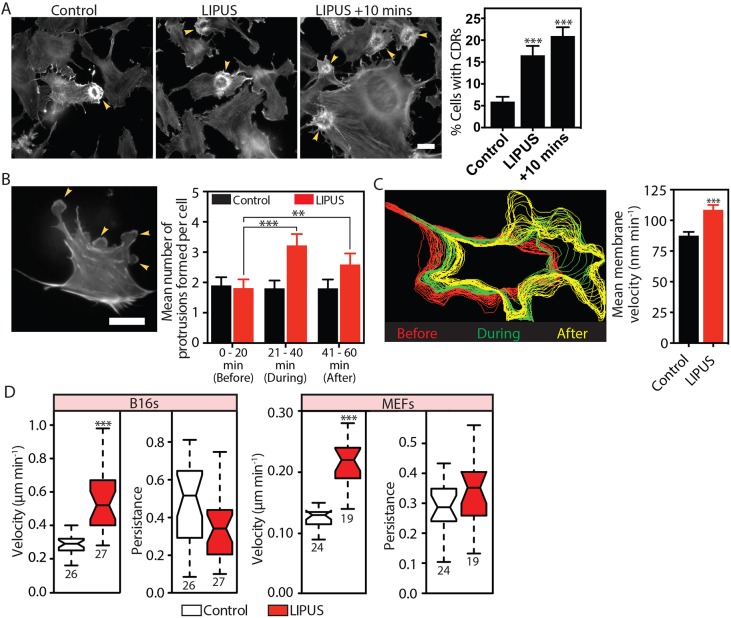
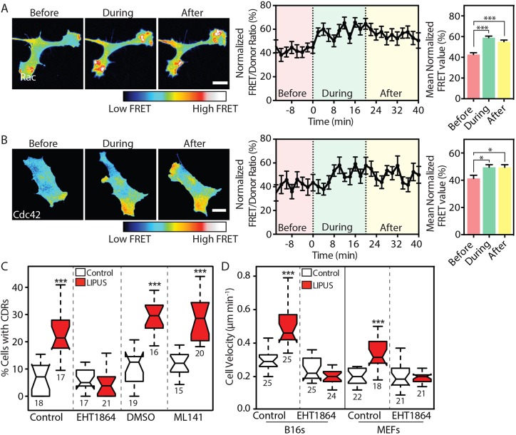
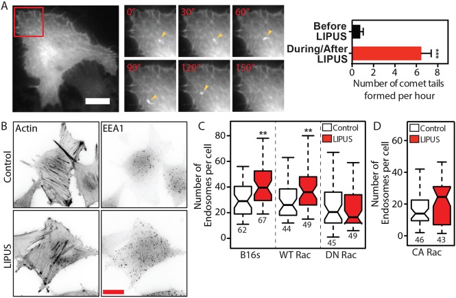
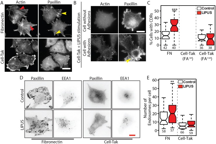
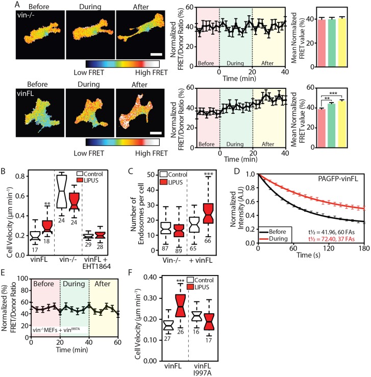
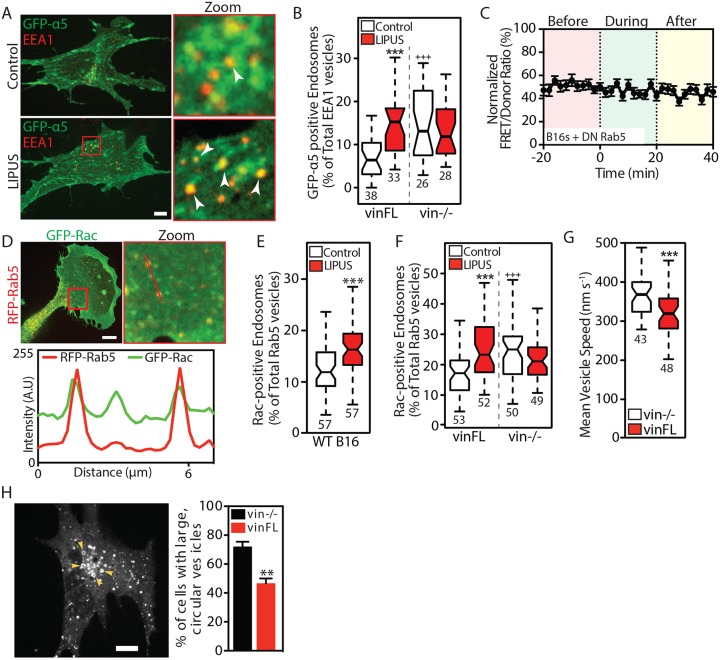
Low-intensity pulsed ultrasound (LIPUS) is a therapy used clinically to promote healing. Using live-cell imaging we show that LIPUS stimulation, acting through integrin-mediated cell-matrix adhesions, rapidly induces Rac1 activation associated with dramatic actin cytoskeleton rearrangements. Our study demonstrates that the mechanosensitive focal adhesion (FA) protein vinculin, and both focal adhesion kinase (FAK, also known as PTK2) and Rab5 (both the Rab5a and Rab5b isoforms) have key roles in regulating these effects. Inhibiting the link of vinculin to the actin-cytoskeleton abolished LIPUS sensing. We show that this vinculin-mediated link was not only critical for Rac1 induction and actin rearrangements, but was also important for the induction of a Rab5-dependent increase in the number of early endosomes. Expression of dominant-negative Rab5, or inhibition of endocytosis with dynasore, also blocked LIPUS-induced Rac1 signalling events. Taken together, our data show that LIPUS is sensed by cell matrix adhesions through vinculin, which in turn modulates a Rab5-Rac1 pathway to control ultrasound-mediated endocytosis and cell motility. Finally, we demonstrate that a similar FAK-Rab5-Rac1 pathway acts to control cell spreading upon fibronectin.

Keywords: Endocytosis; LIPUS; Migration; Rac1; Vinculin.


Conflict of interest statement

Competing interestsA.H. is an employee of Bioventus that manufactures and sells a LIPUS device known as EXOGEN.

Figures

Fig. 1.
Fig. 1.
LIPUS stimulation leads to rapid cytoskeleton rearrangement and increased cell motility. (A) B16 GFP-actin cells were fixed after receiving no LIPUS stimulation (control), immediately after the end of the 20 min stimulation (LIPUS) or 10 min after the end of the stimulation (LIPUS+10 min). Scale bar: 20 µm. Quantification of 60 randomly selected XY positions. Data are mean±s.e.m. from three independent experiments. Note the marked increase in CDRs after LIPUS (see also Movie 1). (B) MEFs expressing LifeAct were imaged for 1 h (Movie 2); LIPUS was started after 20 min. Scale bar: 50 μm. LIPUS stimulation increased the formation of small membrane protrusions, quantified by manual counting. Data are mean±s.e.m. from three independent movies; n=31 (control) and n=27 (LIPUS). (C) Automated detection of the cell periphery and quantification of membrane velocity using QuimP (Bosgraaf et al., 2009). The colour-coded shape outlines indicate representative protrusive activities recorded before (red), during (green), and after (yellow) LIPUS application. Note that the spacing between lines increases due to increases in protrusion velocity of LIPUS-stimulated MEFs compared to nonstimulated controls. Data are mean±s.e.m from three independent movies; n=69 (control) and n=63 (LIPUS). (D) Tracking of LIPUS-stimulated or nonstimulated control cells shows that LIPUS stimulation increases the velocity, but not persistence, of B16 and MEF cells (results are representative of three independent experiments and are presented as described in the Materials and Methods; n numbers are indicated below the plots). **P<0.01, ***P<0.001.
Fig. 2.
Fig. 2.
LIPUS stimulates rapid GTPase activation and endocytosis. (A,B) B16 cells expressing (A) Rac1 or (B) Cdc42 FRET-based activity reporters were imaged before, during and after LIPUS stimulation. Images and quantifications of ratiometric FRET (normalized between 0 and 100 over time) analysis show that LIPUS stimulation leads to rapid activation of both GTPases, and that elevated GTPase activation persists after the stimulation ends. Scale bars: 10 µm. Data are mean±s.e.m. from three independent experiments; n=26 in A and n=17 in B. (C) Pretreatment of B16 cells with the Rac1 inhibitor EHT1864 (10 µM), but not with the Cdc42 inhibitor ML141 (10 µM), blocked LIPUS-induced CDR formation. Measurements are from the indicated numbers of XY positions, representative of three independent experiments. (D) Pretreatment with EHT1864 (10 µM) also blocked LIPUS-induced motility increases in both B16 and MEF cells. Cells were tracked over 16 h to assess cell motility (results are representative of three independent experiments and are presented as described in the Materials and Methods). *P<0.05, ***P<0.001.
Fig. 3.
Fig. 3.
LIPUS increases the number of early endosomes in a Rac1-dependent manner. (A) LIPUS stimulation in serum-starved B16 GFP-actin cells leads to the formation of actin comet-tails (arrowheads). The inset is shown in higher magnification in the right-hand panels at the indicated times (min). Data are mean±s.e.m. from three independent movies; n=17. Scale bar: 20 µm. (B) Immunofluorescence images (colour inverted) of B16 cells with or without LIPUS stimulation, stained for actin or EEA1. Scale bar: 20 µm. (C) Quantification of the number of endosomes in B16 control cells or cells expressing WT Rac1 or DN Rac1. Note that the expression of DN Rac1 blocks the LIPUS-induced increase in early endosomes. (D) Expression of CA Rac1 also prevents any change in the number of EEA1-positive early endosomes after LIPUS stimulation. Data in C and D are from three independent experiments and are presented as described in the Materials and Methods. **P<0.01.
Fig. 4.
Fig. 4.
Cell-ECM adhesions are required for LIPUS-induced CDR formation and increases in early endosomes. (A,B) Cells were seeded on fibronectin or Cell-Tak coated glass and stained for actin and the FA marker paxillin. Cells on fibronectin form FAs (yellow arrowheads), connected to actin stress fibres (red arrowheads), whereas the majority of cells on Cell-Tak did not form any adhesions. In some cases, cells on Cell-Tak were able to form adhesions. Scale bar: 20 µm. (C) LIPUS stimulation of cells on fibronectin (FN) produced an increase in the number of cells with CDRs, but LIPUS stimulation had no effect on CDR formation in cells on Cell-Tak, including those that had formed some small adhesions. (D) Control and LIPUS-treated cells on either fibronectin or Cell-Tak and stained for paxillin and EEA1 (colour inverted). Scale bar: 10 µm. (E) Quantification of the number of early endosomes in LIPUS-stimulated and control cells on FN- or Cell-Tak-coated glass. Note that LIPUS-stimulation had no effect on the number of early endosomes in the cells on Cell-Tak. Data in C and E are from three independent experiments and are presented as described in the Materials and Methods; n numbers are indicated below the plots. **P<0.01, ***P<0.001.
Fig. 5.
Fig. 5.
Vinculin and its actin-binding potential are required for sensing LIPUS stimulation. (A) FRET measurements from vin−/− MEFs expressing the Raichu Rac1 FRET probe without (upper row) or with (lower row) vinFL. An increase in FRET signal in response to LIPUS is only seen in cells coexpressing mCherry-vinFL. Scale bars: 10 µm. Data are mean±s.e.m. from four independent experiments; n=26 (vin−/–) and n=46 (vinFL). (B) LIPUS-stimulated vin−/− MEFs show no change in migration speed. Expression of vinFL rescues LIPUS-enhanced motility, which is blocked by pretreatment with the Rac1 inhibitor EHT1864 (10 µM). (C) The increase in the number of early endosomes after LIPUS stimulation is blocked in vin−/− MEFs, but rescued by expression of vinFL. (D) The turnover of PAGFP-vinculin was assessed using FLAP, before or during LIPUS stimulation. Data are mean±s.e.m. from at least three independent experiments. (E) FRET measurements from vin−/− MEFs coexpressing Raichu Rac1 and mCherry-vinFLI997A. Note the lack of increase in FRET signal in response to LIPUS. Data are mean±s.e.m. from three independent experiments; n=32. (F) Vin–/– MEFs expressing vinFLI997A show no change in motility after LIPUS stimulation. Results in B and F are representative of three independent experiments; results in C are from three independent experiments. Data are presented as described in the Materials and Methods; n numbers are indicated below the plots. **P<0.01, ***P<0.001.
Fig. 6.
Fig. 6.
FAK signalling regulates the cellular responses following mechanosensing of LIPUS by vinculin. (A) B16 cells were stimulated with LIPUS for 0, 2, 5 or 10 min and then lysed. The western blots show an increase in the amount of pFAK-Y397 relative to total FAK, with the maximum increase seen after 10 min of stimulation. The bar graph shows pFAK/FAK ratios from four independent experiments. *P<0.05 (one-way ANOVA). (B) Pretreatment with 3 µM FAKi for 1 h dramatically reduces FAK phosphorylation levels, and also blocks the LIPUS-induced increase in pFAK-Y397 levels (cells were lysed after 10 min of stimulation). The western blots shown are representative of three independent experiments. (C) Raichu Rac1 FRET experiments were performed in cells pretreated with 3 µM FAKi or an equivalent volume of DMSO. FAK inhibition blocks the LIPUS-dependent increase in Rac1 activation. Data are mean±s.e.m. from three independent experiments; n=32 (DMSO) and n=26 (FAKi). (D) FAK inhibition also blocked the increase in the number of EEA1-positive endosomes after LIPUS stimulation (data are from three independent experiments), and (E) blocked the increase in cell velocity seen after LIPUS stimulation (data are representative of three independent experiments). Results are presented as described in the Materials and Methods, n numbers are indicated below the plots). (F) The turnover of PAGFP-vinFL was assessed using FLAP, before or during LIPUS stimulation, in the presence of 3 µM FAKi. In cells undergoing stimulation, the turnover of vinculin is reduced, even when FAK signalling is suppressed. Results are representative of at least three independent experiments. (G) Western blots of pFAK-Y397 and FAK in vin−/– MEFs stimulated with LIPUS for the indicated amount of time. Note that there is no increase in FAK phosphorylation in these cells (blots are representative of four independent experiments). α-Tubulin was used as a loading control. *P<0.05, ***P<0.001.
Fig. 7.
Fig. 7.
Vinculin modulates Rab5-mediated Rac1 activation in response to LIPUS. (A) Confocal images of vin−/– MEFs rescued with CFP-vinculin and coexpressing GFP-α5 integrin, without (top row) or with (bottom row) LIPUS stimulation, fixed and stained for EEA1. Arrowheads indicate colocalization between GFP-α5 and EEA1. Scale bar: 10 µm. (B) Colocalization between GFP-α5 and EEA1 in cells fixed 10 min after the end of LIPUS stimulation. ***P<0.001 against the control; +++P<0.001 against the vinFL control. (C) LIPUS stimulation has no effect on Rac1 activity in B16 cells expressing DN Rab5 (Rab5S34N) (data are mean±s.e.m.; n=37 from three independent experiments). (D) Confocal images of GFP-Rac1 and RFP-Rab5 expressed in B16 cells, with a line profile showing the colocalization of Rac1 and Rab5-positive vesicles. Scale bar: 10 µm. (E) Percentage of Rac1-positive Rab5 vesicles in control (white) and LIPUS-stimulated (red) B16 cells. (F) The recruitment of Rac1 to Rab5-positive vesicles in LIPUS-stimulated MEFs is only seen in vinculin-expressing cells (i.e. is vinculin dependent). Data are mean±s.e.m. from three independent experiments; n numbers are indicated below the plots. ***P<0.001 against the control; +++P<0.001 against the vinFL control. (G) Vesicle dynamics. Note that Rab5-positive vesicles move faster in vin–/– MEFs compared to vin–/– MEFs rescued with vinFL (see Movie 4). (H) Image of a vin–/– MEF expressing RFP-Rab5 (WT). Arrowheads indicate large, circular vesicles. Scale bar: 10 µm. The bar graph shows the (mean±s.e.m., three independent experiments) percentage of vin−/− and vinFL cells with large, circular vesicles. Results in B, E and F are from three independent experiments; results in G are representative of three independent experiments. Data are presented as described in the Materials and Methods; n numbers are indicated below the plots. **P<0.01, ***P<0.001.
Fig. 8.
Fig. 8.
A FAK-Rab5-Rac1 pathway mediates cell spreading and the response to LIPUS. (A) Representative images of B16 cells coexpressing the indicated constructs pretreated in suspension with either DMSO or FAKi for 1 h before being plated onto fibronectin and fixed after 45 min. Scale bar: 20 µm. (B) Measurements of mean cell area and mean cell circularity show that FAKi treatment reduces cell spreading, similar to expression of either Rac1N17 (DN) or Rab5N34 (DN). Data are mean±s.e.m. from three independent experiments; n=46–78. ***P<0.001 against the DMSO control; +++P<0.001 against WT Rac1 and WT Rab5-expressing DMSO control. (C) Representative images of B16 cells expressing Rac1L61 (CA) with either WT Rab5 or Rab5N34. Scale bar: 20 µm. (D) Quantification of the cell area shows that CA Rac1 expression rescues the defects in cell spreading associated with FAKi treatment. Coexpression of Rab5N34 with Rac1L61 reduces cell spreading to a similar extent to FAKi treatment. Data are mean±s.e.m. from three independent experiments; n=50–72. +++P<0.001 against CA Rac1 and WT Rab5-expressing DMSO control. (E) Mean cell area of B16 cells coexpressing the indicated Rac1 and Rab5 constructs, treated in suspension with either DMSO or dynasore. Note that dynasore treatment has a similar effect on cell area as coexpression of Rab5N34. Data are mean±s.e.m. from three independent experiments; n=21–40. ***P<0.001 against DMSO control. (F) B16 cells treated with either EHT1864 (10 µM) or dynasore (80 µM) were stimulated for 10 min with LIPUS to assess pFAK-Y397 levels. Western blots show that inhibition of either Rac1 or dynamin blocks the LIPUS-induced increase in pFAK levels (blots are representative of at least three independent experiments; α-tubulin was used as a loading control). (G) Model of how cells sense and respond to LIPUS: LIPUS stimulation is ‘sensed’ by vinculin at integrin-mediated cell-ECM adhesions, requiring the link between vinculin and the actin cytoskeleton. FAK signalling leads to Rab5-dependent activation of Rac1. Once Rac1 is active, a feedback mechanism promotes further FAK phosphorylation, thereby propagating Rac1 activity. Active Rac1 localizes at Rab5-positive vesicles to facilitate trafficking, promoting rearrangements of the actin cytoskeleton and increased cell motility.

Similar articles

Cited by

References

    1. Alanko J., Mai A., Jacquemet G., Schauer K., Kaukonen R., Saari M., Goud B. and Ivaska J. (2015). Integrin endosomal signalling suppresses anoikis. Nat. Cell Biol. 17, 1412-1421. 10.1038/ncb3250 - DOI - PMC - PubMed
    1. Argadine H., Kinnick R., Bolander M. and Greenleaf J. F. (2005). 1 kHz low power sound stimulates ATDC5 chondrocytes. Proc. IEEE Ultrason Symp. 2, 996-998. 10.1109/ultsym.2005.1603018 - DOI
    1. Atherton P., Stutchbury B., Wang D.-Y., Jethwa D., Tsang R., Meiler-Rodriguez E., Wang P., Bate N., Zent R., Barsukov I. L. et al. (2015). Vinculin controls talin engagement with the actomyosin machinery. Nat. Commun. 6, 10038 10.1038/ncomms10038 - DOI - PMC - PubMed
    1. Atherton P., Stutchbury B., Jethwa D. and Ballestrem C. (2016). Mechanosensitive components of integrin adhesions: role of vinculin. Exp. Cell Res. 343, 21-27. 10.1016/j.yexcr.2015.11.017 - DOI - PMC - PubMed
    1. Bae Y. H., Mui K. L., Hsu B. Y., Liu S.-L., Cretu A., Razinia Z., Xu T., Pure E. and Assoian R. K. (2014). A FAK-Cas-Rac-lamellipodin signaling module transduces extracellular matrix stiffness into mechanosensitive cell cycling. Sci. Signal. 7, ra57 10.1126/scisignal.2004838 - DOI - PMC - PubMed