Review
. 2017 Oct:13:94-162.
doi: 10.1016/j.redox.2017.05.007. Epub 2017 May 18.

European contribution to the study of ROS: A summary of the findings and prospects for the future from the COST action BM1203 (EU-ROS)

Javier Egea  1 Isabel Fabregat  2 Yves M Frapart  3 Pietro Ghezzi  4 Agnes Görlach  5 Thomas Kietzmann  6 Kateryna Kubaichuk  6 Ulla G Knaus  7 Manuela G Lopez  1 Gloria Olaso-Gonzalez  8 Andreas Petry  9 Rainer Schulz  10 Jose Vina  8 Paul Winyard  11 Kahina Abbas  3 Opeyemi S Ademowo  12 Catarina B Afonso  13 Ioanna Andreadou  14 Haike Antelmann  15 Fernando Antunes  16 Mutay Aslan  17 Markus M Bachschmid  18 Rui M Barbosa  19 Vsevolod Belousov  20 Carsten Berndt  21 David Bernlohr  22 Esther Bertrán  2 Alberto Bindoli  23 Serge P Bottari  24 Paula M Brito  25 Guia Carrara  26 Ana I Casas  27 Afroditi Chatzi  28 Niki Chondrogianni  29 Marcus Conrad  30 Marcus S Cooke  31 João G Costa  32 Antonio Cuadrado  33 Pham My-Chan Dang  34 Barbara De Smet  35 Bilge Debelec-Butuner  36 Irundika H K Dias  12 Joe Dan Dunn  37 Amanda J Edson  38 Mariam El Assar  39 Jamel El-Benna  34 Péter Ferdinandy  40 Ana S Fernandes  41 Kari E Fladmark  38 Ulrich Förstermann  42 Rashid Giniatullin  43 Zoltán Giricz  40 Anikó Görbe  40 Helen Griffiths  44 Vaclav Hampl  45 Alina Hanf  46 Jan Herget  45 Pablo Hernansanz-Agustín  47 Melanie Hillion  15 Jingjing Huang  48 Serap Ilikay  49 Pidder Jansen-Dürr  50 Vincent Jaquet  51 Jaap A Joles  52 Balaraman Kalyanaraman  53 Danylo Kaminskyy  54 Mahsa Karbaschi  31 Marina Kleanthous  55 Lars-Oliver Klotz  56 Bato Korac  57 Kemal Sami Korkmaz  58 Rafal Koziel  50 Damir Kračun  9 Karl-Heinz Krause  51 Vladimír Křen  59 Thomas Krieg  60 João Laranjinha  19 Antigone Lazou  61 Huige Li  42 Antonio Martínez-Ruiz  62 Reiko Matsui  18 Gethin J McBean  63 Stuart P Meredith  13 Joris Messens  64 Verónica Miguel  65 Yuliya Mikhed  46 Irina Milisav  66 Lidija Milković  67 Antonio Miranda-Vizuete  68 Miloš Mojović  69 María Monsalve  70 Pierre-Alexis Mouthuy  71 John Mulvey  60 Thomas Münzel  46 Vladimir Muzykantov  72 Isabel T N Nguyen  52 Matthias Oelze  46 Nuno G Oliveira  73 Carlos M Palmeira  74 Nikoletta Papaevgeniou  29 Aleksandra Pavićević  69 Brandán Pedre  64 Fabienne Peyrot  75 Marios Phylactides  55 Gratiela G Pircalabioru  76 Andrew R Pitt  13 Henrik E Poulsen  77 Ignacio Prieto  70 Maria Pia Rigobello  78 Natalia Robledinos-Antón  33 Leocadio Rodríguez-Mañas  79 Anabela P Rolo  74 Francis Rousset  51 Tatjana Ruskovska  80 Nuno Saraiva  41 Shlomo Sasson  81 Katrin Schröder  82 Khrystyna Semen  54 Tamara Seredenina  51 Anastasia Shakirzyanova  43 Geoffrey L Smith  26 Thierry Soldati  37 Bebiana C Sousa  13 Corinne M Spickett  12 Ana Stancic  57 Marie José Stasia  83 Holger Steinbrenner  56 Višnja Stepanić  67 Sebastian Steven  46 Kostas Tokatlidis  28 Erkan Tuncay  84 Belma Turan  84 Fulvio Ursini  85 Jan Vacek  86 Olga Vajnerova  45 Kateřina Valentová  59 Frank Van Breusegem  87 Lokman Varisli  49 Elizabeth A Veal  88 A Suha Yalçın  89 Olha Yelisyeyeva  54 Neven Žarković  71 Martina Zatloukalová  86 Jacek Zielonka  53 Rhian M Touyz  90 Andreas Papapetropoulos  91 Tilman Grune  92 Santiago Lamas  65 Harald H H W Schmidt  27 Fabio Di Lisa  93 Andreas Daiber  94
Affiliations
Review

European contribution to the study of ROS: A summary of the findings and prospects for the future from the COST action BM1203 (EU-ROS)

Javier Egea et al. Redox Biol. 2017 Oct.

Erratum in

  • Corrigendum to "European contribution to the study of ROS: A summary of the findings and prospects for the future from the COST action BM1203 (EU-ROS)" [Redox Biol. 13 (2017) 94-162].
    Egea J, Fabregat I, Frapart YM, Ghezzi P, Görlach A, Kietzmann T, Kubaichuk K, Knaus UG, Lopez MG, Olaso-Gonzalez G, Petry A, Schulz R, Vina J, Winyard P, Abbas K, Ademowo OS, Afonso CB, Andreadou I, Antelmann H, Antunes F, Aslan M, Bachschmid MM, Barbosa RM, Belousov V, Berndt C, Bernlohr D, Bertrán E, Bindoli A, Bottari SP, Brito PM, Carrara G, Casas AI, Chatzi A, Chondrogianni N, Conrad M, Cooke MS, Costa JG, Cuadrado A, My-Chan Dang P, De Smet B, Debelec-Butuner B, Dias IHK, Dunn JD, Edson AJ, El Assar M, El-Benna J, Ferdinandy P, Fernandes AS, Fladmark KE, Förstermann U, Giniatullin R, Giricz Z, Görbe A, Griffiths H, Hampl V, Hanf A, Herget J, Hernansanz-Agustín P, Hillion M, Huang J, Ilikay S, Jansen-Dürr P, Jaquet V, Joles JA, Kalyanaraman B, Kaminskyy D, Karbaschi M, Kleanthous M, Klotz LO, Korac B, Korkmaz KS, Koziel R, Kračun D, Krause KH, Křen V, Krieg T, Laranjinha J, Lazou A, Li H, Martínez-Ruiz A, Matsui R, McBean GJ, Meredith SP, Messens J, Miguel V, Mikhed Y, Milisav I, Milković L, Miranda-Vizuete A, Mojović M, Monsalve M, Mouthuy PA, Mulvey J, Münzel T, Muzykantov V, Nguyen ITN, Oelze M, Oliveira NG, Palmeira CM, Papaevgeniou N, Pavićević A, Pedre B, Peyrot F, Phy… See abstract for full author list ➔ Egea J, et al. Redox Biol. 2018 Apr;14:694-696. doi: 10.1016/j.redox.2017.10.001. Epub 2017 Oct 26. Redox Biol. 2018. PMID: 29107648 Free PMC article. No abstract available.

Abstract

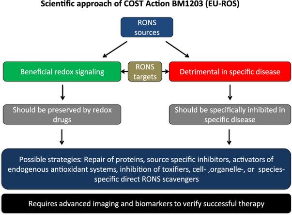
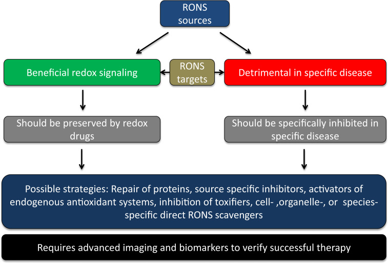
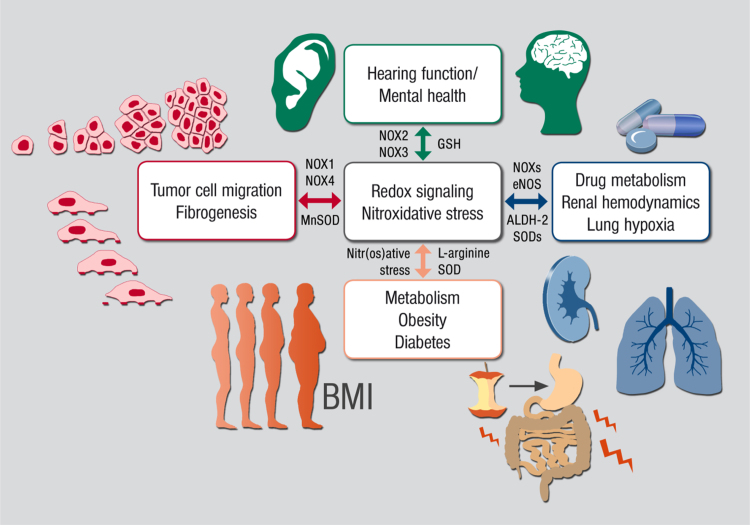
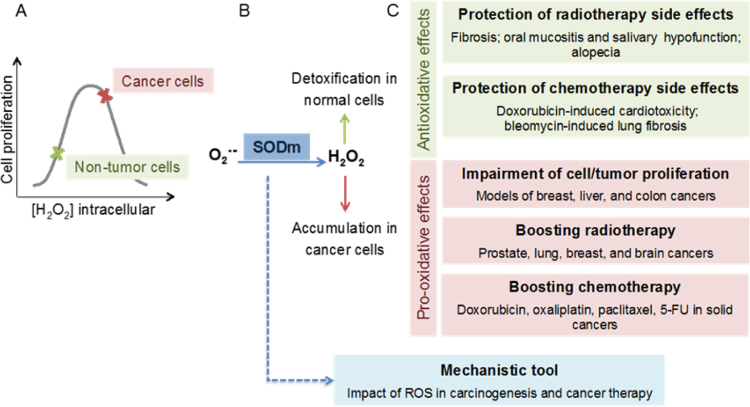
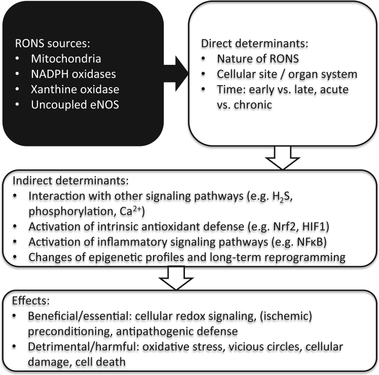
The European Cooperation in Science and Technology (COST) provides an ideal framework to establish multi-disciplinary research networks. COST Action BM1203 (EU-ROS) represents a consortium of researchers from different disciplines who are dedicated to providing new insights and tools for better understanding redox biology and medicine and, in the long run, to finding new therapeutic strategies to target dysregulated redox processes in various diseases. This report highlights the major achievements of EU-ROS as well as research updates and new perspectives arising from its members. The EU-ROS consortium comprised more than 140 active members who worked together for four years on the topics briefly described below. The formation of reactive oxygen and nitrogen species (RONS) is an established hallmark of our aerobic environment and metabolism but RONS also act as messengers via redox regulation of essential cellular processes. The fact that many diseases have been found to be associated with oxidative stress established the theory of oxidative stress as a trigger of diseases that can be corrected by antioxidant therapy. However, while experimental studies support this thesis, clinical studies still generate controversial results, due to complex pathophysiology of oxidative stress in humans. For future improvement of antioxidant therapy and better understanding of redox-associated disease progression detailed knowledge on the sources and targets of RONS formation and discrimination of their detrimental or beneficial roles is required. In order to advance this important area of biology and medicine, highly synergistic approaches combining a variety of diverse and contrasting disciplines are needed.

Keywords: Antioxidants; Oxidative stress; Reactive nitrogen species; Reactive oxygen species; Redox signaling; Redox therapeutics.


Figures

fx1
Graphical abstract
Fig. 1.1
Fig. 1.1
The main concept of our biomedical approach and working scheme within the EU-ROS consortium as explained in detail previously .
Fig. 2.1
Fig. 2.1
(A) Crosstalk between different sources of ROS and RNS (mitochondria, NADPH oxidases, xanthine oxidase and NO synthase). Xanthine oxidase (XO) originates from oxidative stress-mediated conversion of the xanthine dehydrogenase via oxidation of critical thiols in cysteine535/992. NO synthases (mainly eNOS) are uncoupled upon oxidative depletion of tetrahydrobiopterin (BH4), S-glutathionylation (-SSG) and other redox switches. Mitochondrial superoxide/hydrogen peroxide formation may be triggered by oxidative stress from all ROS sources (including other damaged/activated mitochondria) via redox-activation of PKC, MAPK, other kinase pathways and potential involvement of redox-sensitive mitochondrial ATP-sensitive potassium channels (mtKATP) with subsequent p66Shc, monoamine oxidase (MAO), respiratory complex activation or impairment of mitochondrial antioxidant defence. Mitochondrial superoxide/ hydrogen peroxide is released to the cytosol via mitochondrial pores and channels (e.g. redox-sensitive mitochondrial permeability transition pore (mPTP), inner membrane anion channel (IMAC) or aquaporins) or by diffusion due to increased mitochondrial permeability under pro-inflammatory conditions. In the cytosol these species (along with released calcium) cause activation of redox-sensitive protein kinases (PKC) and tyrosine kinases (cSrc) with subsequent activation of NADPH oxidases and amplification of the cellular oxidative stress. Modified from , . With permission of Elsevier. Copyright 2010 & 2015. (B) Redox switches in endothelial nitric oxide synthase (eNOS). X-ray structure of human eNOS with the ironporphyrin (blue), the substrate L-arginine (green), the P450-forming axial iron-thiolate ligand from a cysteine residue (yellow), the cofactor tetrahydrobiopterin (BH4) (purple), the zinc-thiolate complex forming cysteines (red, two from each subunit), and the zinc ion (brown). The boxes represent the ‘‘redox switches’’ in eNOS, such as S-glutathionylation, PKC- and protein tyrosine kinase-2 (PYK-2)–dependent phosphorylation, oxidative BH4 depletion, disruption of the zinc-sulfur cluster, as well as asymmetric dimethylarginine (ADMA) synthesis/degradation, all of which contribute to the regulation of its enzymatic activity. GSH, glutathione; GSSG, glutathione disulfide. The crystal structure was rendered from the protein database entry 3NOS (DOI:10.2210/pdb3nos/pdb) using the PyMOL Molecular Graphics System Version 1.2r1 (DeLano Scientific LLC). Adapted from . With permission of Mary Ann Liebert, Inc. Copyright 2014.
Fig. 3.1
Fig. 3.1
Summary scheme of ROS acting as signaling molecules in different disease settings but also in physiological processes.
Fig. 3.2
Fig. 3.2
(A) Reactive oxygen species can display their regulatory effect on the classical gene regulatory machinery and on epigenetic processes. One of the prominent pathways attributed to oxidative stress is thiol oxidation, which is involved in OxyR, NF-κB and KEAP1 signaling. Oxygen sensing prolyl hydroxylases represent another class of redox-dependent enzymes. For example, epigenetic involvement of ROS has been attributed to oxidative conversion of 5-mC to 5-hmC. (B) ROS impact on mRNA stability at the cytosolic level. Reactive oxygen species are involved in GAPDH signaling by directly altering its structure with the help of GSH or S-nitrosoglutathione (GSNO), and thus activate translation of endothelin-1 (ET-1) mRNA (i). AP-1 thiol redox regulation directly affects the gene regulating factor HuR by stability of its target mRNAs (ii). ROS have been implicated in the regulation of miRNA pathways, altering mRNA stability and their transport inside the cytosol. HRE means hormone response element. With permission of Elsevier and the authors. Copyright 2015.
Fig. 4.1
Fig. 4.1
Redox pathways associated with putative biomarkers of oxidative stress. The processes that lead to oxidative modifications of proteins, lipids, and nucleotides are highly complex. Enzymes, such as XO, NOX, and NOS, can produce ROS and RNS. These ROS can furthermore serve as substrates for other enzymes to generate additional types of ROS, such as the generation of HOCl from H2O2 by MPO. Cellular systems and enzymes, including the GSH and thioredoxin system, together with peroxiredoxins (T/Prx), counterbalance the production of ROS. In addition, increased levels of ROS activate NRF2 to transcribe genes that are involved in counteracting these ROS. Oxidative stress affects cGMP signaling through its effects on nitric oxide (NO) production, scavenging, and on the NO receptor sGC. cGMP, cyclic guanosine monophosphate; GSH, glutathione; H2O2, hydrogen peroxide; HOCl, hypochlorous acid; MPO, myeloperoxidase; NOS, nitric oxide synthase; NOX, NADPH oxidase; RNS, reactive nitrogen species; ROS, reactive oxygen species; sGC, soluble guanylate cyclase; XO, xanthine oxidase. Adpated from . With permission of Mary Ann Liebert, Inc. Copyright 2015.
Fig. 4.2
Fig. 4.2
Cluster analysis of ROS biomarkers in disease. Different diseases were clustered according to described ROS biomarkers in Refs. , , and studies described in this review. Some disease conditions cluster as might be expected, such as ischemia/reperfusion and heart failure, and amyotrophic lateral sclerosis and multiple sclerosis. A comprehensive analysis of ROS markers and pattern analysis in diseases might uncover common disease mechanisms or new measures of disease progression or treatment outcome. Cluster analysis was performed using Genesis software (https://genome.tugraz.at/genesisclient/genesisclient_description.shtml) as described in Mengozzi et al. . Adpated from . With permission of Mary Ann Liebert, Inc. Copyright 2015.
Fig. 5.1
Fig. 5.1
Spectrum of different ROS imaging techniques. In the upper part, different sources of ROS are shown: Mitochondria (mito), lipoxygenases (LPO), monoamine oxidase (MAO), nicotinamide adenine dinucleotide phosphate oxidase (NOX4 and NOX 1/2/5), xanthine oxidase (XO), functional nitric oxide synthases (NOS) and dysfunctional / uncoupled eNOS (u-eNOS). These result in different types of ROS [including superoxide radical (O2•─), hydrogen peroxide (H2O2), hypochlorous acid (HOCl, not shown in the scheme), peroxynitrite anion (ONOO), nitric oxide (NO)] and ROS-induced modifications of GSH, NADPH, proteins, or glucose uptake, which, in turn, are detected by different imaging technologies. Adpated from . With permission of Mary Ann Liebert, Inc. Copyright 2016.
Fig. 5.2
Fig. 5.2
(A) Determination of mtROS triggered NADPH oxidase activation in isolated human neutrophils by electron paramagnetic resonance measurement. Freshly isolated human neutrophils (22x106 PMN/mL) were incubated in PBS with 300 µM Ca2+/Mg2+ and 10 mM DEPMPO for 15 min at 37 °C. Activators and inhibitors of phagocytic NOX were added as shown in the figure. The spectrum for phorbol ester (PDBu)-stimulated PMN is displayed at decreased intensity (1/10). All reactions below the red spectrum contained 20 µM myxothiazol. Incubations were with NOX inhibitor (VAS2870), intracellular calcium chelator (BAPTA-AM), cSrc kinase inhibitor (PP2) and PKC inhibitor (chelerythrine). All spectra were recorded at room temperature in 50 µl glass capillaries (Hirschmann Laborgeräte GmbH, Eberstadt, Germany). EPR conditions: B0 =3300 G, sweep =150 G, sweep time =60 s, modulation =3000 mG, MW power =10 mW, gain =9x10² using a Miniscope MS200 from Magnettech (Berlin, Germany). Representative spectra of mixed DEPMPO-OOH and DEPMPO-OH adduct for 2 independent experiments. Spectra were recorded as described previously . With permission of Mary Ann Liebert, Inc. Copyright 2014. (B) Whole blood Hb-NO levels were determined by electron paramagnetic resonance spectroscopy as a read-out of iNOS activity in endotoxemic (lipopolysaccharide-treated) rats. The EPR measurements were carried out at 77 K using an X-band table-top spectrometer MS400 (Magnettech, Berlin, Germany). The instrument settings were as follows: 10 mW microwave power, 7000 mG amplitude modulation, 100 kHz modulation frequency, 3300 G center field, 300 G sweep width, 60 s sweep time and 3 scans. With permission of Springer-Verlag Berlin Heidelberg. Copyright 2015. (C) Aortic NO formation was measured in untreated control and angiotensin-II infused hypertensive mice by EPR spin trapping using Fe(DETC)2. Each spectrum was measured from one murine aorta. The representative spectra below the bar graph are the mean of all measurements. EPR conditions: B0 =3276 G, sweep =115 G, sweep time =60 s, modulation =7000 mG, MW power =10 mW, gain =9x10² using a Miniscope MS400 from Magnettech (Berlin, Germany). The A23187-stimulated NO signal was absent when the aorta were denuded, L-NAME (200 µM) was added, or when aorta from eNOS-/- mice were used (not shown). With permission of Mary Ann Liebert, Inc. Copyright 2014.
Fig. 6.1
Fig. 6.1
Processes contributing to the increase in ROS levels in various tissues. Mitochondrial pathways are highlighted as prominent sources of ROS, especially in the heart. Besides their involvement in tissue injury, ROS have been described also as mediators of cardiac protection against ischemia/reperfusion damage.
Fig. 6.2
Fig. 6.2
The data show marked increases of [Zn2+]i under either ROS (A) or RNS (B) increases. Bars represent means (±) and *P<0.05 w.r.t. initial value. Inset: Representative electron micrograph images under ZnPT (1-μM) exposure. Magnification: x12,930; bar: 1000 nm; N: nucleus, M: mitochondria, z: Z-line, L: lysosome, arrow: T-tubule, arrow head: sarco/endoplasmic reticulum (SER).
Fig. 6.3
Fig. 6.3
Overview on therapeutic options for the improvement of vascular dysfunction. Targeted antioxidant interventions to alleviate pro-inflammatory activation and oxidative stress in endothelial cells. Endothelial ROS from activated NOX2 enzyme in endosomes are formed in response to cytokine binding to the receptors and ignite signaling cascade of transcription factor NFκB. Targeted delivery of antioxidants, antioxidant enzymes (AOE) and inhibitors of ROS production can be achieved using antibodies and other ligands of endothelial surface determinants including cell adhesion molecules PECAM and ICAM. Surface-bound targeted AOE intercept extracellular ROS, whereas targeted formulations using the same ligands configured in a way permitting internalization into the ROS-signaling endosomes allows interception of pro-inflammatory activation manifested among other characteristics by exposure of inducible cell adhesion molecules – E-selectin, VCAM-1, and ICAM-1 - that can be detected using imaging probes conjugated to the ligands of these molecules. With permission of the publisher. Copyright © 1999–2017 John Wiley & Sons, Inc. All Rights Reserved.
Fig. 6.4
Fig. 6.4
Proposed mechanisms of lipopolysaccharide (LPS)-induced vascular dysfunction and improvement by linagliptin therapy. LPS treatment activates white blood cells (WBC, envisaged by increased oxidative burst), increases serum levels of xanthine oxidase (XO), increases DPP-4 serum activity and activates vascular cells (detected by expression of endothelial adhesion molecules and inducible cyclooxygenase [COX-2]). This leads to the infiltration of WBC to the vascular wall (detected by aortic FACS analysis for myelomonocytic cells, inducible nitric oxide synthase [NOS2], NOX2 and myeloperoxidase [MPO] expression) and oxidative damage of the vasculature (NOX1 expression, ROS formation, 3-nitrotyrosine levels and lipidperoxidation by malondialdehyde [MDA]). Finally, the tissue damage results in smooth muscle constriction and endothelial dysfunction. With permission by Oxford University Press. Copyright © 2012.
Fig. 7.1
Fig. 7.1
Scheme summarizing the main concepts of the free radical theory in aging and development of aging associated diseases and syndromes.
Fig. 7.2
Fig. 7.2
Correlations between mitochondrial oxidative stress (mtROS), mitochondrial DNA (mtDNA) damage and vascular (endothelial) function (ACh-induced maximal relaxation). (A) mtROS formation was plotted for all age-groups and mouse strains versus the corresponding maximal efficacy in response to acetylcholine (ACh). (B) mtROS was plotted for all age-groups and mouse strains versus the corresponding mtDNA damage. ROS were measured using L-012 (100 µM) enhanced chemiluminescence in isolated cardiac mitochondria upon stimulation with succinate (5 mM). r is the correlation coefficient. (C) Hypothetic scheme of aging-induced vascular dysfunction and the role of mitochondria in this process. Aging-induced mitochondrial dysfunction triggers mitochondrial reactive oxygen species (mtROS) formation from respiratory complexes I, II, and III (Q = ubiquinone). Break-down of mtROS is catalyzed by glutathione peroxidase (GPx, for H2O2) or manganese superoxide dismutase (MnSOD), the latter is in turn inhibited by mitochondrial peroxynitrite (ONOO-) formation. mtROS increase the levels of toxic aldehydes and inhibit the mitochondrial aldehyde dehydrogenase (ALDH-2), the detoxifying enzyme of those aldehydes. Increase in mtROS and toxic aldehydes also leads to mtDNA strand breaks which leads to augmented dysfunction in respiratory chain complexes and further increase in mtROS since mtDNA encodes mainly for those respiratory complexes. mtROS also activates mitochondrial permeability transition pore (mPTP), which upon opening releases mtROS to the cytosol leading to protein kinase C (PKC)-dependent NADPH oxidase activation, eNOS uncoupling and finally to endothelial dysfunction . Cytosolic reactive oxygen and nitrogen species (ROS/RNS) in turn were demonstrated to activate KATP channels, which causes alterations in mitochondrial membrane potential (C) and further augments mtROS levels . Effects of rotenone (Rot), cyclosporine A (CsA), diazoxide (Diaz) and glibenclamide (Glib) have been recently demonstrated in related models of vascular dysfunction and oxidative stress, nitroglycerin-induced nitrate tolerance and angiotensin-II triggered hypertension , . With permission of the European Society of Cardiology. All rights reserved. © The Author and Oxford University Press 2008.
Fig. 8.1
Fig. 8.1
Cysteine supply pathways in astrocytes. Cysteine is either taken up in its oxidized form, cystine, from the extracellular medium via the xc- cystine-glutamate exchanger, or generated from methionine via the transsulfuration pathway. Cysteine is the immediate precursor for GSH, which is synthesized by the first two enzymes of the γ-glutamyl cycle, as well as taurine and hydrogen sulpfide. CBS, cystathionine-β-synthase; CDO, cysteine dioxygenase; CSA, cysteine sulfinic acid; CSE, cystathionine-γ-lyase; GCS, glutamate cysteine ligase; GS, glutathione synthase; SAH, S-adenosylhomocysteine; SAM, S-adenosylmethionine.
Fig. 9.1
Fig. 9.1
Summarizing scheme on the contribution of ROS signaling to different physiological and pathophysiological conditions. This section covers an ample spectrum of disorders with the theme of redox signaling and nitroxidative stress as the common modulator of disturbances related to tumor cell migration, fibrogenesis, hearing loss, neuropsychiatric disorders, metabolic syndrome, drug metabolism, renal hemodynamics and lung hypoxia. In the slide the participation of relevant enzymatic pathways (NOXs, SODs, ALDH-2) are indicated. BMI: body mass index. The rest of the abbreviations are defined in the text.
Fig. 9.2
Fig. 9.2
ROS and cell migration. (A) There are several sources of reactive species (RS) whose subcellular distribution dictates the fate and direction of cell migration. In this cartoon they are indicated by Arabic numbers. (B) Key mechanisms involved in the redox-regulation of cell migration. The main effectors participating in cell-cell contact adhesion, gene expression activation, matrix degradation, cytoskeletal remodeling and focal adhesion are indicated.
Fig. 9.3
Fig. 9.3
Structure of the insulin degrading enzyme complex with amyloid-β, sites of redox regulation and major functions as a peptidase. The structure was displayed by the Visual Molecular Dynamics program from the structure PDB ID: 2G47 . Yellow: the sulfur atoms of cysteines; orange: Glu111; red/gold: ATP, light green: amyloid-β fragment. We acknowledge the help of Jure Stojan to prepare the crystal structure image.
Fig. 10.1
Fig. 10.1
Lines of evidence supporting the induction of oxidative stress by Ochratoxin A (OTA). Alterations in biomarkers related with oxidative stress and several antioxidant-based approaches that provide protection against a plethora of OTA-induced toxic effects are indicated. Abbreviations: SOD, superoxide dismutase; CAT, catalase; GSH, glutathione; GST, glutathione S-transferase; GPx, glutathione peroxidase; GR, Glutathione reductase; AP-1, activator protein 1; NRF2, nuclear factor E2-related factor 2; PRDX4, peroxiredoxin-4; VDAC1, voltage-dependent anion channel 1; HO-1, heme-oxygenase 1; DDAH, dimethylarginine dimethylaminohydrolase; TBARS, thiobarbituric acid reactive substances; MDA, malondialdehyde; 4-HNE, 4-hydroxynonenal; FPG, formamidopyrimidine DNA glycosylase.
Fig. 10.2
Fig. 10.2
SODm in cancer therapy. A. Model proposed to describe the opposite effects of intracellular H2O2 concentration on the proliferation of cancer and normal cells. B. SODm generate additional intracellular H2O2, which leads to differential effects in cancer and normal cells. C. Potential applications of SODm in cancer treatment. SODm may protect normal tissues from the adverse side effects of radio and chemotherapy and, conversely, increase the sensitivity of malignant cells to standard radio and chemotherapeutic agents. SODm have also been used as mechanistic tools in the field of redox biology to evaluate the impact of ROS in cancer therapy and in carcinogenesis. (Modified from and book chapter Fernandes et al. Springer International Publishing (2016), Switzerland. DOI 10.1007/978-3-319–30705-3_18).
Fig. 11.1
Fig. 11.1
Who's the bad guy – or which biological source of RONS formation is the most detrimental one? Likely candidates are mitochondrial RONS formation (mitochondrial superoxide/hydrogen peroxide), NADPH oxidases (Nox1, Nox2, Nox4, in humans also Nox3), uncoupled eNOS (uc-eNOS) or xanthine oxidase (XO). The most challenging task for the future is the discrimination between beneficial and detrimental effects of RONS formation and signaling, which is largely determined by the nature of the involved RONS, as well as the time and place they are formed. This concept was put forward previously , , , .

References

    1. Virtual Collection, Emerging concepts in redox biology and oxidative stress, Redox Biol. (18 articles plus editorial), in: Santiago Lamas, Fabio Di Lisa, Andreas Daiber (eds.). 〈https://www.journals.elsevier.com/redox-biology/virtual-collections/eme.... - PMC - PubMed
    1. Forum Issue, Redox medicine, Antioxid. Redox Signal. (9 articles), in: Harald H.H.W. Schmidt, Fabio Di Lisa (eds.) 〈http://online.liebertpub.com/toc/ars/23/14〉.
    1. Daiber A., Di Lisa F., Lamas S. Virtual issue by COST action BM1203 (EU-ROS). Emerging concepts in redox biology and oxidative stress. Redox Biol. 2016;8:439–441. - PMC - PubMed
    1. Themed issue, Redox biology and oxidative stress in health and disease, Br. J. Pharmacol. (16 articles), in: Peter Ferdinandy and Andreas Daiber (eds.). 〈http://onlinelibrary.wiley.com/journal/10.1111/(ISSN)1476–5381/homepage/....
    1. Augusto O., Miyamoto S. Oxygen radicals and related species. In: Pantopoulos K., Schipper H.M., editors. Principles of Free Radical Biomedicine. Nova Science Publishers, Inc; 2011.

Publication types

Substances