. 2017 Jun 2;18(1):114.
doi: 10.1186/s12931-017-0600-3.

Pirfenidone inhibits myofibroblast differentiation and lung fibrosis development during insufficient mitophagy

Affiliations

Pirfenidone inhibits myofibroblast differentiation and lung fibrosis development during insufficient mitophagy

Yusuke Kurita et al. Respir Res. .

Abstract

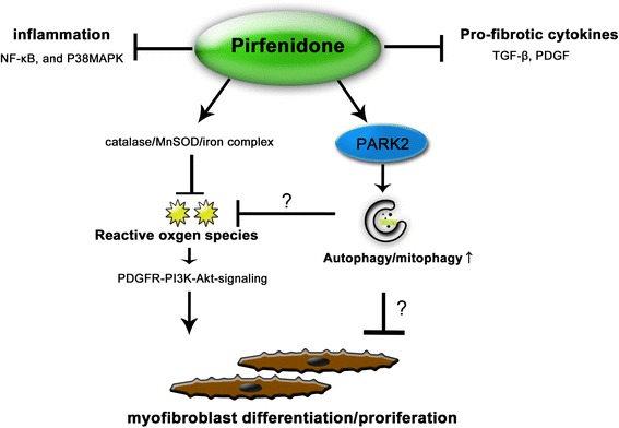
Background: Pirfenidone (PFD) is an anti-fibrotic agent used to treat idiopathic pulmonary fibrosis (IPF), but its precise mechanism of action remains elusive. Accumulation of profibrotic myofibroblasts is a crucial process for fibrotic remodeling in IPF. Recent findings show participation of autophagy/mitophagy, part of the lysosomal degradation machinery, in IPF pathogenesis. Mitophagy has been implicated in myofibroblast differentiation through regulating mitochondrial reactive oxygen species (ROS)-mediated platelet-derived growth factor receptor (PDGFR) activation. In this study, the effect of PFD on autophagy/mitophagy activation in lung fibroblasts (LF) was evaluated, specifically the anti-fibrotic property of PFD for modulation of myofibroblast differentiation during insufficient mitophagy.

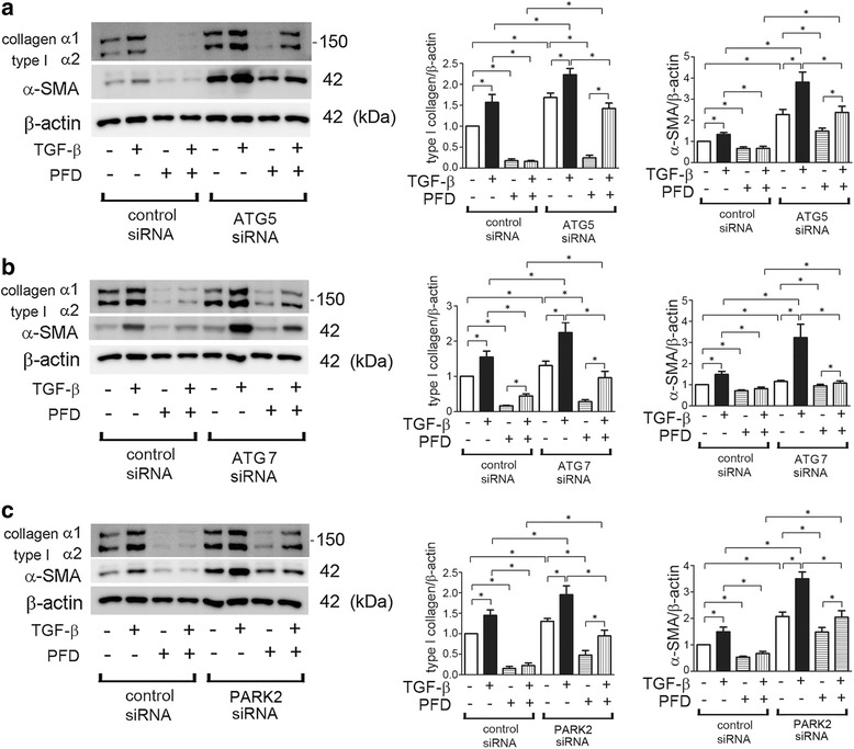
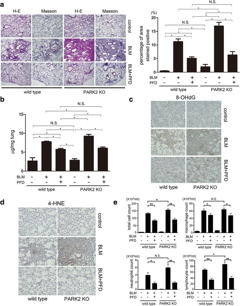
Methods: Transforming growth factor-β (TGF-β)-induced or ATG5, ATG7, and PARK2 knockdown-mediated myofibroblast differentiation in LF were used for in vitro models. The anti-fibrotic role of PFD was examined in a bleomycin (BLM)-induced lung fibrosis model using PARK2 knockout (KO) mice.

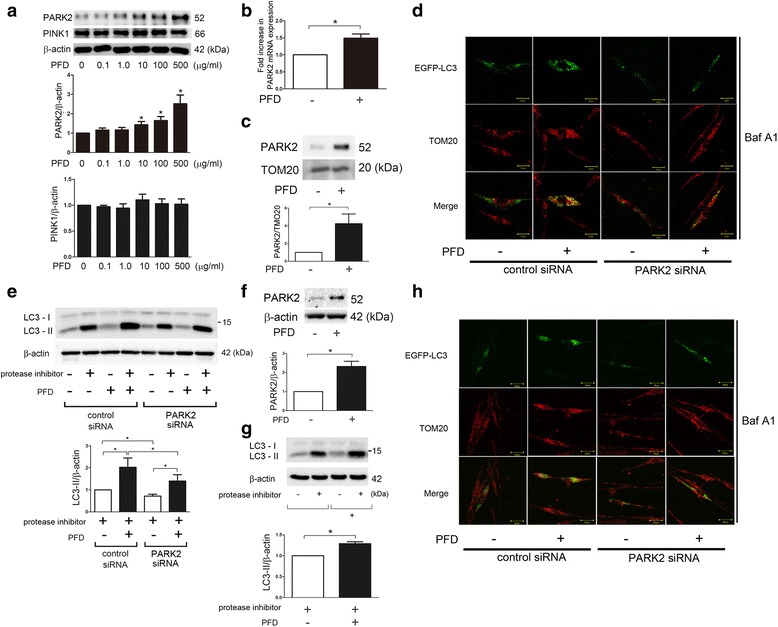
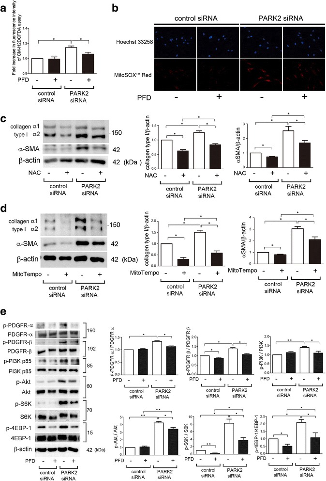
Results: We found that PFD induced autophagy/mitophagy activation via enhanced PARK2 expression, which was partly involved in the inhibition of myofibroblast differentiation in the presence of TGF-β. PFD inhibited the myofibroblast differentiation induced by PARK2 knockdown by reducing mitochondrial ROS and PDGFR-PI3K-Akt activation. BLM-treated PARK2 KO mice demonstrated augmentation of lung fibrosis and oxidative modifications compared to those of BLM-treated wild type mice, which were efficiently attenuated by PFD.

Conclusions: These results suggest that PFD induces PARK2-mediated mitophagy and also inhibits lung fibrosis development in the setting of insufficient mitophagy, which may at least partly explain the anti-fibrotic mechanisms of PFD for IPF treatment.

Keywords: Autophagy; IPF; Mitophagy; Myofibroblast; Pirfenidone.


Figures

Fig. 1
Fig. 1
Autophagy activation by PFD in LF. a Fluorescence microscopic detection of pEGFP-LC3 dot formation in LF: LF were transfected with pEGFP-LC3 and control siRNA, ATG5 siRNA, or ATG7 siRNA. Photomicrographs are taken at the same magnification. (Original magnification, 200X). The lower panel is the percentage of positive cells with more than five dot formations (±SEM) and data was collected from four independent experiments. *p < 0.05. b Western blotting (WB) using anti-LC3 and anti-β-actin of cell lysates from control (lane 1, 2), PFD (500 μg/ml) (lane 3, 4) treated LF. LF were treated with PFD for 24 h and protease inhibitor (E64d 10 μg/ml, pepstatin A 10 μg/ml) treatment was started 6 h before collecting cell lysates. In the lower panel is the average (±SEM) taken from six independent experiments shown as relative expression. *p < 0.05. c WB using anti-LC3 and anti-β-actin of cell lysates from control siRNA (lane 1 ~ 4), ATG5 siRNA (lane 5 ~ 8) transfected LF. PFD (500 μg/ml) treatment was started 48 h post transfection and protein samples were collected after 24 h treatment. Protease inhibitor (E64d 10 μg/ml, pepstatin A 10 μg/ml) treatment was started 6 h before collecting cell lysates. In the right panel is the average (±SEM) taken from five independent experiments shown as relative expression. *p < 0.05. d WB using anti-LC3 and anti-β-actin of cell lysates from control siRNA (lane 1 ~ 4), ATG7 siRNA (lane 5 ~ 8) transfected LF. PFD(500 μg/ml) treatment was started 48 h post transfection and protein samples were collected after 24 h treatment. Protease inhibitor (E64d 10 μg/ml, pepstatin A 10 μg/ml) treatment was started 6 h before collecting cell lysates. In the right panel is the average (±SEM) taken from five independent experiments shown as relative expression. *p < 0.05
Fig. 2
Fig. 2
PARK2-mediated mitophagy activation by PFD in LF. a WB using anti-PARK2, anti-PINK1, and anti-β-actin of cell lysates from control (lane 1) and indicated concentrations of PFD (lane2 ~ 6) treated LF. Protein samples were collected after 24 h treatment with PFD. In the lower panels are the average (±SEM) taken from six independent experiments shown as relative expression. *p < 0.05. b LF were treated with PFD (500 μg/ml) and mRNA samples were collected after 24 h treatment with PFD (n = 4). Real-Time-PCR was performed using primers to PARK2 or β-actin, as a control. PARK2 mRNA expression was normalized to β-actin. Shown is the fold increase (±SEM) relative to control treated cells. *p < 0.05. c WB using anti-PARK2 and anti-TOM20 of cell lysates from PFD (500 μg/ml)-treated LF. Protein samples for mitochondrial fractions were collected after 24 h treatment. The lower panel is the average (±SEM) taken from three independent experiments shown as relative expression. *p < 0.05. d Colocalization analysis of confocal laser scanning microscopic images of TOM20 staining and EGFP-LC3. LF were transfected with pEGFP-LC3 with a concomitant non-silencing control siRNA or PARK2 siRNA. PFD (500 μg/ml) treatment was started 48 h post-transfection. Baf A1 (20 nM) treatment was started 6 h before fixation and LF were fixed after 24 h treatment with PFD. The images are high magnification (400X). (Bar = 20 μm). e WB using anti-LC3 and anti-β-actin of cell lysates from control siRNA (lane 1 ~ 4), PARK2 siRNA (lane 5 ~ 8) transfected LF. PFD (500 μg/ml) treatment was started 48 h post transfection and protein samples were collected after 24 h treatment. Protease inhibitor (E64d 10 μg/ml, pepstatin A 10 μg/ml) treatment was started 6 h before collecting cell lysates. In the right panel is the average (±SEM) taken from four independent experiments shown as relative expression. *p < 0.05. f WB using anti-PARK2 and anti-β-actin of cell lysates from PFD (500 μg/ml)-treated LF isolated from IPF lungs. Protein samples were collected after 24 h treatment. The lower panel is the average (±SEM) taken from six independent experiments shown as relative expression. *p < 0.05. g WB using anti-LC3 and anti-β-actin of cell lysates from control (lane 1, 2), PFD (500 μg/ml) (lane 3, 4) treated LF isolated from IPF lungs. LF were treated with PFD for 24 h and protease inhibitor (E64d 10 μg/ml, pepstatin A 10 μg/ml) treatment was started 6 h before collecting cell lysates. In the lower panel is the average (±SEM) taken from six independent experiments shown as relative expression. *p < 0.05. (H) Colocalization analysis of confocal laser scanning microscopic images of TOM20 staining and EGFP-LC3. LF isolated from IPF lungs were transfected with pEGFP-LC3 with a concomitant non-silencing control siRNA or PARK2 siRNA. PFD (500 μg/ml) treatment was started 48 h post-transfection. Baf A1 (20 nM) treatment was started 6 h before fixation and LF were fixed after 24 h treatment with PFD. The images are high magnification (400X). (Bar = 20 μm)
Fig. 3
Fig. 3
PFD attenuates myofibroblast differentiation during insufficient autopagy/mitophagy in LF. a WB using anti-type I collagen, anti-α-SMA, and anti-β-actin of cell lysates from control siRNA (lane 1 ~ 4), ATG5 siRNA (lane 5 ~ 8) transfected LF. TGF-β (2 ng/ml) and PFD (500 μg/ml) treatment was started 48 h post transfection and protein samples were collected after 24 h treatment. In the right panels are the average (±SEM) taken from five independent experiments shown as relative expression. *p < 0.05. b WB using anti-type I collagen, anti-α-SMA, and anti-β-actin of cell lysates from control siRNA (lane 1 ~ 4), ATG7 siRNA (lane 5 ~ 8) transfected LF. TGF-β (2 ng/ml) and PFD (500 μg/ml) treatment was started 48 h post transfection and protein samples were collected after 48 h treatment. In the right panels are the average (±SEM) taken from five independent experiments shown as relative expression. *p < 0.05. c WB using anti-type I collagen, anti-α-SMA, and anti-β-actin of cell lysates from control siRNA (lane 1 ~ 4), PARK2 siRNA (lane 5 ~ 8) transfected LF. TGF-β (2 ng/ml) and PFD (500 μg/ml) treatment was started 48 h post transfection and protein samples were collected after 24 h treatment. In the right panels are the average (±SEM) taken from five independent experiments shown as relative expression. *p < 0.05
Fig. 4
Fig. 4
PFD attenuates myofibroblast differentiation during insufficient mitophagy via inhibiting PDGFR/PI3K/AKT signaling pathway in LF. a Fluorescence intensity of CM-H2DCFDA staining for intracellular ROS production. PFD (500 μg/ml) treatment was started 48 h post-siRNA transfection and incubation with CM-H2DCFDA (10 μM) was started after 24 h treatment in LF. The fluorescence level in the control siRNA transfected cells without PFD was designated as 1.0. The panel is the average (±SEM) taken from five independent experiments shown as relative expression.* p < 0.05. b Photographs of Hoechst 33258 and MitoSOX Red fluorescence staining in LF. LF were transfected with control siRNA and PARK2 siRNA. PFD (500 μg/ml) treatment was started 48 h post-siRNA transfection and staining was performed 48 h transfection. (Original magnification, 200X) (c) WB using anti-type I collagen, anti-α-SMA, and anti-β-actin of cell lysates from control siRNA (lane 1, 2), PARK2 siRNA (lane 3, 4) transfected LF. NAC (1 mM) treatment was started 48 h post transfection and protein samples were collected after 24 h treatment. In the right panels are the average (±SEM) taken from six independent experiments shown as relative expression. *p < 0.05. d WB using anti-type I collagen, anti-α-SMA, and anti-β-actin of cell lysates from control siRNA (lane 1, 2), PARK2 siRNA (lane 3, 4) transfected LF. MitoTempo (100 μM) treatment was started 48 h post transfection and protein samples were collected after 24 h treatment. In the right panels are the average (±SEM) taken from six independent experiments shown as relative expression. *p < 0.05. e WB using anti-phospho-PDGFR-α (p-PDGFR-α), anti-PDGFR-α, anti-phospho-PDGFR-β (p-PDGFR-β), anti-PDGFR-β, anti-phospho-PI3K p85 (p-PI3K p85), anti-PI3K p85, anti- phospho-AKT (p-AKT), anti-AKT, anti-phopho-S6K (p-S6K), anti-S6K, anti-phospho-4EBP-1 (p-4EBP-1), anti-4EBP-1, and anti-β-actin of cell lysates from control siRNA (lane 1, 2) and PARK2 siRNA(lane 3, 4) transfected LF. PDF (500 μg/ml) treatment was started 48 h post transfection and protein samples were collected after 24 h treatment. In the lower right panels are the average (±SEM) taken from five independent experiments shown as relative expressions. *p < 0.05
Fig. 5
Fig. 5
PFD attenuates bleomycin (BLM)-induced lung fibrosis development in PARK2 KO mice. a Photomicrographs of Hematoxylin & Eosin (H-E) and Masson trichrome staining of mouse lungs at day 21. Original magnification × 200. The right panel shows the average (±SEM) percentage of positively stained areas in Masson trichrome staining quantified using Image J. Treatment groups were composed of vehicle treated wild type (n = 10), BLM-treated wild type (n = 10), BLM-treated wild type with subsequent PFD (n = 10), BLM-treated PARK2 KO (n = 6), and BLM-treated PARK2 KO with subsequent PFD (n = 6). *p < 0.05. N.S.: not statistically significant. b Shown in the panel is the average (±SEM) soluble collagen measurement from Sircol assay using vehicle treated wild type (n = 10), BLM-treated wild type (n = 10), BLM-treated wild type with subsequent PFD (n = 10), BLM-treated PARK2 KO (n = 6), and BLM-treated PARK2 KO with subsequent PFD (n = 6) at day 21. *p < 0.05. N.S.: not statistically significant. c Immunohistochemical staining of 8-hydroxy-2-deoxyguanosine (8-OHdG), oxidized derivative of deoxyguanosine. Original magnification × 200 (d) Immunohistochemical staining of 4-hydroxy-2-nonenal (4-HNE) of lipid peroxidation. Original magnification × 200 (E) Cell counts in broncho-alveolar lavage fluid (BALF). BALF collection was performed at day 21. Cells were counted using a hemocytometer. Differential cell counts in BALF were analyzed from 300 cells stained with Diff-Quick. *p < 0.05, **p < 0.001. N.S.: not statistically significant
Fig. 6
Fig. 6
Hypothetical model of the anti-fibrotic mechanisms of PFD. PFD has previously reported anti-fibrotic mechanisms, including anti-inflammation, anti-pro-fibrotic cytokines, and anti-oxidative properties. Now PARK2-mediated autophay/mitophgy can be included in the anti-fibrotic properties of PFD. Although detailed mechanism remains elusive, PFD attenuates lung fibrosis seen during insufficient mitophagy through regulation of PDGFR-PI3K-Akt signaling by inhibiting mitochondrial ROS production

Similar articles

  • Involvement of PARK2-Mediated Mitophagy in Idiopathic Pulmonary Fibrosis Pathogenesis.
    Kobayashi K, Araya J, Minagawa S, Hara H, Saito N, Kadota T, Sato N, Yoshida M, Tsubouchi K, Kurita Y, Ito S, Fujita Y, Takasaka N, Utsumi H, Yanagisawa H, Hashimoto M, Wakui H, Kojima J, Shimizu K, Numata T, Kawaishi M, Kaneko Y, Asano H, Yamashita M, Odaka M, Morikawa T, Nakayama K, Kuwano K. Kobayashi K, et al. J Immunol. 2016 Jul 15;197(2):504-16. doi: 10.4049/jimmunol.1600265. Epub 2016 Jun 8. J Immunol. 2016. PMID: 27279371
  • Metformin attenuates lung fibrosis development via NOX4 suppression.
    Sato N, Takasaka N, Yoshida M, Tsubouchi K, Minagawa S, Araya J, Saito N, Fujita Y, Kurita Y, Kobayashi K, Ito S, Hara H, Kadota T, Yanagisawa H, Hashimoto M, Utsumi H, Wakui H, Kojima J, Numata T, Kaneko Y, Odaka M, Morikawa T, Nakayama K, Kohrogi H, Kuwano K. Sato N, et al. Respir Res. 2016 Aug 30;17(1):107. doi: 10.1186/s12931-016-0420-x. Respir Res. 2016. PMID: 27576730 Free PMC article.
  • Azithromycin attenuates myofibroblast differentiation and lung fibrosis development through proteasomal degradation of NOX4.
    Tsubouchi K, Araya J, Minagawa S, Hara H, Ichikawa A, Saito N, Kadota T, Sato N, Yoshida M, Kurita Y, Kobayashi K, Ito S, Fujita Y, Utsumi H, Yanagisawa H, Hashimoto M, Wakui H, Yoshii Y, Ishikawa T, Numata T, Kaneko Y, Asano H, Yamashita M, Odaka M, Morikawa T, Nakayama K, Nakanishi Y, Kuwano K. Tsubouchi K, et al. Autophagy. 2017 Aug 3;13(8):1420-1434. doi: 10.1080/15548627.2017.1328348. Epub 2017 Jun 14. Autophagy. 2017. PMID: 28613983 Free PMC article.
  • PINK1-PARK2-mediated mitophagy in COPD and IPF pathogeneses.
    Tsubouchi K, Araya J, Kuwano K. Tsubouchi K, et al. Inflamm Regen. 2018 Oct 24;38:18. doi: 10.1186/s41232-018-0077-6. eCollection 2018. Inflamm Regen. 2018. PMID: 30386443 Free PMC article. Review.
  • Sphingolipids in pulmonary fibrosis.
    Huang LS, Natarajan V. Huang LS, et al. Adv Biol Regul. 2015 Jan;57:55-63. doi: 10.1016/j.jbior.2014.09.008. Epub 2014 Oct 13. Adv Biol Regul. 2015. PMID: 25446881 Free PMC article. Review.

Cited by

References

    1. Selman M, Pardo A. Revealing the pathogenic and aging-related mechanisms of the enigmatic idiopathic pulmonary fibrosis. an integral model. Am J Respir Crit Care Med. 2014;189:1161–1172. doi: 10.1164/rccm.201312-2221PP. - DOI - PubMed
    1. Raghu G, Collard HR, Egan JJ, Martinez FJ, Behr J, Brown KK, Colby TV, Cordier JF, Flaherty KR, Lasky JA, et al. An official ATS/ERS/JRS/ALAT statement: idiopathic pulmonary fibrosis: evidence-based guidelines for diagnosis and management. Am J Respir Crit Care Med. 2011;183:788–824. doi: 10.1164/rccm.2009-040GL. - DOI - PMC - PubMed
    1. Araya J, Nishimura SL. Fibrogenic reactions in lung disease. Annu Rev Pathol. 2010;5:77–98. doi: 10.1146/annurev.pathol.4.110807.092217. - DOI - PubMed
    1. King TE, Jr, Pardo A, Selman M. Idiopathic pulmonary fibrosis. Lancet. 2011;378:1949–1961. doi: 10.1016/S0140-6736(11)60052-4. - DOI - PubMed
    1. Richeldi L, du Bois RM, Raghu G, Azuma A, Brown KK, Costabel U, Cottin V, Flaherty KR, Hansell DM, Inoue Y, et al. Efficacy and safety of nintedanib in idiopathic pulmonary fibrosis. N Engl J Med. 2014;370:2071–2082. doi: 10.1056/NEJMoa1402584. - DOI - PubMed

MeSH terms