. 2017 Jun 2;14(1):111.
doi: 10.1186/s12974-017-0882-x.

Sphingosine 1-phosphate receptor 3 and RhoA signaling mediate inflammatory gene expression in astrocytes

Affiliations

Sphingosine 1-phosphate receptor 3 and RhoA signaling mediate inflammatory gene expression in astrocytes

Stephanie S Dusaban et al. J Neuroinflammation. .

Abstract

Background: Sphingosine 1-phosphate (S1P) signals through G protein-coupled receptors to elicit a wide range of cellular responses. In CNS injury and disease, the blood-brain barrier is compromised, causing leakage of S1P from blood into the brain. S1P can also be locally generated through the enzyme sphingosine kinase-1 (Sphk1). Our previous studies demonstrated that S1P activates inflammation in murine astrocytes. The S1P1 receptor subtype has been most associated with CNS disease, particularly multiple sclerosis. S1P3 is most highly expressed and upregulated on astrocytes, however, thus we explored the involvement of this receptor in inflammatory astrocytic responses.

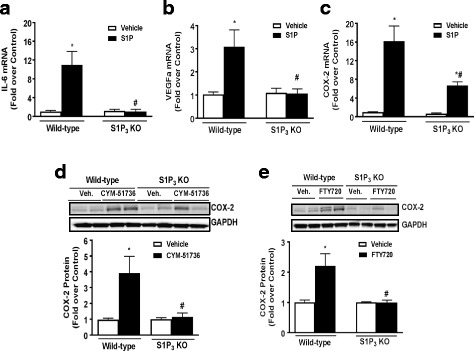
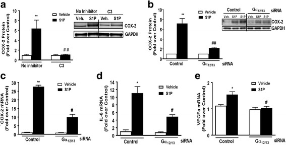
Methods: Astrocytes isolated from wild-type (WT) or S1P3 knockout (KO) mice were treated with S1P3 selective drugs or transfected with short interfering RNA to determine which receptor subtypes mediate S1P-stimulated inflammatory responses. Interleukin-6 (IL-6), and vascular endothelial growth factor A (VEGFa) messenger RNA (mRNA) and cyclooxygenase-2 (COX-2) mRNA and protein were assessed by q-PCR and Western blotting. Activation of RhoA was measured using SRE.L luciferase and RhoA implicated in S1P signaling by knockdown of Gα12/13 proteins or by inhibiting RhoA activation with C3 exoenzyme. Inflammation was simulated by in vitro scratch injury of cultured astrocytes.

Results: S1P3 was highly expressed in astrocytes and further upregulated in response to simulated inflammation. Studies using S1P3 knockdown and S1P3 KO astrocytes demonstrated that S1P3 mediates activation of RhoA and induction of COX-2, IL-6, and VEGFa mRNA, with some contribution from S1P2. S1P induces expression of all of these genes through coupling to the Gα12/13 proteins which activate RhoA. Studies using S1P3 selective agonists/antagonists as well as Fingolimod (FTY720) confirmed that stimulation of S1P3 induces COX-2 expression in astrocytes. Simulated inflammation increased expression of Sphk1 and consequently activated S1P3, demonstrating an autocrine pathway through which S1P is formed and released from astrocytes to regulate COX-2 expression.

Conclusions: S1P3, through its ability to activate RhoA and its upregulation in astrocytes, plays a unique role in inducing inflammatory responses and should be considered as a potentially important therapeutic target for CNS disease progression.

Keywords: Astrocytes; Central nervous system; Inflammation; RhoA; S1P; S1P3.


Figures

Fig. 1
Fig. 1
S1P3 is highly expressed in astrocytes and mediates COX-2 expression. In WT astrocytes, a S1P1, S1P2, and S1P3 mRNA expression was measured by absolute PCR. b Quantification and Western blot of COX-2 protein expression after S1P treatment (0.5 μM, 6 h) following knockdown of S1P1, S1P2, S1P3, and S1P2/3 with siRNA (2 μM). Data shown are mean ± SEM of values from three independent experiments run in triplicate. COX-2 was normalized to GAPDH and expressed relative to control siRNA vehicle treated. *p < 0.05 and **p < 0.01 between vehicle and S1P-treated groups and #p < 0.05 and ##p < 0.01 between control siRNA S1P-treated and S1P receptor siRNA S1P-treated groups. c Quantification and Western blot of COX-2 expression after pretreatment with the S1P2 antagonist JTE-013 for 30 min (1 μM) followed by S1P treatment (0.5 μM, 6 h). Data shown are mean ± SEM of values from three independent samples. COX-2 was normalized to GAPDH and expressed relative to vehicle control. *p < 0.05 between vehicle and S1P-treated groups. d Quantification and Western blot of COX-2 expression after pretreatment with the S1P3 antagonist SPM-354 for 15 min (5 μM) followed by S1P treatment (0.5 μM, 6 h). Data shown are mean ± SEM of values from three independent experiments run in triplicate. COX-2 was normalized to GAPDH and expressed relative to vehicle control. *p < 0.05 between vehicle and S1P-treated groups and #p < 0.05 between control S1P-treated and SPM-454/S1P-treated group
Fig. 2
Fig. 2
S1P3 is required for induction of inflammation in astrocytes. WT and S1P3 KO astrocytes were treated with vehicle or S1P (0.5 μM) for 1 h, and IL-6 (a), VEGFa (b), and COX-2 (c) mRNA expression was measured by q-PCR. IL-6, VEGFa, and COX-2 were normalized to GAPDH, and fold increase expressed relative to the WT or KO vehicle treated. Data shown are mean ± SEM of three independent experiments run in triplicate. *p < 0.01 between vehicle and S1P-treated groups and #p < 0.01 between WT and KO S1P-treated groups. d Quantification and Western blot of COX-2 expression in WT and S1P3 KO astrocytes treated with CYM-51736 (10 μM, 6 h). Data shown are mean ± SEM of values from three independent experiments run in triplicate. COX-2 was normalized to GAPDH and expressed relative to the WT or KO vehicle treated. e Quantification and Western blot of COX-2 expression in FTY720-treated (100 nM, 6 h) WT and S1P3 KO astrocytes. Data shown are mean ± SEM of values from three independent experiments run in triplicate. COX-2 was normalized to GAPDH and expressed relative to the WT or KO vehicle treated. *p < 0.01 between vehicle and treatment groups and #p < 0.01 between WT and KO treatment groups
Fig. 3
Fig. 3
S1P signals through Gα12/13 and RhoA to induce COX-2 expression. a COX-2 protein expression was measured in WT cells pretreated with C3 exoenzyme (0.5 μg/mL) for 4 h prior to vehicle or S1P (0.5 μM) treatment for 6 h. COX-2 was normalized to GAPDH and expressed relative to the averaged ± inhibitor controls. Representative Western blot and data shown are mean ± SEM from three independent experiments run in triplicate. The blot represents a single gel where unnecessary lanes have been removed. **p < 0.01 between vehicle and S1P-treated groups and ##p < 0.01 between S1P-treated ± inhibitor groups. b Quantification and Western blot of COX-2 protein levels after knockdown of Gα12/13 with siRNA (2 μM) followed by S1P treatment (5 μM, 6 h). The blot represents a single gel where unnecessary lanes have been removed. Data shown are mean ± SEM of values from four independent experiments run in triplicate. COX-2 was normalized to GAPDH and expressed relative to the siRNA control. mRNA expression levels of COX-2 (c), IL-6 (d), and VEGFa (e) were measured by q-PCR following knockdown of Gα12/13 with siRNA (2 μM) and S1P treatment (0.5 μM, 1 h). Data shown are mean ± SEM from three independent experiments run in triplicate. COX-2, IL-6, and VEGFa were normalized to GAPDH and fold increase expressed relative to the vehicle-treated control siRNA. *p < 0.05 and **p < 0.01 between vehicle and S1P-treated groups and #p < 0.05 and ##p < 0.01 between control and Gα12/13 S1P-treated groups
Fig. 4
Fig. 4
S1P2, S1P3, and Gα12/13 are required for Rho activation in astrocytes. WT astrocytes were transfected with an SRE.L luciferase reporter construct to assess Rho activation following knockdown with control siRNA or siRNA (2 μM) targeting S1P1, S1P2, S1P3, Gα12/13, or Gαq. Cells were also treated with pertussis toxin (PTX, 100 ng/mL, 24 h) to inhibit Gαi or with the C3 exoenzyme (0.5 μg/mL, 4 h) to inhibit Rho. Following knockdown or pretreatment, cells were stimulated with S1P (0.5 μM, 8 h). Control siRNA treated cells stimulated with S1P were set at 100% response. Data shown are mean ± SEM from four independent experiments run in duplicate. #p < 0.05 between control and siRNA S1P-treated groups
Fig. 5
Fig. 5
S1P3 and Sphk1 are upregulated in response to in vitro scratch injury and mediate COX-2 expression. In WT astrocytes, a S1P1, S1P2, S1P3 and b SphK1 mRNA expression were measured 1 h after scratch injury on culture plates. Data shown are mean ± SEM from three independent experiments run in triplicate. Fold increase is relative to the scratch control for each receptor subtype or Spkh1. *p < 0.05 between control and scratch-treated groups. COX-2 protein levels were measured after 8 h of scratch in c S1P3 KO astrocytes or d WT astrocytes pretreated with SPM-354 (5 μM, 15 min). COX-2 was normalized to GAPDH and expressed relative to the WT or KO controls or the ±inhibitor controls. Data shown are mean ± SEM from three independent experiments run in triplicate. e COX-2 protein levels were measured in WT astrocytes after knockdown of Sphk1 with siRNA (2 μM) or control siRNA followed by scratch for 8 h. COX-2 was normalized to GAPDH and expressed relative to the ±siRNA controls. Data shown are mean ± SEM from three independent experiments run in duplicate. * p < 0.05 and **p < 0.01 between control and scratch-treated groups and #p < 0.05 and ##p < 0.01 between scratch-treated groups of either WT and KO, with or without inhibitor, or siRNA

Similar articles

Cited by

References

    1. Camerer E, Regard JB, Cornelissen I, Srinivasan Y, Duong DN, Palmer D, Pham TH, Wong JS, Pappu R, Coughlin SR. Sphingosine-1-phosphate in the plasma compartment regulates basal and inflammation-induced vascular leak in mice. J Clin Invest. 2009;119:1871–1879. - PMC - PubMed
    1. Anelli V, Bassi R, Tettamanti G, Viani P, Riboni L. Extracellular release of newly synthesized sphingosine-1-phosphate by cerebellar granule cells and astrocytes. J Neurochem. 2005;92:1204–1215. doi: 10.1111/j.1471-4159.2004.02955.x. - DOI - PubMed
    1. Pappu R, Schwab SR, Cornelissen I, Pereira JP, Regard JB, Xu Y, Camerer E, Zheng YW, Huang Y, Cyster JG, Coughlin SR. Promotion of lymphocyte egress into blood and lymph by distinct sources of sphingosine-1-phosphate. Science. 2007;316:295–298. doi: 10.1126/science.1139221. - DOI - PubMed
    1. Yatomi Y, Ohmori T, Rile G, Kazama F, Okamoto H, Sano T, Satoh K, Kume S, Tigyi G, Igarashi Y, Ozaki Y. Sphingosine 1-phosphate as a major bioactive lysophospholipid that is released from platelets and interacts with endothelial cells. Blood. 2000;96:3431–3438. - PubMed
    1. Yatomi Y, Igarashi Y, Yang L, Hisano N, Qi R, Asazuma N, Satoh K, Ozaki Y, Kume S. Sphingosine 1-phosphate, a bioactive sphingolipid abundantly stored in platelets, is a normal constituent of human plasma and serum. J Biochem. 1997;121:969–973. doi: 10.1093/oxfordjournals.jbchem.a021681. - DOI - PubMed

Publication types

MeSH terms