. 2017 Aug:508:199-212.
doi: 10.1016/j.virol.2017.05.016. Epub 2017 May 31.

Glycosylation and oligomeric state of envelope protein might influence HIV-1 virion capture by α4β7 integrin

Affiliations

Glycosylation and oligomeric state of envelope protein might influence HIV-1 virion capture by α4β7 integrin

Subhash Chand et al. Virology. 2017 Aug.

Abstract

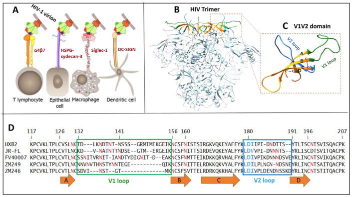
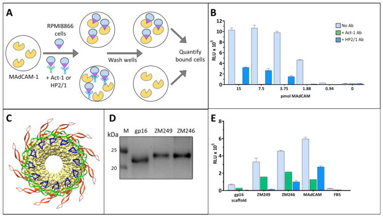
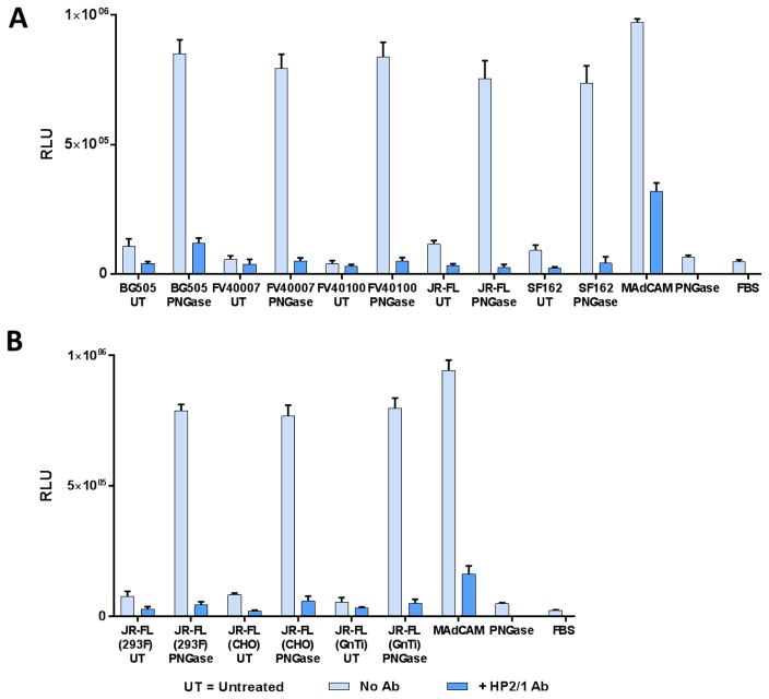
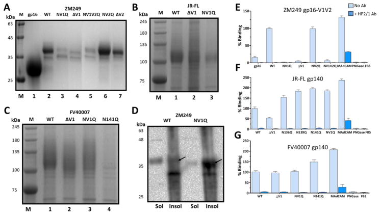
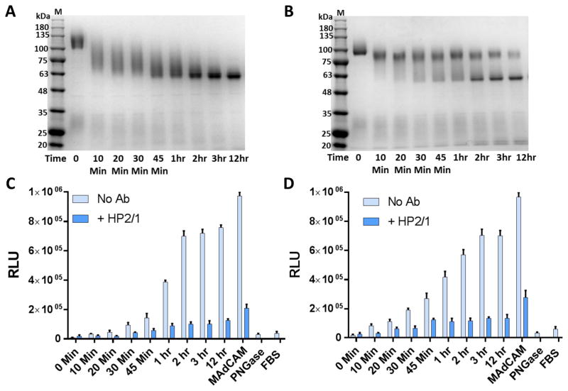
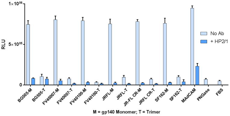
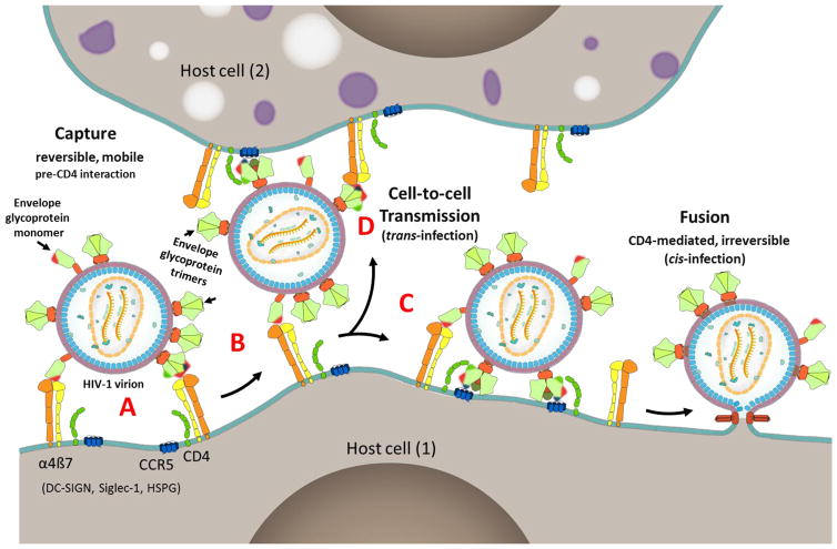
The α4ß7 integrin present on host cells recognizes the V1V2 domain of the HIV-1 envelope protein. This interaction might be involved in virus transmission. Administration of α4ß7-specific antibodies inhibit acquisition of SIV in a macaque challenge model. But the molecular details of V1V2: α4ß7 interaction are unknown and its importance in HIV-1 infection remains controversial. Our biochemical and mutational analyses show that glycosylation is a key modulator of V1V2 conformation and binding to α4ß7. Partially glycosylated, but not fully glycosylated, envelope proteins are preferred substrates for α4ß7 binding. Surprisingly, monomers of the envelope protein bound strongly to α4ß7 whereas trimers bound poorly. Our results suggest that a conformationally flexible V1V2 domain allows binding of the HIV-1 virion to the α4ß7 integrin, which might impart selectivity for the poorly glycosylated HIV-1 envelope containing monomers to be more efficiently captured by α4ß7 integrin present on mucosal cells at the time of HIV-1 transmission.

Keywords: Envelope glycoprotein; HIV vaccine; HIV-1; V1V2 domain; Virus capture; Virus entry; α4β7 integrin.


Conflict of interest statement

Conflicts of interest

The authors declare that they have no conflicts of interest with the contents of this article.

Figures

Fig. 1
Fig. 1. Structure and function of the V1V2 domain of HIV-1 envelope glycoprotein
A, Attachment of HIV-1 virion to host cells may be mediated by interaction of Env V1V2 domain with different surface molecules (attachment factors) present on different host cells. Siglec-1 binds sialic acid moieties on glycans in the V1V2 region. HSPG presented on sydecan-3 binds the V3 loop and has a binding site in the C-strand of the V1V2 region. DC-SIGN binds glycans on gp120 and enhancement of virus infection can be modulated by the V1V2 length, the overall V3 charge, and N-linked glycosylation patterns. One Env trimer of HIV-1 virion is shown; V1V2 domain is shown in red, CD4 binding site in green, and V3 domain in blue. B, Structure of the HIV-1 trimer (PDB: 4NCO (Ringe et al., 2013) showing the V1V2 domains at the apex. C, V1V2 domain is enlarged. V1 loop is shown in green, β-strands labeled A–D in orange, and V2 loop in blue. The residues missing in the structure are modeled using Swiss-Model web-server by homology with PDB:4NCO. D, Sequence alignment of the V1V2 domains used in this study. The β-strands are denoted as orange arrows. Potential N-linked glycosylation sites (NXT/S) are shown in red. The variable loops are boxed (colors correspond to C). The conserved LDI/V tripeptide is highlighted in blue. The numbers correspond to amino acids in HXB2 envelope glycoprotein.
Fig. 2
Fig. 2. gp16-V1V2 scaffold and gp140 protomer proteins binding to CH58, CH59 and PG9
A, CH58 binding residues in V2 sequence (156–187) of JR-FL, FV40007, Zm249 are denoted with blue letters. B, CH59 binding residues in V2 sequence (164–187) of JR-FL, FV40007, Zm249 is denoted with orange letters. C–E gp16-V1V2 scaffold and gp140 protomer proteins binding to CH58 C, CH59 D, and PG9 E. The proteins used in binding are gp16 expressed in E. coli (pink line), Zm249 gp16-V1V2 expressed in E. coli (red line), gp16 expressed in GnTI (khaki line), Zm249 gp16-V1V2 expressed in GnTI (black line), JR-FL gp140 expressed in 293F (blue line), and FV40007 gp140 expressed in 293F (green line). Binding data are representative of at least three independent assays, done in triplicate.
Fig. 3
Fig. 3. Specificity of interaction between the V1V2 domain and the α4β7 integrin
A, A schematic representation of the α4β7 binding assay. Wells of a 96-well plate are coated with the MAdCAM-1 and blocked with FBS to prevent nonspecific binding. The α4β7-expressing RPMI cells are added to the wells and the extent of binding is quantified by the luciferase-based CellTiterGlo kit (Promega). See Materials and Methods for more details. B, α4β7 binding of MAdCAM-1 over a range of concentrations. Binding of MAdCAM-1 (light blue bars) is specific as shown by the inhibition of binding by α4β7-specific mAbs, Act-1 (green bars) and HP2/1 (dark blue bars). C, A model depicting the multimeric display of the V1V2 domain (colored as in Fig. 1B) fused to the dodecameric phage T4 gp16-scaffold (yellow). The V1V2 domain is shown in ribbons and gp16 in surface view. D, SDS-PAGE (4–20% gradient) analysis of the E. coli-produced gp16 and gp16-V1V2 proteins. Lane M represents molecular weight markers. E, α4β7 binding activity of the proteins shown in D. Specificity of V1V2 binding to α4β7 was determined by the decrease in relative luminescence in the presence of the α4β7-specific mAbs, Act-1 (green bars) and HP2/1 (dark blue bars). Light blue bars show α4β7 binding activity in the absence of any mAb inhibitor. Data are shown as mean +/− SEM and are representative of at least three independent assays and three different preparations of proteins, done in triplicate.
Fig. 4
Fig. 4. Deglycosylated, but not glycosylated, V1V2 domain binds α4β7
A, α4β7 binding of ZM249 gp16-V1V2 with (PNGase) and without (untreated) PNGase treatment. B, SDS-PAGE (4–20% gradient) analysis of ZM249 gp16-V1V2 treated with PNGase in the presence (+) or absence (−) of SDS. C, Native-PAGE (4–16% gradient) analysis of JR-FL gp140 and ZM249 gp16-V1V2 with and without PNGase treatment. D, SDS-PAGE (4–20% gradient) analysis of JR-FL and FV40007 gp140s treated with PNGase, with and without SDS. E and F α4β7 binding activity of FV40007 E and JR-FL F, gp140s with and without PNGase treatment. Lanes M represents molecular weight markers. α4β7 binding assays in the absence of mAb inhibitor (light blue bars) or in the presence of Act-1 mAb (green bars) or HP2/1 mAb (dark blue bars) were performed as described in the Fig. 3 legend. Binding was expressed as percent of PNGase-treated gp140 from FV40007 E or JR-FL F. Data are shown as mean +/− SEM and are representative of at least three independent assays and three different preparations of proteins, done in triplicate.
Fig. 5
Fig. 5. The α4β7 integrin binds to deglycosylated (poorly glycosylated) gp140 HIV-1 envelope protein, regardless of the type of glycosylation or the HIV-1 subtype
A, α4β7 binding with gp140 proteins of five strains of HIV-1 (BG505, FV40007, FV40100, JR-FL, and SF162). B, α4β7 binding activity of the JR-FL gp140 proteins produced in three different cell lines (293F, CHO, and GnTI). α4β7 binding activity in the absence of any mAb inhibitor was shown in light blue bars. Specificity of V1V2 binding to α4β7 was determined by the decrease in relative luminescence in the presence of the α4β7-specific HP2/1 mAb (dark blue bars). Data are shown as mean +/− SEM and are representative of at least three independent assays and three different preparations of proteins, done in triplicate.
Fig. 6
Fig. 6. Glycosylation of V1V2 domain modulates protein conformation, processing, and α4β7 binding
A–C, SDS-PAGE (12%) analysis of the glycosylation mutants of Zm249 gp16-V1V2 A, JR-FL gp140 B, and FV40007 gp140 C. D, Western blot analysis using gp16-antibodies of the soluble secreted protein (sol) and the insoluble cell pellet (insol) following transfection with WT or various glycosylation mutants of ZM249 gp16-V1V2. E–G, α4β7 binding activity of the glycosylation mutants of Zm249 gp16-V1V2 E, FV40007 gp140 F, and JR-FL gp140 G after treatment with PNGase under non-denaturing conditions (no SDS). Lanes M represents molecular weight markers. NV1Q and NV2Q refer to mutants in which all asparagine residues were mutated to glutamine residues in the V1 or V2 loop, respectively, and NV1V2Q refers to the mutant in which all asparagine residues were mutated to glutamine residues in both the V1 and the V2 loops. ΔV1 refers to the deletion of the entire V1 loop. α4β7 binding assays in the absence (light blue bars) or presence (dark blue bars) of HP2/1 mAb were performed as described in the Fig. 3 legend. Binding was expressed as percent of PNGase-treated: ZM249 gp16-V1V2 E, WT JR-FL gp140 F, or WT FV40007 gp140 G. Data are shown as mean +/− SEM and are representative of at least three independent assays and three different preparations of proteins, done in triplicate.
Fig. 7
Fig. 7. Degree of deglycosylation correlates with α4β7 binding
A and B, SDS-PAGE (4–20% gradient) analysis of FV40007 gp140 A, and JR-FL gp140 B. gp140 Env monomers were treated with PNGase for different time intervals (0 Min to 12 hr). C and D, Binding of α4β7 to FV40007 gp140 C, and JR-FL gp140 D after deglycosylation for the time intervals indicated in A and B. Lanes M represent molecular weight markers. α4β7 binding assays in the absence of mAb inhibitor (light blue bars) or in the presence of HP2/1 mAb (dark blue bars) were performed as described in the Fig. 3 legend. Data are shown as mean +/− SEM and are representative of at least three independent assays done in triplicate.
Fig. 8
Fig. 8. Monomers, but not trimers, of gp140 bind to α4β7
α4β7 binding activity of the monomers and trimers of BG505, FV40007, FV40100, JR-FL, JR-FL CR and SF162 before and after PNGase treatment under non-denaturing conditions (see Materials and Methods section for additional details). “M” represents monomers. “T” represents trimers. “CR” represents cleavage resistant (uncleaved). All except JR-FL were cleaved. See Supplementary file 1: Supplementary Fig. S5A–D for the purification profiles of monomers and trimers. α4β7 binding in the absence of mAb inhibitor (light blue bars) or in the presence of HP2/1 mAb (dark blue bars) were performed as described in the Fig. 3 legend. Data are shown as mean +/− SEM and are representative of at least three independent assays done in triplicate.
Fig. 9
Fig. 9. A model for HIV-1 virion capture
A, HIV-1 virion attaches to host cell (1) through reversible multipoint attachment between the V1V2 domain and α4β7 integrin, leading to virus capture. Other cell surface molecules such as DC-SIGN, Siglec-1, and HSPG (Fig. 1A) might also serve as attachment factors leading to virus capture by a variety of host cells. Poorly glycosylated envelope and presence of monomers of the envelope protein enhance virion capture by α4β7 integrin. Patches of red, green, and blue shown on the interacting envelope proteins represent V1V2 domain, CD4 binding site, and V3 domain, which serve as binding sites for α4β7 integrin, CD4 receptor, and CCR5 receptor, respectively. B and C, Capture allows the virus to move on the cell surface and find its primary receptor CD4 and co-receptor CCR5 while remained bound to the host cell. The curvature of the virion might also be a factor in reaching the receptors that may otherwise be buried in the surface maze (C). A series of conformational changes follow resulting in membrane fusion and entry (cis-infection). D, Alternatively, the virus engages with the primary and co-receptors present on another host cell (2) resulting in cell-to-cell transmission (trans-infection).

Similar articles

Cited by

References

    1. AlSalmi W, Mahalingam M, Ananthaswamy N, Hamlin C, Flores D, Gao G, Rao VB. A New Approach to Produce HIV-1 Envelope Trimers: BOTH CLEAVAGE AND PROPER GLYCOSYLATION ARE ESSENTIAL TO GENERATE AUTHENTIC TRIMERS. J Biol Chem. 2015;290(32):19780–19795. doi: 10.1074/jbc.M115.656611. - DOI - PMC - PubMed
    1. Ansari AA, Reimann KA, Mayne AE, Takahashi Y, Stephenson ST, Wang R, … Villinger F. Blocking of alpha4beta7 gut-homing integrin during acute infection leads to decreased plasma and gastrointestinal tissue viral loads in simian immunodeficiency virus-infected rhesus macaques. J Immunol. 2011;186(2):1044–1059. doi: 10.4049/jimmunol.1003052. - DOI - PMC - PubMed
    1. Arthos J, Cicala C, Martinelli E, Macleod K, Van Ryk D, Wei D, … Fauci AS. HIV-1 envelope protein binds to and signals through integrin alpha4beta7, the gut mucosal homing receptor for peripheral T cells. Nat Immunol. 2008;9(3):301–309. doi: 10.1038/ni1566. - DOI - PubMed
    1. Beddows S, Schulke N, Kirschner M, Barnes K, Franti M, Michael E, … Moore JP. Evaluating the immunogenicity of a disulfide-stabilized, cleaved, trimeric form of the envelope glycoprotein complex of human immunodeficiency virus type 1. J Virol. 2005;79(14):8812–8827. doi: 10.1128/JVI.79.14.8812-8827.2005. - DOI - PMC - PubMed
    1. Berlin C, Berg EL, Briskin MJ, Andrew DP, Kilshaw PJ, Holzmann B, … Butcher EC. Alpha 4 beta 7 integrin mediates lymphocyte binding to the mucosal vascular addressin MAdCAM-1. Cell. 1993;74(1):185–195. - PubMed

Publication types

MeSH terms