. 2017 Aug:161:174-192.
doi: 10.1016/j.exer.2017.05.012. Epub 2017 May 31.

Sox2 regulates Müller glia reprogramming and proliferation in the regenerating zebrafish retina via Lin28 and Ascl1a

Affiliations

Sox2 regulates Müller glia reprogramming and proliferation in the regenerating zebrafish retina via Lin28 and Ascl1a

Ryne A Gorsuch et al. Exp Eye Res. 2017 Aug.

Abstract

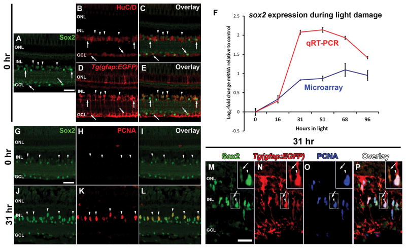
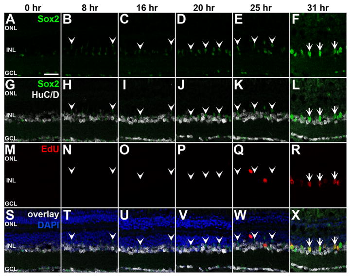
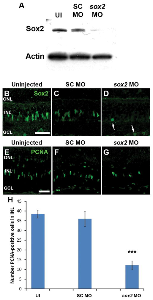
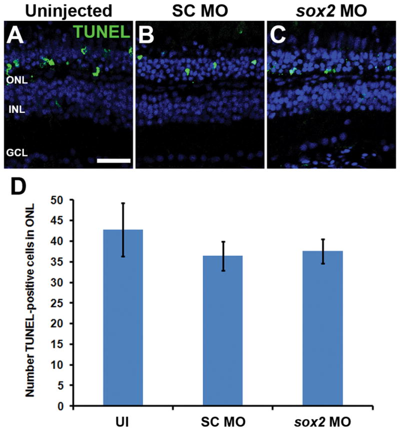
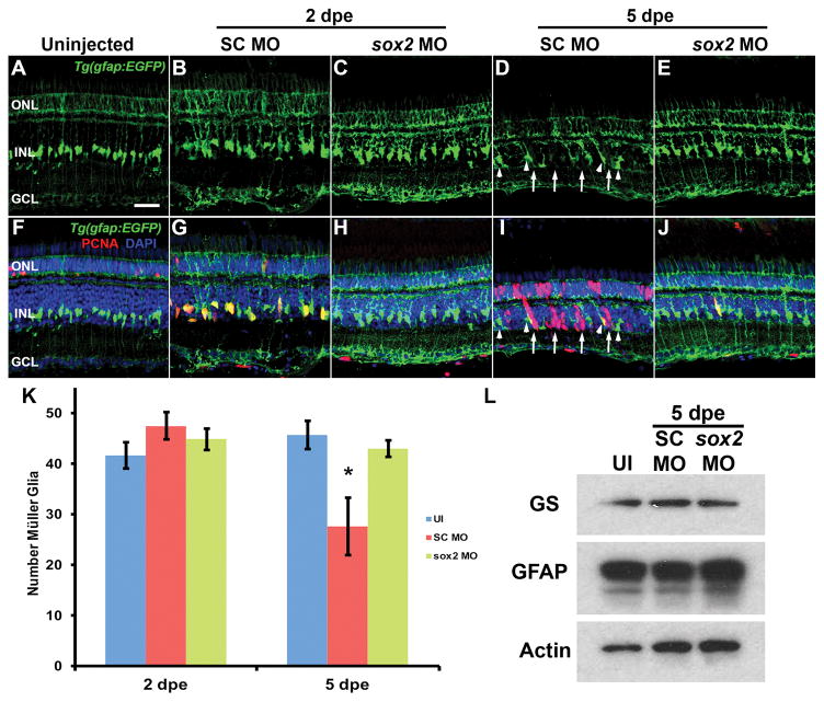
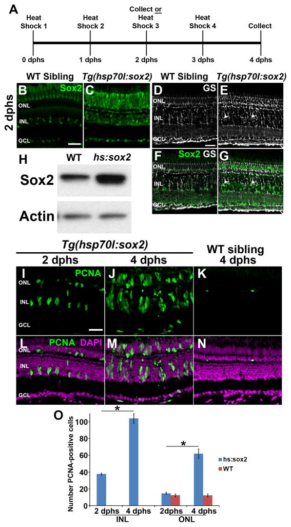
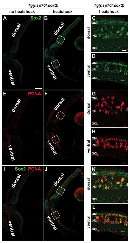
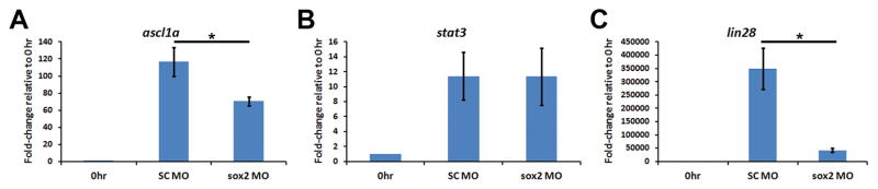
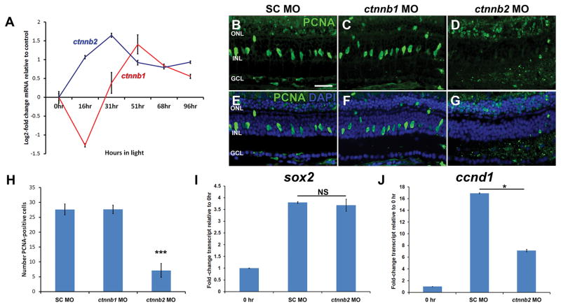
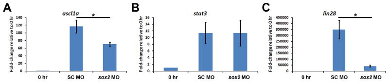
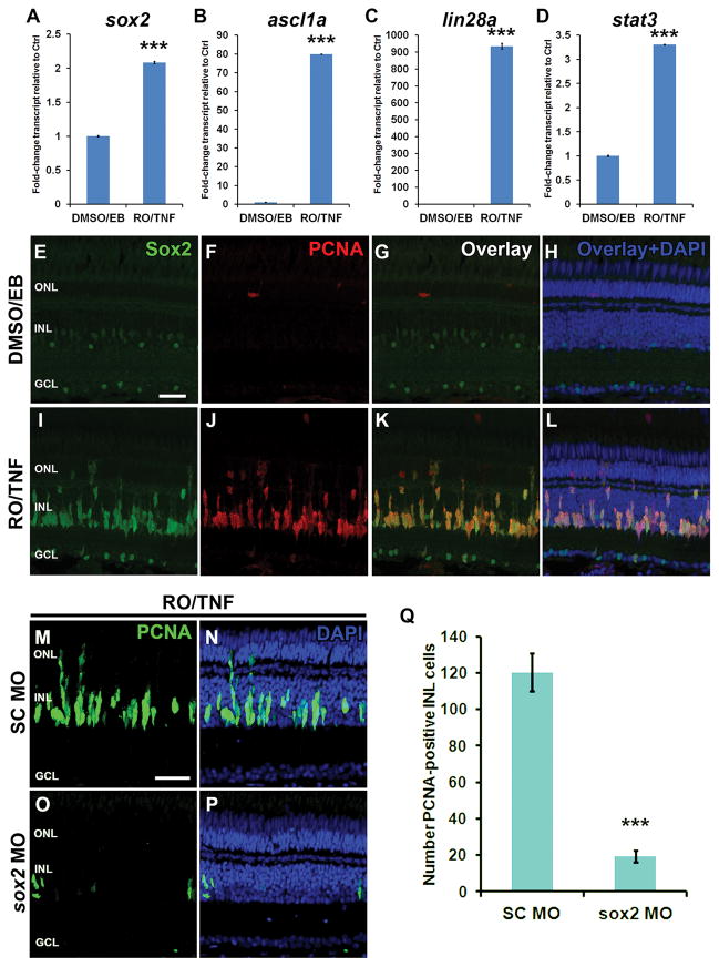
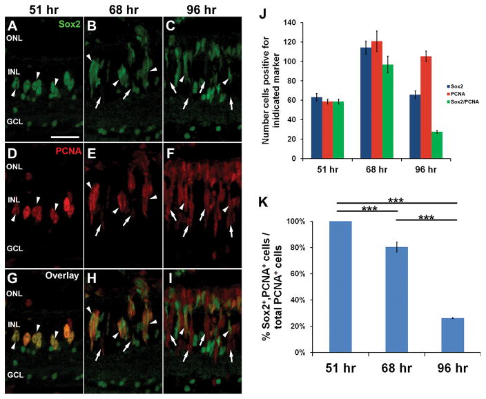
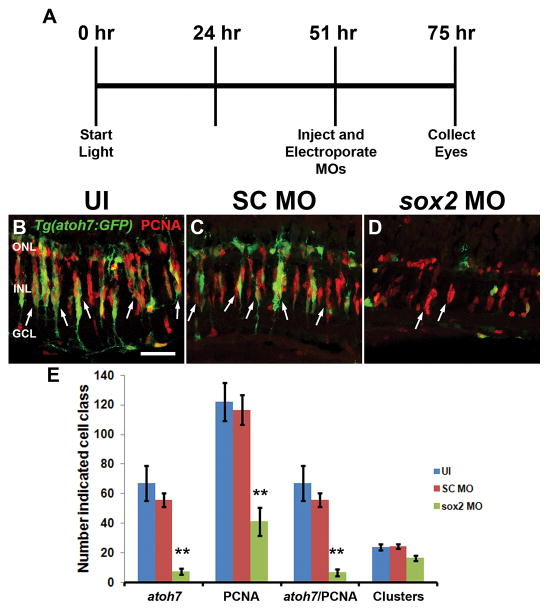
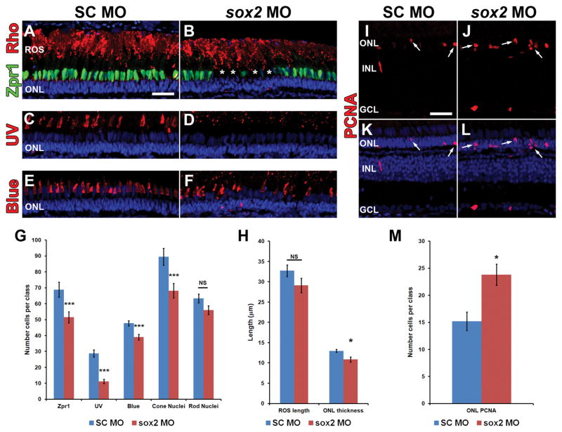
Sox2 is a well-established neuronal stem cell-associated transcription factor that regulates neural development and adult neurogenesis in vertebrates, and is one of the critical genes used to reprogram differentiated cells into induced pluripotent stem cells. We examined if Sox2 was involved in the early reprogramming-like events that Müller glia undergo as they upregulate many pluripotency- and neural stem cell-associated genes required for proliferation in light-damaged adult zebrafish retinas. In the undamaged adult zebrafish retina, Sox2 is expressed in Müller glia and a subset of amacrine cells, similar to other vertebrates. Following 31 h of light damage, Sox2 expression significantly increased in proliferating Müller glia. Morpholino-mediated knockdown of Sox2 expression resulted in decreased numbers of proliferating Müller glia, while induced overexpression of Sox2 stimulated Müller glia proliferation in the absence of retinal damage. Thus, Sox2 is necessary and sufficient for Müller glia proliferation. We investigated the role of Wnt/β-catenin signaling, which is a known regulator of sox2 expression during vertebrate retinal development. While β-catenin 2, but not β-catenin 1, was necessary for Müller glia proliferation, neither β-catenin paralog was required for sox2 expression following retinal damage. Sox2 expression was also necessary for ascl1a (neurogenic) and lin28a (reprogramming) expression, but not stat3 expression following retinal damage. Furthermore, Sox2 was required for Müller glial-derived neuronal progenitor cell amplification and expression of the pro-neural marker Tg(atoh7:EGFP). Finally, loss of Sox2 expression prevented complete regeneration of cone photoreceptors. This study is the first to identify a functional role for Sox2 during Müller glial-based regeneration of the vertebrate retina.

Keywords: Müller glia; Neuronal progenitor cell; Regeneration; Sox2; β-catenin.


Conflict of interest statement

Conflict of Interest: The authors declare no competing financial interests.

Figures

Figure 1
Figure 1. Sox2 expression is dynamic in the regenerating zebrafish retina
Cryosections of undamaged adult albino;Tg(gfap:EGFP) zebrafish retinas were prepared and labeled with anti-Sox2 antiserum (A–E) and either the amacrine and ganglion cell marker HuC/D (B,C) or anti-EGFP to label Müller glia (D,E). Sox2 co-labeled with HuC/D-positive round nuclei in the INL and GCL (A–C; arrows) and fusiform Müller glia nuclei in the INL (A, D, E, arrowheads). RNA was isolated from adult zebrafish retinas at 0, 16, 31, 51, 68 and 96 hours of constant intense light. Total cDNA was prepared and sox2 expression analyzed via qRT-PCR (F). After 31 hours of constant light, sox2 expression increased > 2 log2-fold, and remained elevated throughout the remainder of the time course. This increased sox2 expression is consistent with our previous microarray data (Kassen et al., 2007; F). Retinal cryosections from undamaged (0 h, G–I) or 31 hour light-damaged eyes (J–L) revealed low levels of Sox2 expression in Müller glia of the undamaged retina (G and I, arrowheads) and greatly increased expression in PCNA-positive INL cells following 31 hours of light (J–L, arrowheads). The acquisition settings for panels G–L were equivalent and set for an optimal dynamic range for Sox2 expression at the 31 hour time point (J and L). Co-labeling of Sox2, PCNA and EGFP in Tg(gfap:EGFP) transgenic zebrafish retinas (M–P), which express EGFP in the Müller glia, revealed elevated Sox2 expression in PCNA-positive Müller glia compared to PCNA-negative Müller glia (arrowheads and arrows, respectively) after 31 hours of light. Inset corresponds to the boxed region in each panel and contains both a PCNA-positive and PCNA-negative Müller glia (arrowhead and arrow, respectively). qRT-PCR, quantitative real-time polymerase chain reaction; PCNA, proliferating cell nuclear antigen; GCL, ganglion cell layer; INL, inner nuclear layer; ONL, outer nuclear layer. Scale bars in panels A, G and M are 25 μm and are the same for panels B–E, H–L and N–P, respectively.
Figure 2
Figure 2. Sox2 expression increases in the light-damaged retina prior to Müller glia proliferation
Dark-adapted adult albino zebrafish were placed in constant intense light and one hour before eyes were fixed at 0, 8, 16, 20, 25 and 31 hours zebrafish were intravitreally injected with 1 mg/ml of EdU. Cryosections were labeled for Sox2 (A–L, S–X), HuC/D (G–L, S–X), EdU (M–X) and DAPI (S–X). Sox2 protein expression in HuC/D-negative cells (i.e. Müller glia) began to increase at 20 hours of light-treatment compared to undamaged retinas (0 h) and continued to increase in expression at subsequent timepoints, while EdU was only incorporated into cells beginning at 25 hours of light-treatment. Notably, the EdU-positive cells were Sox2-positive and HuC/D-negative, consistent with them representing Müller glia. Arrowheads mark Sox2-positive cells that are HuC/D-negative and EdU-negative, while arrows identify Sox2-positive cells that are HuC/D-negative and EdU-positive. PCNA, proliferating cell nuclear antigen; GCL, ganglion cell layer; INL, inner nuclear layer; ONL, outer nuclear layer. Scale bar in A, 25 μm and is the same for panels B–X.
Figure 3
Figure 3. Sox2 is required for Müller glia proliferation in the light-damaged zebrafish retina
Immunoblots were performed on protein lysates prepared from 72 hours postfertilization (hpf) embryos that were either uninjected (UI), or injected with standard control morpholino (SC MO) or anti-sox2 morpholino (sox2 MO) at the 1–4 cell stage (A). These blots revealed a dramatic reduction of Sox2 expression in the sox2 MO embryos compared to controls. Actin was used as a loading control. Adult eyes were either uninjected or injected and electroporated with SC MO or sox2 MO immediately prior to 36 hours of constant light damage. Retinal sections were labeled with either Sox2 antibodies (B–D) or PCNA antibodies (E–G). Sox2 expression was dramatically knocked down in the majority of the Müller glia and amacrine cells in sox2 MO retinas (D) relative to control retinas (B, C). Very few round Sox2-positive nuclei were observed in sox2 MO retinas (D, arrows). The sox2 MO retinas (G, H) also contained significantly fewer PCNA-positive cells relative to either control (E–F and H; ANOVA analysis, p = 6.68 × 10⁻⁸, Tukey’s post-hoc test p = 0.001, n ≥ 9). PCNA, proliferating cell nuclear antigen; GCL, ganglion cell layer; INL, inner nuclear layer; ONL, outer nuclear layer, UI, uninjected. Scale bars in panels B and E are 25 μm and are the same for panels C–D and F–G, respectively.
Figure 4
Figure 4. Sox2 knockdown does not affect light-induced photoreceptor cell death
Adult albino zebrafish eyes were either uninjected (UI) or injected and electroporated with either standard control morpholino (SC MO) or anti-sox2 morpholino (sox2 MO). After 16 hours of constant light treatment, retinal cryosections were analyzed for cell death by TUNEL. The TUNEL signal in the ONL of uninjected, SC morphant and sox2 morphant retinas (A–C) was not statistically different (D; ANOVA p = 0.589, n = 5). Retinas were stained with DAPI to detect the nuclear layers (A–C). TUNEL, terminal deoxynucleotidyl transferase dUTP nick end labeling; GCL, ganglion cell layer; INL, inner nuclear layer; ONL, outer nuclear layer, UI, uninjected. Scale bar in panel A is 25 μm and is the same for panels B and C.
Figure 5
Figure 5. Sox2 is not required to maintain Müller glia identity in the undamaged zebrafish retina
Eyes of adult albino;Tg(gfap:EGFP) zebrafish were either uninjected (A, F) or injected and electroporated with either the Standard Control morpholino (SC MO; B, D, G, I) or anti-sox2 morpholino (sox2 MO; C, E, H, J) and placed back in a standard light-dark cycle to recover. Retinal sections were immunolabeled for GFP (A–J) and PCNA (F–J) after either 2 (B, C, G, H) or 5 days post electroporation (dpe, D, E, I, J). At both time points, no differences in the morphology of Müller glia were observed between sox2 morphant retinas, SC morphant retinas and uninjected control retinas. There was no significant difference between the number of Müller glia in all three groups at 2 dpe (K; ANOVA p = 0.293, n = 5). At 5 dpe, the SC morphant retinas contained significantly fewer GFP-positive Müller glia relative to both the uninjected control and sox2 morphant retinas (K; ANOVA p = 0.0025 and Tukey’s post-hoc test p = 0.006 between UI and SC MO, p = 0.023 for SC MO and sox2 MO, n = 5), due to a subset of Müller glia reentering the cell cycle and producing clusters of PCNA-positive neuronal progenitor cells in the sox2 morphant (I, arrows; arrowheads indicate PCNA-negative GFP-positive Müller glia). The damage response is also evident by the presence of PCNA-positive Müller glia in the SC MO retina at 2 dpe (G) and the hypertrophied GFP-positive Müller glial processes in all the electroporated retinas (B–E, G–J) relative to the uninjected retinas. Retinas were stained with DAPI to detect the nuclear layers (F–J). At 5 dpe, immunoblots of retinal lysates confirmed that there was not a global reduction in the number of Müller glia based on the relatively equivalent expression of the Müller glia proteins, Glutamine Synthetase (GS) and Glial Acidic Fibrillary Protein (GFAP) in uninjected (UI), SC MO or sox2 MO retinas (L). PCNA, proliferating cell nuclear antigen; GCL, ganglion cell layer; INL, inner nuclear layer; ONL, outer nuclear layer, UI, uninjected. Scale bar in panel A is 25 μm and is the same for panels B–J.
Figure 6
Figure 6. Overexpression of Sox2 can induce Müller glia proliferation in the undamaged zebrafish retina
Tg(hsp70l:sox2)x21 transgenic zebrafish or wild-type (WT) sibling control zebrafish were exposed to one hour of heat shock daily at 38°C for either two or four days (A). After 2 days post-initial heat shock (2 dphs), retinal sections were immunolabeled for Sox2 (B, C, F and G) and the Müller glia marker, glutamine synthetase (GS, D–G). Sox2 expression in undamaged wild-type siblings (B) was low while Tg(hsp70l:sox2)x21 retinas exhibited strong ubiquitous expression (C). Co-labeling with glutamine synthetase revealed increased expression of Sox2 in Müller glia in Tg(hsp70l:sox2)x21 retinas (E,G) compared to wild-type sibling (D, F). Immunoblots of protein lysates confirmed that increased Sox2 expression in Tg(hsp70l:sox2)x21 retinas persisted 24 hours after the second heat shock relative to WT sibling retinas that were identically heat shocked (H). Immunolabeling of Tg(hsp70l:sox2)x21 retinas at 2 dphs (I and L) and 4 dphs (J and M) revealed significantly greater numbers of PCNA-positive cells from 2 dphs to 4 dphs in both the INL (O; ANOVA p = 8.85 × 10⁻¹⁶, Tukey’s post-hoc test p = 0.001, n = 4) and ONL cells (O; ANOVA p = 5.72 × 10⁻¹⁰, Tukey’s post-hoc test p = 0.001 n = 4) Retinas were stained with DAPI to detect the nuclear layers (L–N). dpHS, day post heatshock; GCL, ganglion cell layer; GS, glutamine synthetase, INL, inner nuclear layer; ONL, outer nuclear layer; PCNA, proliferating cell nuclear antigen; WT, wildtype. Scale bars in panels B, D, and I are 25 μm and are the same for panels C, E–G, and J–N, respectively.
Figure 7
Figure 7. Heat-shock-induced ectopic expression in Tg(hsp70l:sox2)x21 zebrafish of Sox2 and PCNA across the entire retinal section
Single confocal images of a retinal section from a Tg(hsp70l:sox2)x21 zebrafish that was either not heat-shocked (A, E, I) or heat shocked daily at 38°C for 3 days (B, F, J). Retinal cryosections were immunolabeled for Sox2 (A–D and I–L) and PCNA (E–L). The non-heat shocked fish exhibited a low basal level of Sox2 expression across the retina, while the heat-shocked Tg(hsp70l:sox2)x21 fish exhibited increased levels of Sox2 expression that were non-uniform across the retina, with higher levels in the central dorsal and ventral retina and decreased expression towards the retinal margins. Additionally, PCNA expression was restricted to the regions containing increased Sox2 expression in the INL of both the ventral and central dorsal retina. The boxed regions in B, F, and J represent the magnified dorsal and ventral images in C, D, G, H, K, and L. GCL, ganglion cell layer; INL, inner nuclear layer; ONL, outer nuclear layer. The scale bar in A, 200 μm and is the same for B, E, F, I, and J. The scale bar in C, 25 μm and is the same for D, G, H, K, and L.
Figure 8
Figure 8. Ectopic Sox2 expression does not induce cell death or expansion of non-quiescent Müller glia
Tg(hsp70l:sox2)x21 transgenic zebrafish or wild-type (WT) sibling control zebrafish were exposed to one hour of heat shock daily at 38°C for either two or four days. Retinal sections were analyzed for cell death using TUNEL assays (A–J). DNase I-treated AB control retinal sections displayed uibiquitous TUNEL-positive cells throughout cell nuclei in the retina (E and J). TUNEL-positive cells were not observed in either Tg(hsp70l:sox2) (C–D, H–I) or wild-type (A–B, F–G) sibling control zebrafish at either time point analyzed. Tg(hsp70l:sox2) transgenic zebrafish were injected with EdU 4 hours prior to beginning the 2 day heat shock regimen (K). Retinal sections revealed no PCNA-positive cells that were also EdU-positive (L–N). The occasional EdU-positive cells in the ONL (L–N, arrowhead) likely represent cycling rod precursor cells. Retinas were stained with DAPI to detect the nuclear layers (F–J, N). TUNEL, terminal deoxynucleotidyl transferase dUTP nick end labeling; PCNA, proliferating cell nuclear antigen; EdU, 5-ethynyl-2′-deoxyuridine; GCL, ganglion cell layer; INL, inner nuclear layer; ONL, outer nuclear layer. Scale bars in panels A and L are 25 μm and are the same for panels B–J and M–N, respectively.
Figure 9
Figure 9. β-catenin 2 is required for Müller glia proliferation, but does not regulate sox2 expression
RNA was isolated from dark-adapted albino zebrafish retinas either immediately prior to (0 hr) or following 16, 31, 51, 68 or 96 hours of constant light treatment. qRT-PCR revealed differential expression of ctnnb2 and ctnnb1 (A). Adult albino zebrafish retinas were injected and electroporated with either standard control morpholino (SC MO), anti-ctnnb1 morpholino (ctnnb1 MO), or anti-ctnnb2 morpholino (ctnnb2 MO) immediately prior to light treatment. After 31 hours of constant light, multiple PCNA-positive cells were observed in the INL of SC morphant (B and E) and ctnnb1 morphant (C and F) retinas, while significantly fewer PCNA-positive cells were observed in the INL of ctnnb2 morphant retinas (D, G, H; ANOVA p = 4.23 × 10⁻⁸, Tukey’s post-hoc test p = 0.001, n = 9). Retinas were stained with DAPI to detect the nuclear layers (E–G). RNA isolated after 31 hours of constant light was analyzed by qRT-PCR and revealed a similarly large increase in sox2 expression in both SC and ctnnb2 morphant retinas relative to the 0 hour control, but not a significant difference between the SC and ctnnb2 morphant at 31 hours (I; Student’s t-test p = 0.07, n = 3). To independently assay the quality of the mRNA, qRT-PCR confirmed cyclin D1 (ccnd1) expression was significantly reduced in ctnnb2 morphant retinas relative to SC morphant after 31 hours of light (J; Student’s t-test p = 3.11 × 10⁻⁷, n = 3). PCNA, proliferating cell nuclear antigen; GCL, ganglion cell layer; INL, inner nuclear layer; NS, not significant; ONL, outer nuclear layer. Scale bar in panel B is 25 μm and is the same for panels C–G.
Figure 10
Figure 10. Sox2 regulates ascl1a and lin28a, but not stat3, expression in the regenerating zebrafish retina
Adult albino zebrafish eyes were either uninjected, or injected and electroporated with either standard control morpholino (SC MO) or anti-sox2 morpholino (sox2 MO) immediately prior to the onset of light treatment. Total RNA was isolated from retinas after 31 hours of constant light and expression of ascl1a, stat3, and lin28a were assayed via qRT-PCR. Knockdown of Sox2 expression had no effect on stat3 expression (B; Student’s t-test, p > 0.9, n = 3). In contrast, expression of both ascl1a (A; Student’s t-test, p < 0.0001, n = 3) and lin28a (C; Student’s t-test, p = 0.001, n = 3) decreased significantly in sox2 morphant retinas relative to SC morphant retinas. The 0 hour control is shown for each transcript to simply demonstrate the extent of increased gene expression in the control at 31 hours.
Figure 11
Figure 11. Sox2 regulates Müller glia proliferation in the undamaged retina following TNFα exposure and Notch inhibition
Adult AB zebrafish were intraperitoneally (ip) injected with the γ-secretase inhibitor RO4929097 (RO) and left eyes were intravitreally injected with recombinant zebrafish soluble TNFα (TNF). Control fish were injected ip with 10% DMSO and left eyes were intravitreally injected with the Ni-NTA native elution buffer (EB) used for TNFα purification. Injections were carried out every 12 hours for 3 days. RNA was purified from left eyes and the sox2 (A), ascl1a (B), lin28a (C), or stat3 (D) gene expression levels were determined by qRT-PCR. Compared to control eyes (DMSO/EB), expression of all four genes increased following treatment with RO4929097 and TNFα (RO/TNF; Student’s t-tests, sox2 p = 6.4 × 10⁻⁷, ascl1a p = 5.9 × 10⁻⁹, lin28a p = 3.3 × 10⁻⁶, stat3 p = 2.5 × 10⁻⁶, n=3). Immunocytochemistry confirmed that the RO4929097 and TNFα treatment (RO/TNF) was sufficient to induce increased Sox2 protein expression in PCNA-positive cells relative to buffer (DMSO/EB) injected eyes (E–L). Immediately prior to the RO4929097 and TNFα or control injections, either standard control morpholino (SC MO) or anti-sox2 morpholino (sox2 MO) were injected and electroporated into eyes. After the three-day injection regimen, retinal sections were labeled with anti-PCNA antibodies (M–P). The sox2 morphant retina (O–Q) possessed significantly fewer PCNA-positive cells relative to the SC morphant retinas (M, N, Q; Student’s t-test, p = 8.5×10⁻⁷, n = 7). Retinas were stained with DAPI to detect the nuclear layers (H, L, N, P). PCNA, proliferating cell nuclear antigen; GCL, ganglion cell layer; INL, inner nuclear layer; ONL, outer nuclear layer. Scale bars in panels E and M are 25 μm and are the same for panels F–L and N–P, respectively.
Figure 12
Figure 12. Sox2 expression dynamics in early and late-stage neuronal progenitor cells
Dark-adapted adult albino zebrafish were exposed to constant intense light for either 51, 68 or 96 hours and retinal cryosections were labeled with antibodies to Sox2 (A–C, G–I) and PCNA (D–I). After 51 hours of constant light, many small clusters of INL cells were observed expressing both PCNA and Sox2 (A,D,G, arrowheads). After 68 hours, most PCNA-positive cells in the INL still expressed Sox2 (B,E,H, arrowheads), but some cells within these clusters did not express Sox2 (B,E,H, arrows). At 96 hours, few cells expressed both markers (C,F,I, arrowheads), as the majority of PCNA-positive cells did not express Sox2 (C,F,I, arrows). The number of INL cells expressing Sox2, PCNA, or those expressing both markers (J) and the percentages of all of the PCNA-positive INL cells that also express Sox2 at each time point was determined (K). We observed a significant reduction in the percentage of PCNA-positive cells that also express Sox2 from 51 hours through 96 hours of light treatment (K; ANOVA p = 3.71 × 10⁻¹¹, for 51 to 68 hours, Tukey’s post-hoc test p = 0.001, n = 5, for 68 to 96 hours, Tukey’s post-hoc test p = 0.001, n = 5). PCNA, proliferating cell nuclear antigen; GCL, ganglion cell layer; INL, inner nuclear layer; ONL, outer nuclear layer. Scale bar in panel A is 25 μm and is the same for panels B–I.
Figure 13
Figure 13. Sox2 regulates NPC amplification and neuronal commitment
Following 51 hours of constant intense light treatment, adult albino; Tg(atoh7:EGFP) zebrafish eyes were either uninjected (UI) or injected and electroporated with either standard control morpholino (SC MO) or anti-sox2 morpholino (sox2 MO). Fish were placed back into constant light for an additional 24 hours (75 hours of total light exposure) before eyes were collected for analysis (A). Retinal sections were labeled with antibodies to GFP and PCNA (B–D). The sox2 morphant retinas (D, E) contained significantly fewer PCNA-positive cells in the INL relative to either uninjected or SC morphant retinas (B–C, E; ANOVA p = 5.19 × 10⁻⁴, Tukey’s post-hoc test p < 0.01, n ≥ 5). Additionally, significantly fewer EGFP-positive cells (atoh7) were present in the sox2 morphant retinas compared to either uninjected or SC morphant retinas (E; ANOVA p = 5.20 × 10⁻⁴, Tukey’s post-hoc test p < 0.01, n = 5). There was also a slight, but marginally significant decrease in the number of PCNA-positive cell clusters (B–D, arrows; one cluster is indicative of one initial Müller glial division) in sox2 morphants relative to either uninjected or SC morphant retinas (E; ANOVA p = 0.04, Tukey’s post-hoc test p = 0.06, n ≥ 5). PCNA, proliferating cell nuclear antigen; GCL, ganglion cell layer; INL, inner nuclear layer; ONL, outer nuclear layer, UI, uninjected. Scale bar in panel B is 25 μm and is the same for panels C and D.
Figure 14
Figure 14. Sox2 is required for optimal regeneration of cones following light-damage
Adult albino zebrafish retinas were injected and electroporated with either standard control morpholino (SC MO) or anti-sox2 morpholino (sox2 MO) immediately prior to light treatment. Following the standard four days of constant light, fish were returned to a normal light/dark cycle to recover. After 21 days of recovery, retinal sections were prepared and labeled with photoreceptor markers Zpr1 (A–B, green), Rhodopsin (A–B, red), UV opsin (C–D, red), and Blue opsin (E–F, red). Fewer cones of all classes were observed in sox2 morphant retinas (B, D, F) compared to controls (A, C, E). Cones were often missing in small patches throughout the sox2 morphant retinas (B, asterisk). Rhodopsin labeled rod outer segments (ROS) appeared to be slightly shorter in length in sox2 morphant retinas (B) compared to controls (A). Quantification of photoreceptor markers (G) indicated a significant reduction in the numbers of cells expressing Zpr1 (Student’s t-test, p = 0.009, n = 10), Blue opsin (Student’s t-test, p = 0.003, n ≥ 8), and UV opsin (Student’s t-test, p = 8.37 × 10⁻⁶, n = 8), and cone nuclei (Student’s t-test, p = 0.007, n = 10) in sox2 morphants relative to control retinas. While the average thickness of the ONL was significantly reduced in sox2 morphant retinas compared to controls (H; Student’s t-test, p = 0.0127, n = 10), the length of ROS (H; Student’s t-test, p = 0.136, n = 10) and number of rod nuclei per 100 μm (G; Student’s t-test, p = 0.074, n = 10) was not statistically altered between groups. Retinas labeled with antibodies to PCNA (I–L, arrows), displayed a significant increase in ONL proliferation (M; Student’s t-test, p = 0.011, n = 10) in sox2 morphant retinas compared to controls. Retinas were stained with DAPI to detect the nuclear layers (A–F, K, L). PCNA, proliferating cell nuclear antigen; GCL, ganglion cell layer; INL, inn and I are 25 μm and are the same for panels B–F and J–L.

References

    1. Agathocleous M, Iordanova I, Willardsen MI, Xue XY, Vetter ML, Harris WA, Moore KB. A directional Wnt/beta-catenin-Sox2-proneural pathway regulates the transition from proliferation to differentiation in the Xenopus retina. Development. 2009;136:3289–3299. - PMC - PubMed
    1. Amador-Arjona A, Cimadamore F, Huang CT, Wright R, Lewis S, Gage FH, Terskikh AV. SOX2 primes the epigenetic landscape in neural precursors enabling proper gene activation during hippocampal neurogenesis. Proc Natl Acad Sci U S A. 2015;112:1936–45. - PMC - PubMed
    1. Bachleda AR, Pevny LH, Weiss ER. Sox2-deficient Muller glia disrupt the structural and functional maturation of the mammalian retina. IOVS. 2016;57:1488–1499. - PMC - PubMed
    1. Bellipanni G, Varga M, Maegawa S, Imai Y, Kelly C, Myers AP, Chu F, Talbot WS, Weinberg ES. Essential and opposing roles of zebrafish β-catenins in the formation of dorsal axial structures and neurectoderm. Development. 2006;133:1299–1309. - PubMed
    1. Bernardos RL, Barthel LK, Meyers JR, Raymond PA. Late-stage neuronal progenitors in the retina are radial Muller glia that function as retinal stem cells. J Neurosci. 2007;27:7028–7040. - PMC - PubMed

MeSH terms