. 2017 Aug:146:31-41.
doi: 10.1016/j.mod.2017.05.004. Epub 2017 May 31.

SOX7 expression is critically required in FLK1-expressing cells for vasculogenesis and angiogenesis during mouse embryonic development

Affiliations

SOX7 expression is critically required in FLK1-expressing cells for vasculogenesis and angiogenesis during mouse embryonic development

Andrew J Lilly et al. Mech Dev. 2017 Aug.

Abstract

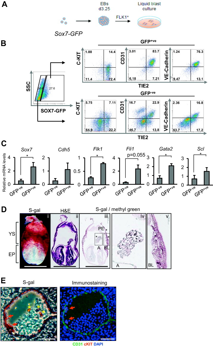
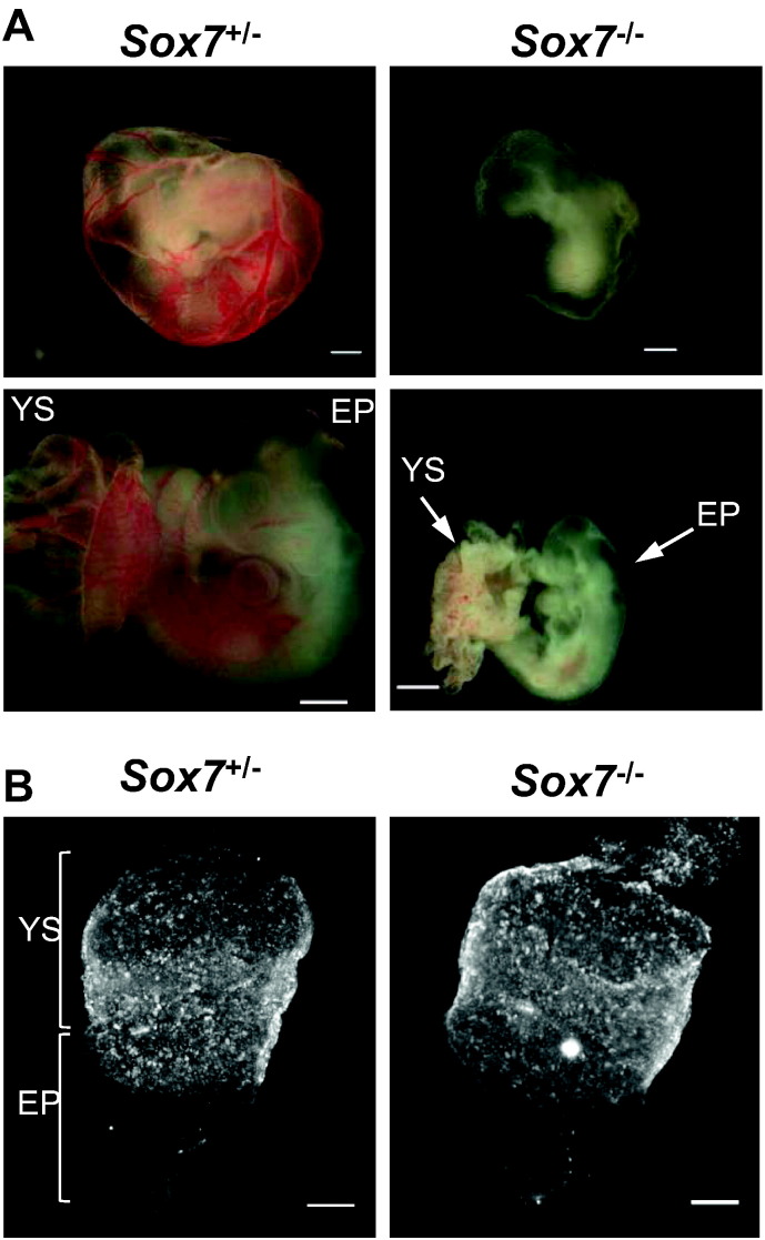
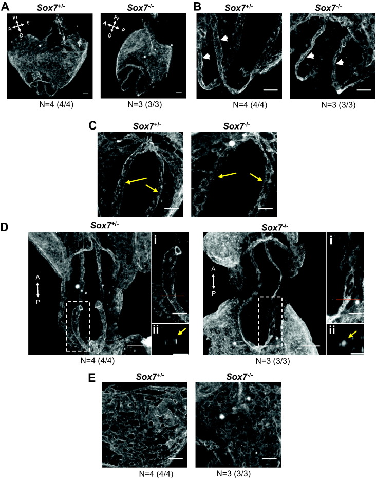
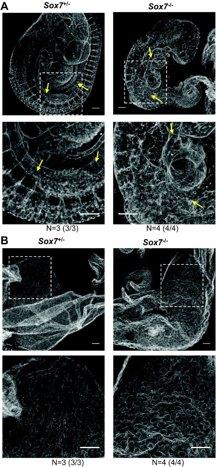
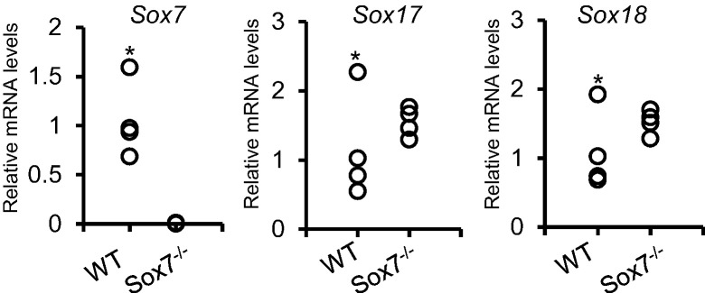
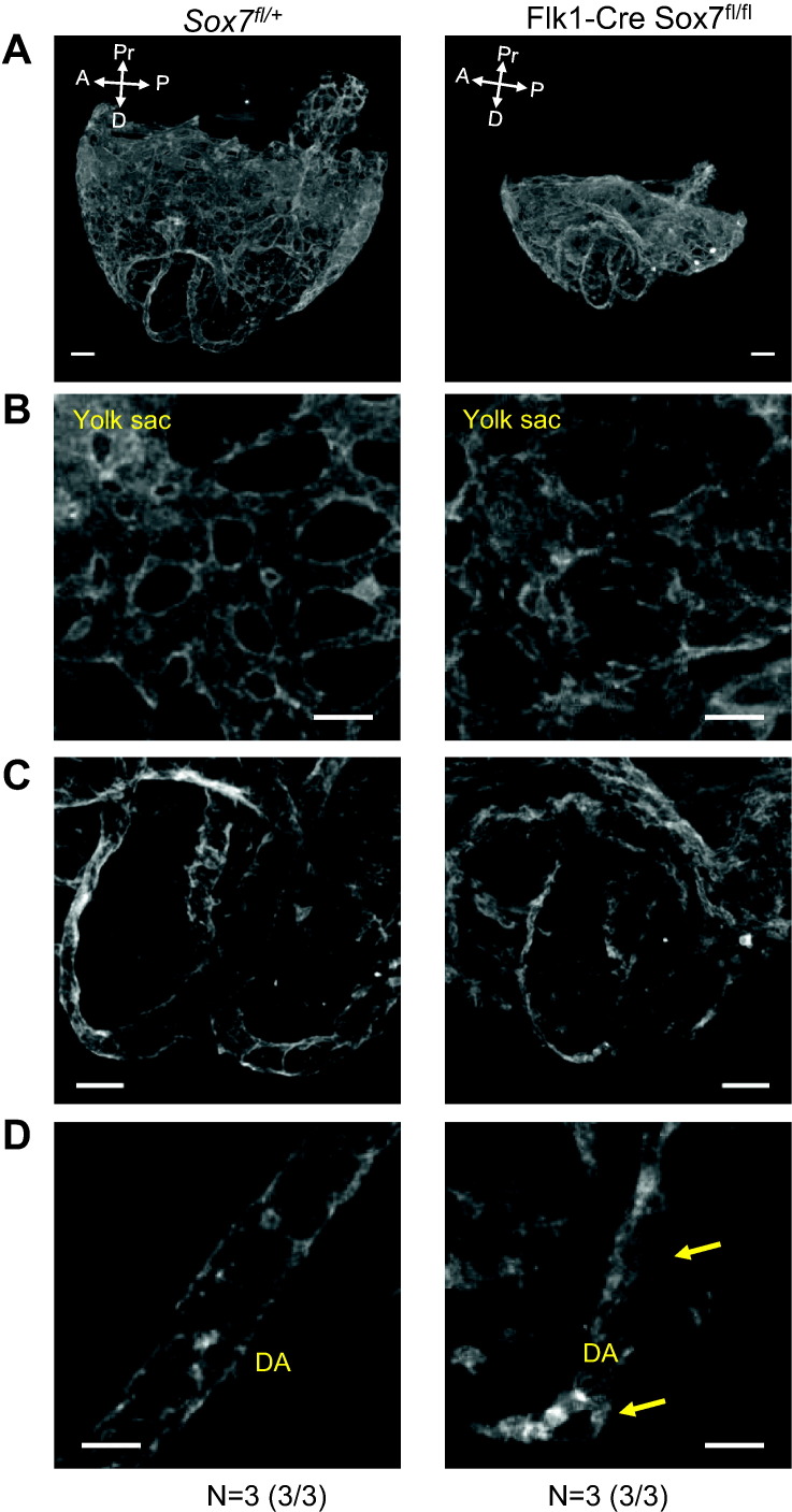
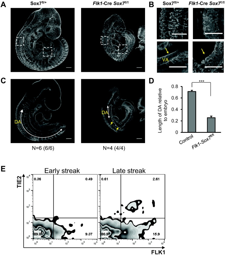
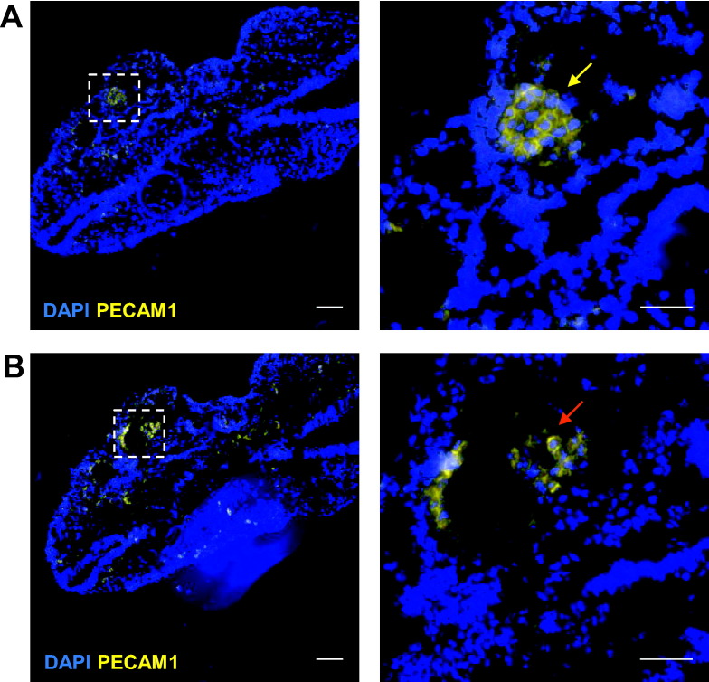
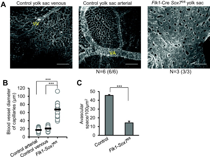
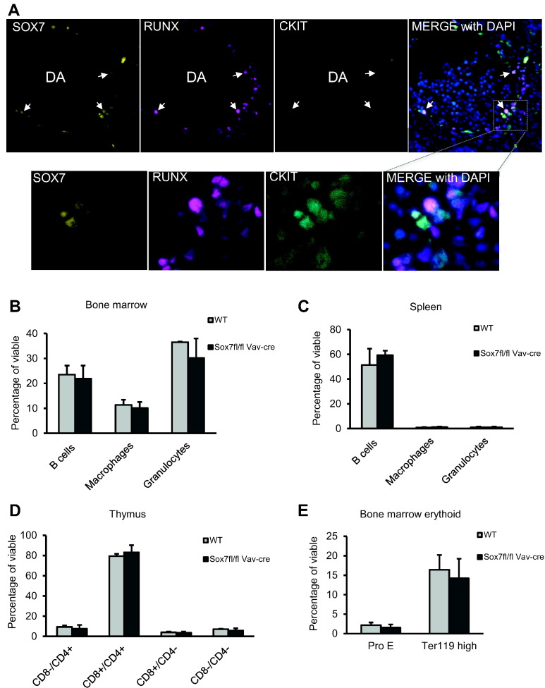
The transcriptional program that regulates the differentiation of endothelial precursor cells into a highly organized vascular network is still poorly understood. Here we explore the role of SOX7 during this process, performing a detailed analysis of the vascular defects resulting from either a complete deficiency in Sox7 expression or from the conditional deletion of Sox7 in FLK1-expressing cells. We analysed the consequence of Sox7 deficiency from E7.5 onward to determine from which stage of development the effect of Sox7 deficiency can be observed. We show that while Sox7 is expressed at the onset of endothelial specification from mesoderm, Sox7 deficiency does not impact the emergence of the first endothelial progenitors. However, by E8.5, clear signs of defective vascular development are already observed with the presence of highly unorganised endothelial cords rather than distinct paired dorsal aorta. By E10.5, both Sox7 complete knockout and FLK1-specific deletion of Sox7 lead to widespread vascular defects. In contrast, while SOX7 is expressed in the earliest specified blood progenitors, the VAV-specific deletion of Sox7 does not affect the hematopoietic system. Together, our data reveal the unique role of SOX7 in vasculogenesis and angiogenesis during embryonic development.

Keywords: Endothelium; SOXF; Vascular development.


Figures

Fig. 1
Fig. 1
SOX7 is expressed at the onset of endothelial differentiation from mesodermal precursors. (A) FLK1+ cells were sorted from day 3.25 Sox7-Gfp embryoid bodies (EBs) and cultured in 2D culture. (B) Flow cytometry analysis of SOX7-GFP+ and SOX-GFP fractions at day 2 of culture. Data are representative of 3 independent experiments. (C) QRT-PCR analysis for the expression of the indicated genes in sorted SOX7-GFP+ and SOX7-GFP fractions at day 2 of the culture. Error bars indicate ± SEM (n = 3 independent experiments), Student's paired two-tailed t-test. (D) E7.5 Sox7LacZ/WT embryos: (i) whole mount S-gal staining, (ii) hematoxylin and eosin (H&E) staining on section, (iii) S-gal and methyl green staining on section, (iv) close-up image of allantois from the S-gal staining, (v) close-up image of blood island from the S-gal staining. YS: yolk sac, EP: embryo proper, A: allantois, BL: blood island, PE: primitive endoderm. Scale bars: 500 μm. (E) E10.5 Sox7LacZ/WT embryos. Left panel: S-gal staining on a section from dorsal aorta. Right: Immunostaining on the following section. Red arrows indicate emerging hematopoietic clusters. Yellow asterisks indicate SOX7::S-gal+ blood cells. Scale bars: 50 μm.
Fig. 2
Fig. 2
SOX7 deficient embryos show dramatic growth delay at E10.5 but generate primordial PECAM1+ cells at E7.5. (A) Light microscope images of heterozygous (Sox7+/−) and Sox7 knockout (Sox7−/−) embryos at E10.5. Top panel: images of embryos embedded within their yolk sacs, bottom panel: images of embryo proper and yolk sac. (B) Whole mount PECAM1 staining of heterozygous (Sox7+/−) and Sox7 knockout (Sox7−/−) yolk sacs embryos at E7.5. YS: yolk sac, EP: embryo proper.
Fig. 3
Fig. 3
SOX7 deficient embryos show defects in the dorsal aorta and vascular plexus of the yolk sac at E8.5. (A) Whole mount PECAM1 staining of heterozygous (Sox7+/−) and Sox7 knockout (Sox7−/−) embryos and yolk sacs at E8.5 (5–6 somite pairs). 3D projection of the embryo embedded within its yolk sac. White arrows indicate anterior (A), posterior (P), distal (D) and proximal (Pr) axes. (B) Anterior view of the dorsal aorta (white arrowheads). (C) Posterior tip of the dorsal aorta (yellow arrows). (D) Whole mount PECAM1 staining of Sox7+/− and Sox7−/ E8.5 embryos (3–5 somite pairs). The boxes denote area of magnification: (i) magnified view of dorsal aorta, red bar denotes cross section area, (ii) cross section of dorsal aorta lumen. (E) Details of the yolk sac vasculature. Scale bars: 100 μm. Data shown are representative of at least 3 embryos with 100% penetrance of the phenotype observed for knockout embryos.
Fig. 4
Fig. 4
By E10.5 SOX7 deficient embryos have profound and widespread defects in vascular development. Whole mount PECAM1 staining of heterozygous (Sox7+/−) and Sox7 knockout (Sox7−/−) embryos at E10.5. (A) Embryo proper, white boxes indicate areas of magnification. Yellow arrows indicate dorsal aorta. (B) Yolk sac vasculature, white boxes indicate areas of magnification. Scale bars: 250 μm.
Fig. 5
Fig. 5
Change in the expression levels of Sox17 and Sox18 in SOX7 deficient embryos. PCR analysis of Sox7, Sox17 and Sox18 expression levels in wild type (WT) and Sox7 knockout (Sox7−/−) embryos at E10 (n = 4).* Denotes the same WT embryo with overall higher levels of Sox7, Sox17 and Sox18.
Fig. 6
Fig. 6
Flk1-Cre Sox7fl/fl embryos have defects in the dorsal aorta at E8.5. Whole mount PECAM1 staining of control (Sox7fl/+) and Flk1-Cre Sox7fl/fl embryos and yolk sac at E8.5 (5–7 somite pairs). (A) 3D projection of embryos embedded within their yolk sacs, white arrows indicated anterior (A), posterior (P), distal (D) and proximal (Pr) axes. (B) Details of the yolk sac vasculature. (C) Paired dorsal aorta region. (D) Magnified view of a posterior region of a single dorsal aorta. Yellow arrows indicate malformation in the dorsal aorta. Scale bars 100 μm (A and C), 50 μm (B and D). Data are representative of at least three embryos with 100% penetrance of the phenotype observed for knockout embryos.
Fig. 7
Fig. 7
Flk1-Cre Sox7fl/fl embryos have severe and widespread vascular defects at E10.5. Whole mount PECAM1 staining of Sox7fl/+ and Flk1-Cre Sox7fl/fl embryos at E10.5. (A) 3D projection of embryo proper, white boxes indicate areas of magnification. Data shown are representative of 4 embryos. (B) Top panel: organization of capillaries in posterior region, bottom panel: vitelline artery (VA). (C) Sagittal slices through the embryo proper. DA: dorsal aorta, white arrow indicates length of functional dorsal aorta; yellow arrows indicate malformation of the DA. (D) Mean length of DA relative to embryo ± SEM, n = 6 control (Sox7fl/+ and Sox7fl/fl) versus n = 4 Flk1-Cre Sox7fl/fl embryos. (E) Expression of FLK1 and TIE2 in wild type gastrulating embryos at early streak (left panel) and late streak (right panel) stages measured by flow cytometry.
Fig. 8
Fig. 8
Flk1-Cre Sox7fl/fl embryos have lumenization defects in blood vessels. (A-B) PECAM1 staining of E9.5 Flk1-Cre Sox7fl/fl embryo sections. Left panels: 10 × objective, scale bars: 100 μm; right panels: 40 × objective, scale bars: 50 μm. Boxes denote areas of magnification. Top and bottom panels are two subsequent sections of the embryo. Yellow arrow: nonlumenized section of a blood vessel, red arrow: lumenized section of the same blood vessel.
Fig. 9
Fig. 9
Complete absence of remodelling in the yolk sac of Flk1-Cre Sox7fl/fl embryos. (A) Flat mounted E10.5 yolk sacs stained for PECAM1. Control yolk sacs have venous and arterial systems and large blood vessels such as a vitelline vein (VV) and a vitelline artery (VA). Data shown are representative of 3 embryos. (B) Diameter of capillaries in a control yolk sac (Sox7fl/+), compared with a Flk1-Cre Sox7fl/fl yolk sac. Data are presented as mean diameter (black bar), n = 30 capillaries. (C) Avascular space of capillaries/100μm2 in control versus Flk1-Cre Sox7fl/fl yolk sacs. Data are presented as mean ± SEM, n = 6 control and n = 3 Flk1-Cre Sox7fl/fl yolk sacs. All statistical analyses are Student's two-tailed t-test. Scale bars = 500 μm.
Fig. 10
Fig. 10
Vav-Cre Sox7fl/fl mice live to adulthood and have no hematopoietic defects. (A) Immuno-staining of E10.5 wild embryo transversal section. White arrows indicate example of cells co-expressing SOX7, RUNX and cKIT. Upper panels are 40 × magnification; lower panels are 100 × magnification. DA: dorsal aorta. (B and E) Bone marrow, (C) spleen and (D) thymus harvested from aged matched adult WT and Vav-Cre Sox7fl/fl mice and analysed by flow cytometry, gating first on the viable cells. B cells: CD19+ and/or B220+; macrophages: CD11b+ and F4/80+; granulocytes: CD11b+ and GR1+; Pro erythroblast (Pro E): Ter119med and CD71+; maturing erythroid cells: Ter119high. Data are presented as mean ± SE, n = 3 mice in each group. No differences were observed between WT and Vav-Cre Sox7fl/fl populations (Student's paired two-tailed t-test).

Similar articles

Cited by

References

    1. de Boer J., Williams A., Skavdis G., Harker N., Coles M., Tolaini M., Norton T., Williams K., Roderick K., Potocnik A.J., Kioussis D. Transgenic mice with hematopoietic and lymphoid specific expression of Cre. Eur. J. Immunol. 2003;33:314–325. - PubMed
    1. Chung M.I., Ma A.C., Fung T.K., Leung A.Y. Characterization of Sry-related HMG box group F genes in zebrafish hematopoiesis. Exp. Hematol. 2011;39(986–998) - PubMed
    1. Corada M., Orsenigo F., Morini M.F., Pitulescu M.E., Bhat G., Nyqvist D., Breviario F., Conti V., Briot A., Iruela-Arispe M.L., Adams R.H., Dejana E. Sox17 is indispensable for acquisition and maintenance of arterial identity. Nat. Commun. 2013;4:2609. - PMC - PubMed
    1. De Val S., Black B.L. Transcriptional control of endothelial cell development. Dev. Cell. 2009;16:180–195. - PMC - PubMed
    1. Drake C.J., Fleming P.A. Vasculogenesis in the day 6.5 to 9.5 mouse embryo. Blood. 2000;95:1671–1679. - PubMed

Publication types

Substances

LinkOut - more resources