Review
. 2018 Jan;1860(1):102-123.
doi: 10.1016/j.bbamem.2017.05.019. Epub 2017 Jun 1.

Electrical synapses in mammalian CNS: Past eras, present focus and future directions

Affiliations
Review

Electrical synapses in mammalian CNS: Past eras, present focus and future directions

James I Nagy et al. Biochim Biophys Acta Biomembr. 2018 Jan.

Abstract

Gap junctions provide the basis for electrical synapses between neurons. Early studies in well-defined circuits in lower vertebrates laid the foundation for understanding various properties conferred by electrical synaptic transmission. Knowledge surrounding electrical synapses in mammalian systems unfolded first with evidence indicating the presence of gap junctions between neurons in various brain regions, but with little appreciation of their functional roles. Beginning at about the turn of this century, new approaches were applied to scrutinize electrical synapses, revealing the prevalence of neuronal gap junctions, the connexin protein composition of many of those junctions, and the myriad diverse neural systems in which they occur in the mammalian CNS. Subsequent progress indicated that electrical synapses constitute key elements in synaptic circuitry, govern the collective activity of ensembles of electrically coupled neurons, and in part orchestrate the synchronized neuronal network activity and rhythmic oscillations that underlie fundamental integrative processes. This article is part of a Special Issue entitled: Gap Junction Proteins edited by Jean Claude Herve.

Keywords: Cell localization; Connexins; Mixed chemical/electrical synapses; Neuronal gap junctions; Protein composition; Ultrastructural diversity.


Figures

Fig. 1
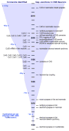
Fig. 1. Time line of significant technical advances and discoveries regarding electrical synapses in vertebrate species
The blue overlay is a graphical representation of the previous 2632 publications that have investigated electrical synapses by a diversity of approaches in a variety of invertebrate and vertebrate species, with average number of publications per year for each decade indicated by numbered blue dots on the left margin. The center column indicates years from the point electrical synapses were discovered to the present. The left side lists the discovery dates for all mammalian connexins, with the first neuronal connexin identified towards the end of the 1990’s. The right side indicates landmark discoveries regarding the structure, function, and connexin composition of neuronal gap junctions and the development of methods used to study electrical synapses. Note the dense concentration of significant event around the turn of the century.
Fig. 2
Fig. 2. Correlation of immunofluorescence and FRIL methods used to identify, quantify, map, and immunocytochemically characterize the wide variety of Cx36-containing gap junctions in adult rat retina
A: Confocal image showing Cx36-puncta (green) vs. Nissl stained (red) neurons in a full-thickness section of rat retina. B: Correlative FRIL image showing exact locations of photomapped immunogold-labeled gap junctions. Numbered red triangles = locations of string gap junctions (all in sublaminae S2 and S1). Numbered blue circles = plaque gap junctions (in S2–S5). C: Higher magnification of boxed area in B. This crystalline plaque gap junction, mapped to sublamina S4, contains ca. 300 connexons that were immunogold double-labeled for Cx36 by 6-nm and 18-nm gold beads. D: Small string gap junction, mapped to S2, contains ca. 30 connexons labeled by 18-nm gold beads. E: Tiny crystalline plaque gap junction mapped to S2; 11 connexons are labeled by a single 12-nm gold bead. Based on quantitative FRIL analysis [84] showing that virtually all string gap junctions (mostly <100 connexons) and many small plaque gap junctions were abundant in S2 of both rats and mice (where others had found few or no gap junctions), we calculated that conventional immunofluorescence imaging had not revealed gap junctions smaller than ca. 100 connexons, which comprise the majority of gap junctions in retina, particularly in S1 and S2. F–G: For correlation, we compared immunofluorescence imaging protocols in which photomultiplier gain was reduced to avoid autofluorescence “noise” in tissue sections (F), with images in which photomultiplier gain was set at increased (optimum) gain in the same 8-μm section of retina (G). In F, relatively few puncta were detected in sublamina S2, where FRIL had revealed a high density of string and miniature plaque gap junctions (<100 connexons). With photomultiplier gain increased, many additional puncta were detected in the same area of S2 (G; circles), but without significantly increasing background autofluorescence. In contrast, the number of puncta visible in ON sublamina S4 (F; square) (where small gap junctions were rare by FRIL) was only slightly increased (G; square box), thereby allowing the first semi-quantitative immunofluorescence detection of this new class of abundant gap junctions containing 30–100 connexons. (Those smaller than 30 connexons remain undetectable by immunofluorescence in tissue slices.) F–G may be viewed as a stereo pair to visualize the 3-D distribution of small to large puncta in the various sublaminae. Offset arrows at C reveal gap junctions at top and bottom of section. RPE = retinal pigment epithelium; OS and IS = outer and inner segments of rods and cones; ONL = outer nuclear layer; OPL = outer plexiform layer; INL = inner nuclear layer; IPL = inner plexiform layer; GCL = ganglion cell layer; Ax = axons on the anterior surface of the retina. Numbers at left margins indicate approximate locations of sublaminae S1–S5. All images modified from [84], with permission. Calibration bars in all FRIL images are 0.1 μm unless otherwise indicated.
Fig. 3
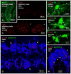
Fig. 3. Immunofluorescence labeling of Cx36 in various CNS regions of adult mouse and rat
A: Main olfactory bulb of mouse, showing overview of a fluorescence Nissl-stained whole transverse section of the olfactory bulb. B,C: Higher magnification images showing labeling of Cx36 in an area (B) of the mitral cell layer (MCL) indicated by the boxed region to the right in A, and labeling of Cx36 in an area (C) of the glomerular cell layer (GCL) shown by the boxed region to the left in A. Detection of Cx36 appears exclusively punctate in both the mitral cell layer and glomeruli, which correspond to sites of heavily-concentrated neuronal gap junctions. D–F: Double immunofluorescence labeling for Cx36 and parvalbumin (PV) in the cerebral cortex (D) and the striatum (E), and double labelling for Cx36 and tyrosine hydroxylase (TH) in the hypothalamic arcuate nucleus (F), showing PV-positive and TH-positive neurons (arrowheads) with Cx36-puncta distributed on their somata and/or processes (arrows). G,H: Double immunofluorescence labeling for Cx36 and the motoneuron marker peripherin, showing closely packed peripherin-positive motoneurons in the sexually dimorphic dorsomedial nucleus (DMN) (G, arrows) in a horizontal section of the lumbosacral region of rat spinal cord, with Cx36-puncta surrounding the motoneuronal somata and localized along motoneuron dendrites. Higher magnification shows peripherin-positive motoneurons (H, arrowheads) displaying Cx36-puncta at their somatic appositions (H, arrows).
Fig. 4
Fig. 4. FRIL images of a Cx36 immunogold-labeled reticular gap junction on neuronal soma in adult rat suprachiasmatic nucleus
A: Neuron cell body, revealing its nucleus (N, with abundant nuclear pores), cross-fractured cytoplasm (*), and its somatic plasma membrane containing an immunogold-labeled gap junction (inscribed box). Note the complete absence of “background” labeling. Double-ended arrow traces continuity of cytoplasm, as maintained by the carbon support film, even in the absence of an electron-dense platinum coating. B: High magnification of the boxed area in A, showing a distinctive reticular gap junction, characterized by the presence of connexon-free voids within the junction (modified from Fig. 3 in [96] with permission). Both images originally published as stereo pairs to facilitate 3-D analysis.)
Fig. 5
Fig. 5. Visualized paired recordings of electrically coupled cells in the mesencephalic trigeminal nucleus (MesV)
A: Infrared-differential interference contrast (IR-DIC) image of two contiguous MesV neurons during a simultaneous whole-cell recording (electrode 1 and electrode 2). B: Simultaneous recordings from a pair of strongly coupled MesV neurons. Voltage responses to 200 ms hyperpolarizing ( 400 pA) current pulses injected either in cell 1 (red trace) or cell 2 (black trace) evoke corresponding coupling potentials (asterisks). Rebound depolarization after hyperpolarizing pulses causes cell firing. Unpublished image and recordings kindly provided by S. Curti (UdelaR, Montevideo, Uruguay).
Fig. 6
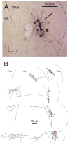
Fig. 6. Tracer coupling reveals the organization of functional compartments formed by electrical synapses
A: Cluster of neurons in the thalamic reticular nucleus following intracellular injection of Neurobiotin (0.5% in the internal solution) during a whole cell recording. While the injected cells appear completely filled and labeled by Neurobiotin, only the cell bodies and proximal process are Neurobiotin-positive in the coupled cells. B: Dye coupling reveals that clusters can be spherical or elongated (compare the extension and spread of clusters), suggesting differences in coupling architecture. These clusters project to different regions of the ventrobasal (VB) and posterior medial (POm) nuclei of the thalamus. Panels A and B are modified from Lee et al, 2014 [253], with permission.
Fig. 7
Fig. 7. Matched double-replica FRIL (MDR-FRIL) showing separate gap junctional domains labeled solely for Cx36 adjacent to domains labeled solely for Cx45
The image shows a single gap junction viewed after capture of both its hemiplaques in apposing membranes, with one hemiplaque showing areas of particles (A, arrow) and pits (A, arrowhead) and the other in corresponding areas showing instead pits (B, arrowhead) and particles (B, arrow), separating the regions where the membrane fracture skips from one apposed membrane to the other (B, double arrowhead). This and six other matched gap junctions displayed separate domains of Cx36 localization in one hemiplaque opposite Cx36 localization in the apposed hemiplaque, and similarly, Cx45 localization in the same hemiplaque opposite labeling for Cx45 in the apposed hemiplaque. Combined with data from 60 other unmatched hemiplaques containing both Cx45 and Cx36, we concluded that most/all retinal neurons expressing Cx45 also express Cx36, with both connexins co-existing in both apposed hemiplaques. Thus in retina, these two connexins form bi-homotypic (Cx36:Cx36 + Cx45:Cx45) gap junctions [136], rather than forming heterotypic Cx36:Cx45 gap junctions, as previously proposed (references cited in [136]). (Modified from Fig. 6 in [136], with permission.)
Fig. 8
Fig. 8. Seven basic morphologies of neuronal gap junctions in retina and hippocampus immunogold labeled for Cx36
A: Large-diameter crystalline plaque gap junction from ON lamina of adult rat retina (ca. 900 connexons); B: E-face image of small-diameter non-crystalline plaque gap junction in ON sublamina of rat retina (ca. 90 connexons immunogold labeled with two sizes of gold beads. C: Reticular gap junction from adult rat hippocampus. D: Large multi-strand ribbon gap junction (ca. 490 connexons; pink overlay) in the OFF sublamina of adult rat retina (S2) labeled for Cx36 with two sizes of gold beads. E. Portion of E-face image of large compound string gap junction in the OFF sublamina S2 (previously unpublished image). F: “Meandering” gap junction in rat hippocampus, consisting of 25 connexons labeled for Cx36 beneath their E-face pits. G: Dispersed clusters of connexons in adult rat hippocampus (unpublished image). (A and B, modified form Fig. 3 in [84]; C and F, modified from Fig. 11 in [273]), D, modified from Fig. 5A in [84]; all with permission.

References

    1. Furshpan EJ, Potter DD. Transmission at the giant motor synapses of the crayfish. J Physiol (London) 1959;145:289–325. - PMC - PubMed
    1. Bennett MVL, Crain SM, Grundfest H. Electrophysiology of Supramedullary Neurons in Spheroides maculatus III. Organization of the supramedulary neurons. J Gen Physiol. 1959;43:221–250. - PMC - PubMed
    1. Bennett MVL, Aljure E, Nakajima Y, Pappas GD. Electrotonic junctions between teleost spinal neurons: Electrophysiology and ultrastructure. Science. 1963;141:262–264. - PubMed
    1. Robertson JD. The occurrence of a subunit pattern in the unit membranes of club endings in Mauthner cell synapses in goldfish brains. J Cell Biol. 1963;19:201–221. - PMC - PubMed
    1. Robertson JD, Bodenheimer TS, Stage DE. The ultrastructure of Mauthner cell synapses and nodes in goldfish brains. J Cell Biol. 1963;19:159–199. - PMC - PubMed

Publication types

LinkOut - more resources