

Protein kinase D signaling in cancer: A friend or foe?
- PMID: 28577984
- PMCID: PMC5548606
- DOI: 10.1016/j.bbcan.2017.05.008
Protein kinase D signaling in cancer: A friend or foe?
Abstract
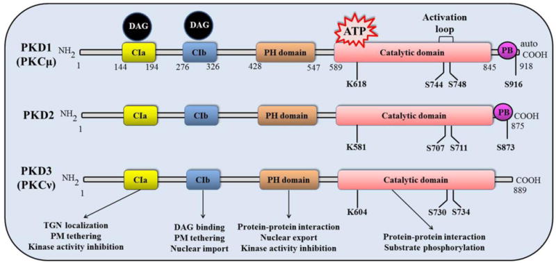
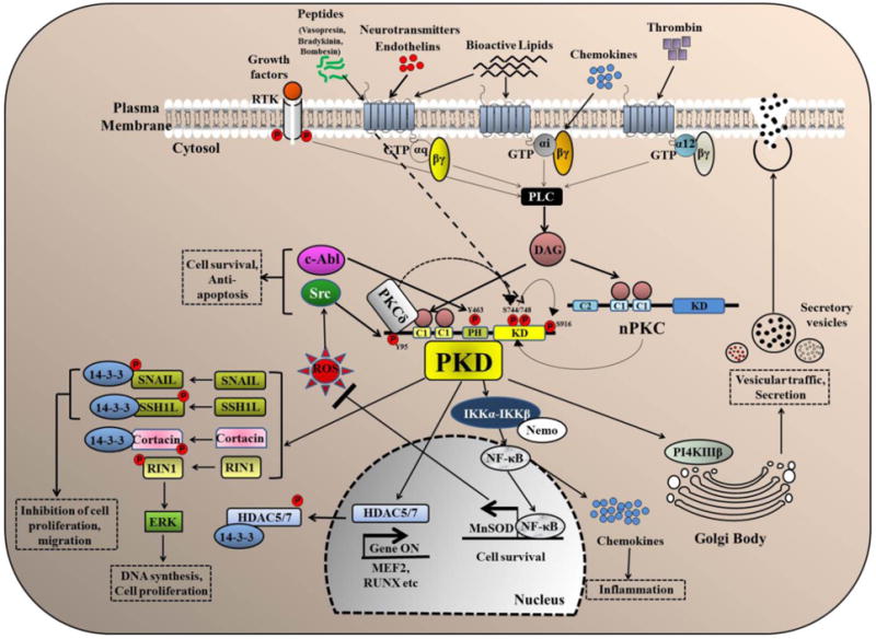
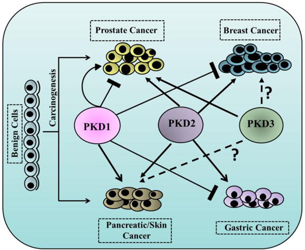
Protein kinase D is a family of evolutionarily conserved serine/threonine kinases that belongs to the Ca++/Calmodulin-dependent kinase superfamily. Signal transduction pathways mediated by PKD can be triggered by a variety of stimuli including G protein-coupled receptor agonists, growth factors, hormones, and cellular stresses. The regulatory mechanisms and physiological roles of PKD have been well documented including cell proliferation, survival, migration, angiogenesis, regulation of gene expression, and protein/membrane trafficking. However, its precise roles in disease progression, especially in cancer, remain elusive. A plethora of studies documented the cell- and tissue-specific expressions and functions of PKD in various cancer-associated biological processes, while the causes of the differential effects of PKD have not been thoroughly investigated. In this review, we have discussed the structural-functional properties, activation mechanisms, signaling pathways and physiological functions of PKD in the context of human cancer. Additionally, we have provided a comprehensive review of the reported tumor promoting or tumor suppressive functions of PKD in several major cancer types and discussed the discrepancies that have been raised on PKD as a major regulator of malignant transformation.
Keywords: Cancer; Function; PKD; Regulation; Signaling mechanisms; Structure.
Copyright © 2017 Elsevier B.V. All rights reserved.
Conflict of interest statement
The authors declare no conflict of interest
Figures
References
-
- Manning G, Whyte DB, Martinez R, Hunter T, Sudarsanam S. The protein kinase complement of the human genome. Science. 2002;298:1912–1934. - PubMed
-
- Johannes FJ, Prestle J, Eis S, Oberhagemann P, Pfizenmaier K. PKCu is a novel, atypical member of the protein kinase C family. J Biol Chem. 1994;269:6140–6148. - PubMed
-
- Cano A, Perez-Moreno MA, Rodrigo I, Locascio A, Blanco MJ, del Barrio MG, Portillo F, Nieto MA. The transcription factor snail controls epithelial-mesenchymal transitions by repressing E-cadherin expression. Nat Cell Biol. 2000;2:76–83. - PubMed
-
- Hayashi A, Seki N, Hattori A, Kozuma S, Saito T. PKCnu, a new member of the protein kinase C family, composes a fourth subfamily with PKCmu. Biochim Biophys Acta. 1999;1450:99–106. - PubMed
Publication types
MeSH terms
Substances
Grants and funding
LinkOut - more resources
Full Text Sources
Other Literature Sources
