. 2017 Jul 21;292(29):12054-12064.
doi: 10.1074/jbc.M117.790568. Epub 2017 Jun 3.

Docosahexaenoic acid preserves visual function by maintaining correct disc morphology in retinal photoreceptor cells

Affiliations

Docosahexaenoic acid preserves visual function by maintaining correct disc morphology in retinal photoreceptor cells

Hideo Shindou et al. J Biol Chem. .

Abstract

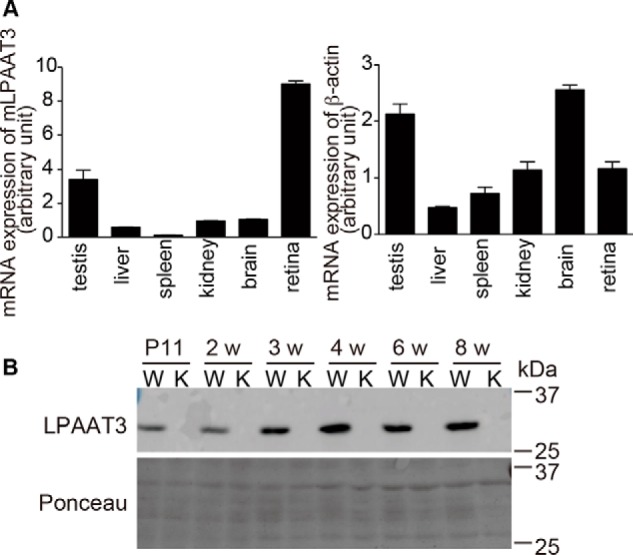
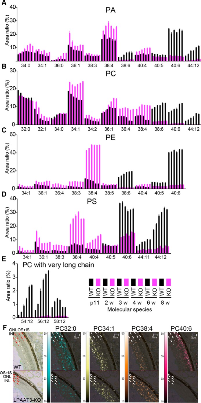
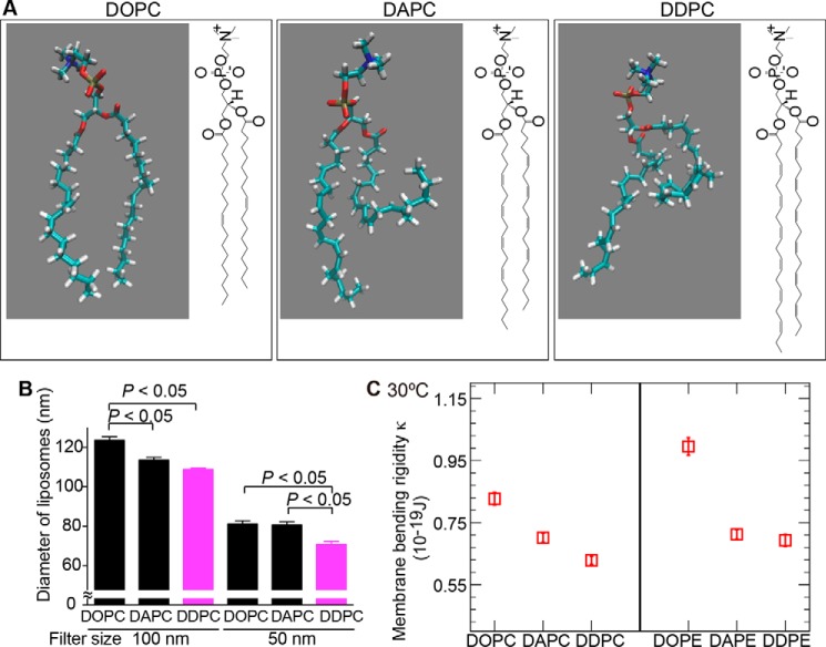
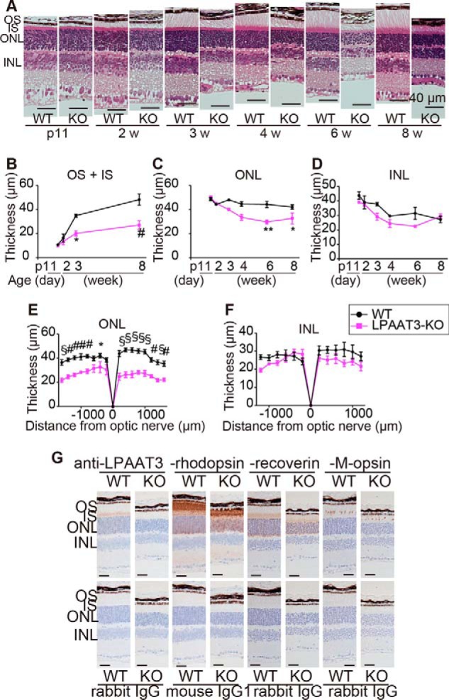
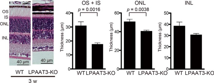
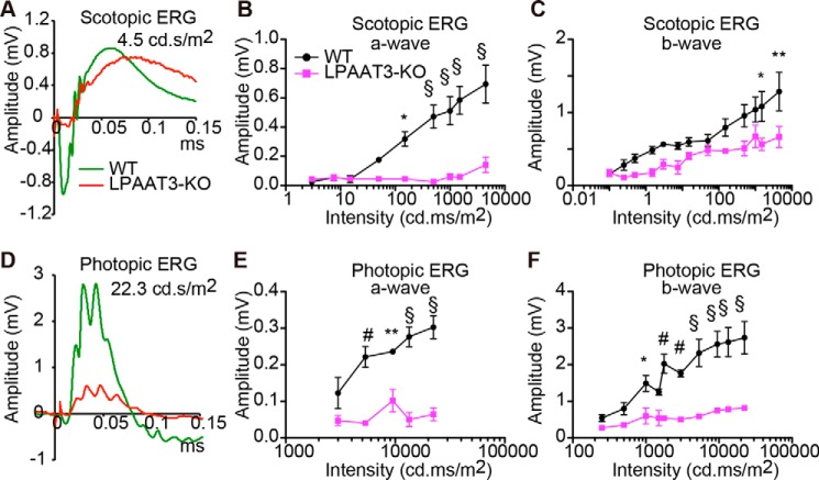
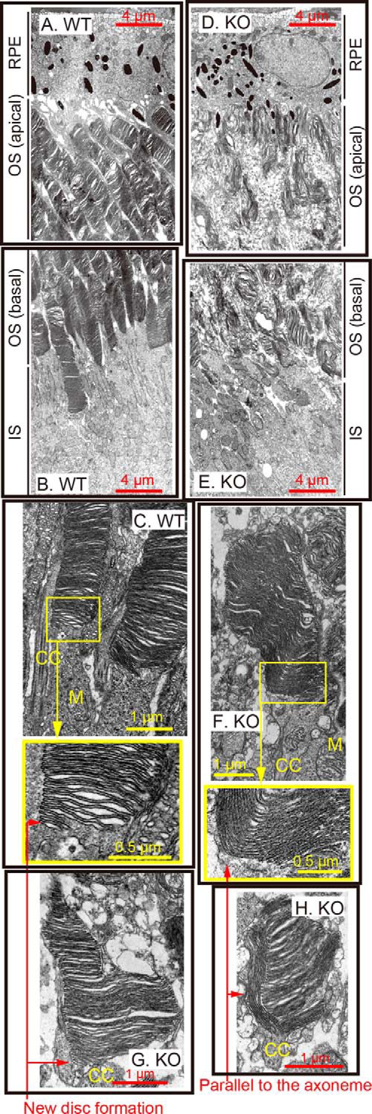
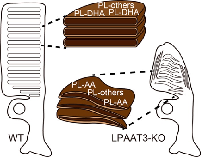
Docosahexaenoic acid (DHA) has essential roles in photoreceptor cells in the retina and is therefore crucial to healthy vision. Although the influence of dietary DHA on visual acuity is well known and the retina has an abundance of DHA-containing phospholipids (PL-DHA), the mechanisms associated with DHA's effects on visual function are unknown. We previously identified lysophosphatidic acid acyltransferase 3 (LPAAT3) as a PL-DHA biosynthetic enzyme. Here, using comprehensive phospholipid analyses and imaging mass spectroscopy, we found that LPAAT3 is expressed in the inner segment of photoreceptor cells and that PL-DHA disappears from the outer segment in the LPAAT3-knock-out mice. Dynamic light-scattering analysis of liposomes and molecular dynamics simulations revealed that the physical characteristics of DHA reduced membrane-bending rigidity. Following loss of PL-DHA, LPAAT3-knock-out mice exhibited abnormalities in the retinal layers, such as incomplete elongation of the outer segment and decreased thickness of the outer nuclear layers and impaired visual function, as well as disordered disc morphology in photoreceptor cells. Our results indicate that PL-DHA contributes to visual function by maintaining the disc shape in photoreceptor cells and that this is a function of DHA in the retina. This study thus provides the reason why DHA is required for visual acuity and may help inform approaches for overcoming retinal disorders associated with DHA deficiency or dysfunction.

Keywords: DHA; LPAAT3; glycerophospholipid; lysophospholipid acyltransferase; membrane biophysics; membrane lipid; phospholipid turnover; retinal degeneration.


Conflict of interest statement

The authors declare that they have no conflicts of interest with the contents of this article

Figures

Figure 1.
Figure 1.
High expression of LPAAT3 in the retina. A, LPAAT3 mRNA levels in the tissues from 8-week-old mice were measured by quantitative-PCR (left). β-Actin level was used as a control (right). LPAAT3 was highly expressed in the retinas. Results are expressed as the mean ± S.E. of three independent experiments. B, LPAAT3 protein levels were detected by Western blot analysis using an anti-LPAAT3 antibody. LPAAT3 levels increased on an age-dependent basis. LPAAT3 was indeed disrupted in LPAAT3-KO retina. Three independent experiments were performed with similar results. W, WT; K, LPAAT3-KO.
Figure 2.
Figure 2.
Attenuation of PL-DHA in LPAAT3-KO retinas. PA (A), PC (B), PE (C), PS (D), and PC with very long chain fatty acids (E) were measured by comprehensive phospholipid analysis and indicated by the area ratio (y axis, 100% indicates sum of the detected signals for each phospholipid). The x axis indicates the summation of fatty acid information at the sn-1 and sn-2 positions (number of carbon and double bonds, i.e. 34:0) in the phospholipids. 38:6, 40:6, and 44:12 in all phospholipids and very long chain in PC may contain DHA, which were suppressed in LPAAT3-KO mice. By contrast, 36:4, 38:4, and 40:4 may contain AA and were increased in LPAAT3-KO mice. 34:1 and 36:1 thought to have oleic acid were increased. These data were obtained from p11 to 8-week-old mice; WT (black) and LPAAT3-KO (magenta). Results are expressed as the mean ± S.E. of four independent experiments. F, PC localization of 8-week-old mice (WT, upper; KO, lower) was observed by imaging mass microscope. PC32:0, PC34:1, PC38:4, and PC40:6 are supposed to have C16:0 (palmitic acid), C18:1 (oleic acid), C20:4 (AA), and C22:6 (DHA), respectively. The signals of PC40:6 were disappeared in the OS of LPAAT3-KO retina. Imaging data of phospholipids were merged with captured light images. Scale bar, 70 μm. Two independent experiments were performed with similar results.
Figure 3.
Figure 3.
Alternation of phospholipid physical properties by fatty acids. A, single-molecule illustrations and structures of DDPC, DAPC, and DOPC. B, liposome sizes were measured by DLS analysis. The sizes of DDPC liposomes were smaller than those of DOPC and DAPC following passage through a 50 nm pore size filter. Results are expressed as the mean ± S.E. of three independent experiments. p < 0.05; one-way ANOVA, Tukey's multiple comparison test. C, κ was calculated by MD simulations. The κ value for DDPC was lower than that for DOPC and DAPC. DDPE also showed similar results. Statistical errors are estimated using the block average method (53).
Figure 4.
Figure 4.
Loss of retinal layers of LPAAT3-KO mice. Retinal structure (A) and their thicknesses (B–F) are shown. OS + IS and ONL were thinner in LPAAT3-KO retina. Three independent experiments were performed with similar results (A). Results are expressed as the mean ± S.E. of three (p11 to 6 weeks (w)) or five (8 weeks) independent experiments (B–F). *, p < 0.05; **, p < 0.01; #, p < 0.001; §, p < 0.0001; two-way ANOVA, Bonferroni test. G, immunohistochemistry was performed to detect LPAAT3, rhodopsin, recoverin, and M-opsin in 6-week-old mice. Diaminobenzidine (brown) signals indicate each protein. Scale bar, 100 μm. Independent experiments from three mice were performed with similar results.
Figure 5.
Figure 5.
No influence of phototoxicity on retinal disruption in the LPAAT3-KO mice. Mice at 1–3 weeks old were maintained under dark conditions for 2 weeks, and their retinal layers were analyzed. OS/IS and ONL from LPAAT3-KO retinas were lower than those of WT retinas, with similar results to those under normal light/dark cycle conditions. LPAAT3-KO mice were not influenced by phototoxicity under the conditions. Four independent experiments were performed with similar results (hematoxylin and eosin staining). Results are expressed as the mean ± S.E. p value, Mann-Whitney U test. Experimental numbers of OS/IS were n = 5 in WT and n = 8 in LPAAT3-KO retinas from each four mice. ONL and INL were n = 8 from four retinas from each mouse.
Figure 6.
Figure 6.
Disorder of visual function of LPAAT3-KO mice. ERG amplitudes at the indicated light intensities are shown at scotopic (A–C) and photopic (D–F) responses. A and D, independent experiments from three 8-week-old mice were performed with similar results. B, C, E, and F, results are expressed as the mean ± S.E. of three independent experiments. *, p < 0.05; **, p < 0.01; #, p < 0.001; §, p < 0.0001; two-way ANOVA, Bonferroni test.
Figure 7.
Figure 7.
Disc disruption in LPAAT3-KO. Retinal structures of WT (A–C) and LPAAT3-KO (D–H) mice were observed by TEM. Abnormal disc morphogenesis was observed in LPAAT3-KO retinas. Independent experiments from three mice were performed with similar results. CC, connecting cilium; M, mitochondria.
Figure 8.
Figure 8.
Model figures of DHA roles. Model figures of proposed DHA roles in photoreceptor cells. Low levels of PL-DHA in LPAAT3-KO-induced abnormal disc morphology/organization, which was not rescued by increased PL-AA levels.

References

    1. Benolken R. M., Anderson R. E., and Wheeler T. G. (1973) Membrane fatty acids associated with the electrical response in visual excitation. Science 182, 1253–1254 - PubMed
    1. Jastrzebska B., Debinski A., Filipek S., and Palczewski K. (2011) Role of membrane integrity on G protein-coupled receptors: rhodopsin stability and function. Prog. Lipid Res. 50, 267–277 - PMC - PubMed
    1. Jeffrey B. G., and Neuringer M. (2009) Age-related decline in rod phototransduction sensitivity in rhesus monkeys fed an n-3 fatty acid-deficient diet. Invest. Ophthalmol. Vis. Sci. 50, 4360–4367 - PMC - PubMed
    1. Neuringer M., Connor W. E., Van Petten C., and Barstad L. (1984) Dietary ω-3 fatty acid deficiency and visual loss in infant rhesus monkeys. J. Clin. Invest. 73, 272–276 - PMC - PubMed
    1. Antonny B., Vanni S., Shindou H., and Ferreira T. (2015) From zero to six double bonds: phospholipid unsaturation and organelle function. Trends Cell Biol. 25, 427–436 - PubMed

Publication types

MeSH terms

LinkOut - more resources