. 2017 Jun 2;7(1):2695.
doi: 10.1038/s41598-017-02705-0.

An appropriate Wnt/β-catenin expression level during the remodeling phase is required for improved bone fracture healing in mice

Affiliations

An appropriate Wnt/β-catenin expression level during the remodeling phase is required for improved bone fracture healing in mice

Quanwei Bao et al. Sci Rep. .

Abstract

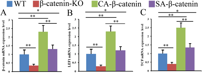
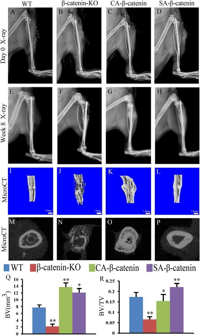
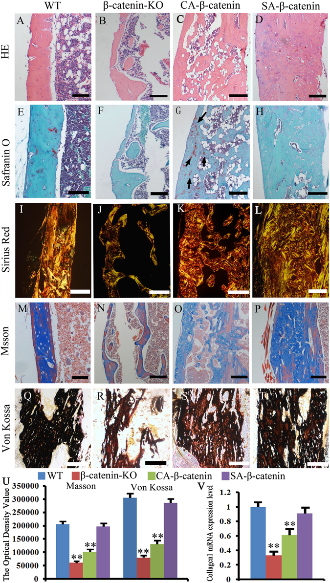
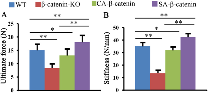
Accumulating evidence demonstrates that the Wnt/β-catenin signaling pathway plays a dominant role in bone repair. However, the role of Wnt/β-catenin signaling in the remodeling phase during bone fracture healing is currently unknown. In the present study, β-catenin was activated at different levels or deleted in mice at the late stage of fracture healing, and the effects on healing quality were investigated. Deletion of β-catenin disturbed bone remodeling, as confirmed by increased bone resorption and decreased bone formation, and significantly decreased bone strength compared with wildtype mice. In addition, the constitutive activation of β-catenin significantly increased the bone mass and delayed the bone remodeling process, resulting in slightly impaired bone strength. In contrast, a slight activation of β-catenin significantly increased bone formation and slightly hindered bone resorption. These effects lead to improved bone fracture healing quality compared with wildtype mice. In summary, the present study provides the first demonstration showing that Wnt/β-catenin signaling should be maintained at a slightly activated level during the late stage of fracture healing to ensure better bone fracture repair.


Conflict of interest statement

The authors declare that they have no competing interests.

Figures

Figure 1
Figure 1
Wnt/β-catenin signaling pathway was deleted or activated in different levels at fracture sites in WT, β-catenin-KO, CA-β-catenin and SA-β-catenin mice. The mRNA expression level of β-catenin, TCF and LEF1 were examined by RT-PCR (AC). The values are expressed as the means ± SDs. (n = 6/group, *p < 0.05, **p < 0.01).
Figure 2
Figure 2
X-ray radiology and MicroCT examinations revealed that slight activation of β-catenin in the remodeling phases could improve healing quality. Representative X-ray radiographs of fracture sites in WT, β-catenin-KO, CA-β-catenin and SA-β-catenin mice at day 0 (AD) and week 8 (EH) post-fracture. (IL) and (MP) Representative 3-D and 2-D transverse reconstructive μCT-images in WT, β-catenin-KO, CA-β-catenin and SA-β-catenin mice at 8weeks post-fracture. (Q and R) Quantitative BV and BV/TV in WT, β-catenin-KO, CA-β-catenin and SA-β-catenin mice at week 8 post-fracture. The values are expressed as the means ± SDs. *p < 0.05, **p < 0.01 (Scale bar = 1 mm in I–L, n = 6/group).
Figure 3
Figure 3
Histological examination revealed that SA-β-catenin mice have better histological morphology at week 8 post-fracture. (AD) Representative HE staining of the fracture site in WT, β-catenin-KO, CA-β-catenin and SA-β-catenin mice. (EH) Representative Fast Green/Safranin O staining images of the fracture site in each group. (IL) Representative sirius red staining images of the fracture site in each group. (MP) Representative Masson staining images of the fracture site in each group. (QT) Representative Von Kossa staining images of the fracture site in each group. (U) The optical density of Masson staining and Von Kossa staining images of the fracture site in each group. (V) mRNA expression level of collagen I at the fracture site in each group. The values are expressed as the means ± SDs. *p < 0.05, **p < 0.01. (arrows in G: the cartilage matrix, Scale bar = 100 µm in A–H and M–P, 50 µm in I–L and Q–T, n = 6/group).
Figure 4
Figure 4
Slight activation of β-catenin in the remodeling phases enhanced bone formation without obvious hindering bone resorption. (AF) The ALP, OSX, Rux2, RANKL, OPG mRNA expression levels and RANKL/OPG ratio in WT, β-catenin-KO, CA-β-catenin and SA-β-catenin mice. (GK) Representative TRAP staining of tibia fracture calluses in each group and the quantitative data. (L and M) The serum concentration of ALP and CTX in each group. The values are expressed as the means ± SDs. *p < 0.05, **p < 0.01 (Scale bar = 25 µm in GJ, n = 6/group).
Figure 5
Figure 5
Mechanical test revealed that better bone fracture healing quality was obtained in SA-β-catenin mice. The ultimate load (A) and stiffness (B) of tibia fracture calluses in WT, β-catenin-KO, CA-β-catenin mice and SA-β-catenin mice. The values are expressed as the means ± SDs. *p < 0.05, **p < 0.01 (n = 6/group).

References

    1. Komatsu DE, Warden SJ. The control of fracture healing and its therapeutic targeting: improving upon nature. Journal of cellular biochemistry. 2010;109:302–311. - PubMed
    1. Schindeler A, McDonald MM, Bokko P, Little DG. Bone remodeling during fracture repair: The cellular picture. Seminars in cell & developmental biology. 2008;19:459–466. doi: 10.1016/j.semcdb.2008.07.004. - DOI - PubMed
    1. Beier EE, et al. Inhibition of beta-catenin signaling by Pb leads to incomplete fracture healing. J Orthop Res. 2014;32:1397–405. doi: 10.1002/jor.22677. - DOI - PMC - PubMed
    1. Chen Y, Alman BA. Wnt pathway, an essential role in bone regeneration. Journal of cellular biochemistry. 2009;106:353–362. doi: 10.1002/jcb.22020. - DOI - PubMed
    1. Agholme F, Aspenberg P. Wnt signaling and orthopedics, an overview. Acta orthopaedica. 2011;82:125–130. doi: 10.3109/17453674.2011.572252. - DOI - PMC - PubMed

Publication types

LinkOut - more resources