. 2017 Jun 2;7(1):2701.
doi: 10.1038/s41598-017-02953-0.

Ancestral protein resurrection and engineering opportunities of the mamba aminergic toxins

Affiliations

Ancestral protein resurrection and engineering opportunities of the mamba aminergic toxins

Guillaume Blanchet et al. Sci Rep. .

Abstract

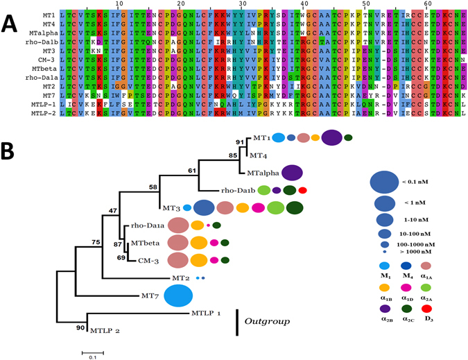
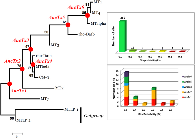
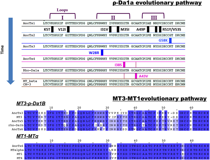
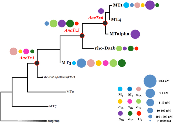
Mamba venoms contain a multiplicity of three-finger fold aminergic toxins known to interact with various α-adrenergic, muscarinic and dopaminergic receptors with different pharmacological profiles. In order to generate novel functions on this structural scaffold and to avoid the daunting task of producing and screening an overwhelming number of variants generated by a classical protein engineering strategy, we accepted the challenge of resurrecting ancestral proteins, likely to have possessed functional properties. This innovative approach that exploits molecular evolution models to efficiently guide protein engineering, has allowed us to generate a small library of six ancestral toxin (AncTx) variants and associate their pharmacological profiles to key functional substitutions. Among these variants, we identified AncTx1 as the most α1A-adrenoceptor selective peptide known to date and AncTx5 as the most potent inhibitor of the three α2 adrenoceptor subtypes. Three positions in the ρ-Da1a evolutionary pathway, positions 28, 38 and 43 have been identified as key modulators of the affinities for the α1 and α2C adrenoceptor subtypes. Here, we present a first attempt at rational engineering of the aminergic toxins, revealing an epistasis phenomenon.


Conflict of interest statement

The authors declare that they have no competing interests.

Figures

Figure 1
Figure 1
Sequence alignment and phylogenetic tree of aminergic toxins. (A) Sequence alignment of the aminergic toxin family. Color display is settled in order to emphasize amino acid conservation along all toxins. (B) Maximum likelihood tree and associated function of the aminergic toxins. The best evolutionary model established by MEGA5 was the Dayhoff model using a discrete Gamma distribution (+G). Bootstraps value higher than 50% are shown. MTLP-1 and 2 were used as out-group. Toxin’s functions are also illustrated by circles identifying the receptor targeted (color) and the corresponding Ki value (circle size), as described in the right legend, at the right side of each toxin name.
Figure 2
Figure 2
Maximum likelihood tree of the aminergic toxins. The best evolutionary model established by MEGA5 was the Dayhoff model using a discrete Gamma distribution (+G). Bootstraps value higher than 50% are shown. MTLP-1 and 2 were used as out-group. Six ancestral sequences are figured on the nodes of the aminergic toxins tree. Quality of the reconstruction is shown on right side upper panel. For each site, a probability P is given. In total 390 sites were reconstructed. Among them, only 31 showed a P < 0.9, highlighting a good quality of the reconstruction. As shown in the lower panel, a high proportion of these ambiguous sites are presented in the sequences of AncTx1 and 2. The ambiguities were resolved by chosen the residues with the best probability. Due to the high divergence of MT7 and MT2 in term of sequence and function, this choice leads AncTx1 and 2 to be closer of AncTx3 and ρ-Da1a than MT7/MT2.
Figure 3
Figure 3
Amino acid substitutions in the aminergic toxins and their ancestors from the two evolutionary pathways. Sequences are following the time/tree topology of the aminergic toxins. MT7 and MT2 sequences are not shown in the ρ-Da1a pathway due to divergences in sequence and function. Amino acid substitutions are highlighted in the first pathway. MT1-MT3 evolutionary pathway is also illustrated. Color display is settled in order to emphasize amino acid conservation along the toxins. Stronger blue color indicates residue conservation among all sequences.
Figure 4
Figure 4
X-ray structure of AncTx1 (variant W28R-I38S). (A) Three-finger fold structure of AncTx1 variant. (B) superimposition of AncTx1 variant structure with other aminergic toxins from mamba, MT1 (PDB id: 4DO8), MT2 (PDB id: 1FF4), MT7 (PDB id: 2VLW) and ρ-Da1a (PDB id: 4IYE). (C) Superimposition of AncTx1W28R-I38S and MT2 loop 2. (D) structural variations at the tip of loop 1 of aminergic toxins highlighting a large kink in MT7 and MT2. The RMSD from C-α for core residues (white background) shows that the backbones for all the toxins superimpose well, but the agreement deteriorates if loop residues are also added to the calculation (light blue background).
Figure 5
Figure 5
Maximum likelihood tree and associated function of the aminergic toxins from ρ-Da1a evolutionary pathway. Bootstraps value higher than 50% are shown. Toxin’s functions are illustrated by circles identifying the receptor targeted (color) and the corresponding Ki value (circle size), as described in the right legend, at the right side of each toxin name.
Figure 6
Figure 6
Affinity and structure analysis of CM-3, AncTx4 and ρ-Da1a toxins on adrenoceptors. (A) pKi values are reported for CM-3, AncTx4 and ρ-Da1a on the 4 adrenoceptors α1A, α1B, α1D and α2C. (B) The X-ray structure of ρ-Da1a K34A (code PDB: 4IYE), the position 43 is in the top of the third loop quite far and on the opposite side compare to the substitution S38I. The lateral chain of Ala43 is surrounded by the phenyl ring of Phe25, the side chain of Leu23, and by oxygen atoms from Cys42 and Thr45 main chains. This region of the globular core is indeed compact. Hence the substitution A43V has to reorganize the packing to accommodate the bigger lateral chain of the valine.
Figure 7
Figure 7
Affinity constants of AncTx1 and AncTx3 variants on adrenoceptors. pKi values are reported for AncTx3, AncTx4, ρ-Da1a, AncTx1 and its 2 variants (AncTx1 I38S and W28R-I38S) on the 4 adrenoceptors α1A, α1B, α1D and α2C. The 12-times increase in selectivity for α1A between ρ-Da1a and AncTx1 is shown. AncTx4 and ρ-Da1a are here represented as AncTx3 variants (W28R and W28R-I38S respectively), like I38S and W28R-I38S for AncTx1, in order to compare the effect of these substitutions in AncTx3 and AncTx1.
Figure 8
Figure 8
Maximum likelihood tree and associated function of the aminergic toxins from MT3-MT1 evolutionary pathway. Bootstraps value higher than 50% are shown. Toxin’s functions are illustrated by circles identifying the receptor targeted (color) and the corresponding Ki value (circle size), as described in the right legend, at the right side of each toxin name.

References

    1. Casewell NR, Wuster W, Vonk FJ, Harrison RA, Fry BG. Complex cocktails: the evolutionary novelty of venoms. Trends Ecol. Evol. 2013;28:219–229. doi: 10.1016/j.tree.2012.10.020. - DOI - PubMed
    1. Fry BG, et al. The toxicogenomic multiverse: convergent recruitment of proteins into animal venoms. Annu. Rev. Genomics Hum. Genet. 2009;10:483–511. doi: 10.1146/annurev.genom.9.081307.164356. - DOI - PubMed
    1. King, G. F. Venoms to Drugs - Venom as a source for the development of human therapeutics. 1–306 (Royal Society of Chemistry, 2015).
    1. King GF. Venoms as a platform for human drugs: translating toxins into therapeutics. Expert Opin. Biol. Ther. 2011;11:1469–1484. doi: 10.1517/14712598.2011.621940. - DOI - PubMed
    1. McCleary RJ, Kini RM. Non-enzymatic proteins from snake venoms: a gold mine of pharmacological tools and drug leads. Toxicon. 2013;62:56–74. doi: 10.1016/j.toxicon.2012.09.008. - DOI - PubMed

LinkOut - more resources