

A Potential Role of Esophageal Cancer Related Gene-4 for Atrial Fibrillation
- PMID: 28578429
- PMCID: PMC5457405
- DOI: 10.1038/s41598-017-02902-x
A Potential Role of Esophageal Cancer Related Gene-4 for Atrial Fibrillation
Abstract
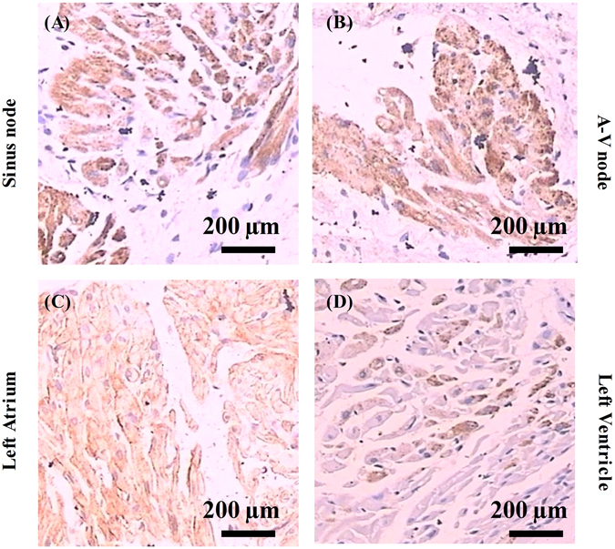
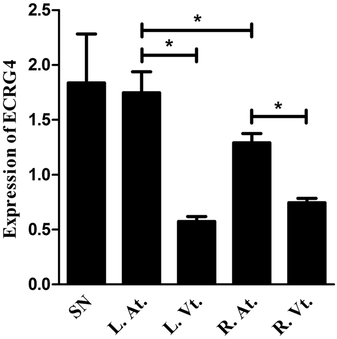
Epidemiological studies have shown a strong correlation between tumor and AF. However, the molecular link between tumor and AF remains unknown. ECRG4, a tumor suppressor gene that is expressed in the A-V node and in sporadic ventricular myocytes, inhibits tumorigenesis and monitors tissue homeostasis by functioning as a 'sentinel' molecule gauging inflammatory and cell proliferative responses. To explore the potential physiological function of Ecrg4 in heart, we evaluated its distribution in heart, analyzed its expression in patients with persistent AF and in a canine AF model, and dissected the molecular events downstream of Ecrg4. The results showed that the level of Ecrg4 expression is homogenously high in atria and the conduction systems and in sporadic ventricular myocytes. Importantly, the expression of Ecrg4 was significantly decreased in atrial appendages of AF patients than patients with SR. Moreover, in rapid pacing canine AF models, the expression of ECRG4 in atria was significantly decreased compared to that of the controls. Mechanistically, knockdown ECRG4 in atrial myocytes significantly shortened the APDs, inhibited the expression of Gja1, and activated pro-inflammatory cascades and genes involved in cardiac remodeling. These results suggest that Ecrg4 may play a critical role in the pathogenesis of AF.
Conflict of interest statement
The authors declare that they have no competing interests.
Figures


Similar articles
-
Cross-talk between macrophages and atrial myocytes in atrial fibrillation.Basic Res Cardiol. 2016 Nov;111(6):63. doi: 10.1007/s00395-016-0584-z. Epub 2016 Sep 22. Basic Res Cardiol. 2016. PMID: 27660282 Free PMC article.
-
Increased Susceptibility to Atrial Fibrillation Secondary to Atrial Fibrosis in Transgenic Goats Expressing Transforming Growth Factor-β1.J Cardiovasc Electrophysiol. 2016 Oct;27(10):1220-1229. doi: 10.1111/jce.13049. Epub 2016 Aug 30. J Cardiovasc Electrophysiol. 2016. PMID: 27447370 Free PMC article.
-
Mapping genetic changes in the cAMP-signaling cascade in human atria.J Mol Cell Cardiol. 2021 Jun;155:10-20. doi: 10.1016/j.yjmcc.2021.02.006. Epub 2021 Feb 22. J Mol Cell Cardiol. 2021. PMID: 33631188
-
Noncoding RNAs in Atrial Fibrillation: Current Status and Prospect.J Cardiovasc Pharmacol. 2020 Jan;75(1):10-17. doi: 10.1097/FJC.0000000000000762. J Cardiovasc Pharmacol. 2020. PMID: 31895877 Review.
-
Atrial fibrosis and the mechanisms of atrial fibrillation.Heart Rhythm. 2007 Mar;4(3 Suppl):S24-7. doi: 10.1016/j.hrthm.2006.12.040. Epub 2006 Dec 28. Heart Rhythm. 2007. PMID: 17336879 Free PMC article. Review.
Cited by
-
Cardiac Expression of Esophageal Cancer-Related Gene-4 is Regulated by Sp1 and is a Potential Early Target of Doxorubicin-Induced Cardiotoxicity.Cardiovasc Toxicol. 2022 May;22(5):404-418. doi: 10.1007/s12012-022-09722-0. Epub 2022 Feb 7. Cardiovasc Toxicol. 2022. PMID: 35129819
-
Potential Target Genes in the Development of Atrial Fibrillation: A Comprehensive Bioinformatics Analysis.Med Sci Monit. 2021 Mar 20;27:e928366. doi: 10.12659/MSM.928366. Med Sci Monit. 2021. PMID: 33741890 Free PMC article.
-
Esophageal Cancer-Related Gene-4 Contributes to Lipopolysaccharide-Induced Ion Channel Dysfunction in hiPSC-Derived Cardiomyocytes.J Inflamm Res. 2024 Dec 3;17:10183-10197. doi: 10.2147/JIR.S470828. eCollection 2024. J Inflamm Res. 2024. PMID: 39649417 Free PMC article.
-
Potential functions of esophageal cancer-related gene-4 in the cardiovascular system.Front Med. 2019 Dec;13(6):639-645. doi: 10.1007/s11684-019-0701-0. Epub 2019 Aug 29. Front Med. 2019. PMID: 31468282 Review.
-
ECRG4: a new potential target in precision medicine.Front Med. 2019 Oct;13(5):540-546. doi: 10.1007/s11684-018-0637-9. Epub 2018 Jul 12. Front Med. 2019. PMID: 30003403 Review.
References
-
- Lazzerini PE, Capecchi PL, Galeazzi M, Laghi-Pasini F. Biologic drugs and arrhythmic risk in chronic inflammatory arthritis: the good and the bad. Immunologic research. 2016 - PubMed
Publication types
MeSH terms
Substances
LinkOut - more resources
Full Text Sources
Other Literature Sources
Medical
Research Materials
