. 2017 Jul;121(3):271-278.
doi: 10.1016/j.ymgme.2017.05.015. Epub 2017 May 22.

Psychosine, a marker of Krabbe phenotype and treatment effect

Affiliations

Psychosine, a marker of Krabbe phenotype and treatment effect

M L Escolar et al. Mol Genet Metab. 2017 Jul.

Abstract

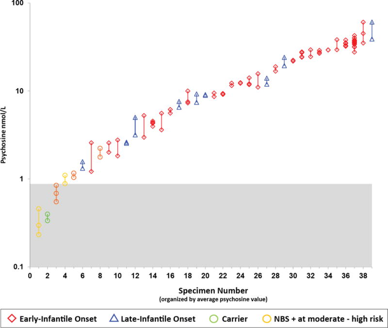
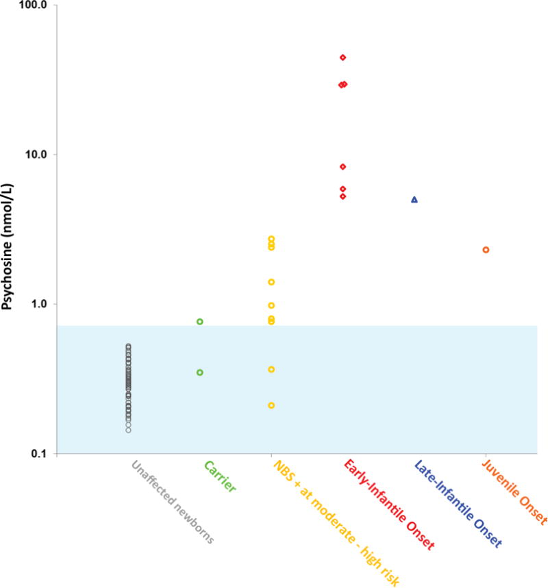
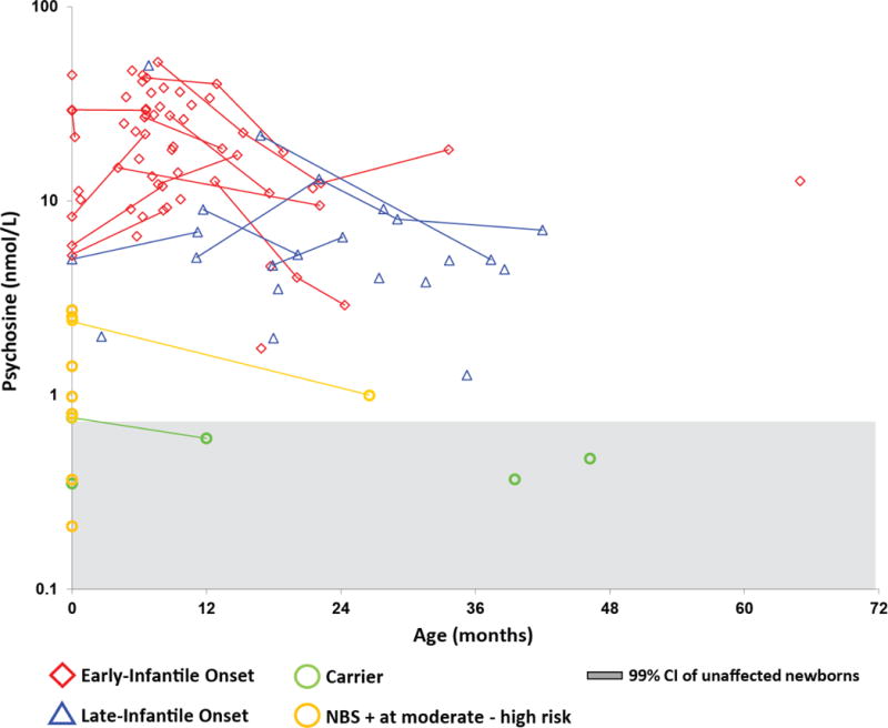
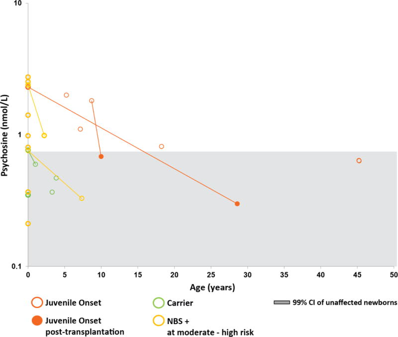
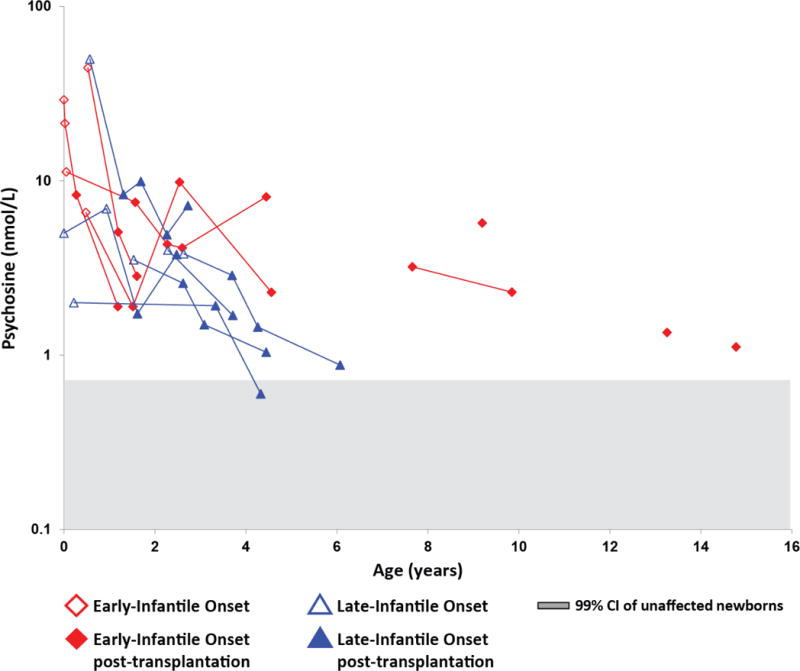
Newborn screening (NBS) for Krabbe disease, a rare neurodegenerative disorder caused by deficient galactocerebrosidase (GALC) enzyme activity, has recently been implemented in a number of US states. However, the spectrum of phenotypic manifestations associated with deficient GALC activity complicates the management of screen-positive newborns and underscores the need to identify clinically relevant biomarkers. Earlier studies with a small number of patients identified psychosine, a substrate of the GALC enzyme, as a potential biomarker for Krabbe disease. In this study, we provide, for the first time, longitudinal data on dried blood spot (DBS) psychosine concentrations in different Krabbe disease phenotypes for both untreated patients and those treated with hematopoietic stem cell transplantation (HSCT). Our cohort included patients previously identified by NBS to be at high risk to develop Krabbe disease. Substantially elevated DBS psychosine concentration during the newborn period was found to be a highly specific marker for infantile Krabbe disease. This finding supports the use of DBS psychosine concentration as a second-tier NBS test to aid in the identification of patients who require urgent evaluation for HSCT. In addition, longitudinal assessments showed that both natural disease progression and treatment with HSCT were associated with decreases in DBS psychosine concentrations. Based on these findings we provide recommendations for the interpretation of psychosine concentrations in DBS specimens collected during the first year of life. Future studies should aim to better delineate the relationship between DBS psychosine concentration and disease onset in patients with later-onset forms of Krabbe disease.

Keywords: Galactosylsphingosine; Globoid cell leukodystrophy; Krabbe disease; Newborn screening; Psychosine; Tandem mass spectrometry.


Conflict of interest statement

Conflicts of Interest

The authors declare no conflict of interest.

Figures

Fig. 1
Fig. 1
Within-specimen variability in DBS psychosine concentrations. Psychosine concentrations were plotted on a log scale for each DBS specimen, each of which was tested 2–20 times. Vertical lines connect replicates from the same specimen. The red diamonds represent EIKD patients; blue triangles represent LIKD patients; green circles represent sibling carriers; gold circles represent newborn screen-positive (NBS+) patients classified as moderate to high risk of developing KD. Each test was carried out using a separate 3-mm punch from the same DBS or from multiple DBS from the same bleed.
Fig. 2
Fig. 2
Psychosine concentrations in newborn DBS specimens. The blue shaded area is the estimated 8 standard deviation range (mean ± 4 SDs), which should include >99.9% of the unaffected population. This estimate is based on the distribution of the 75 unaffected newborns (gray). The mean psychosine concentration of one of the carrier DBS specimens (green) was similar to that of the unaffected newborns but was elevated in the other specimen. The newborn screen-positive (NBS+) infants considered to be at moderate to high risk based on GALC enzyme activity levels and mutational analysis are shown in gold. Two of these infants had psychosine levels within the range of the unaffected newborns, whereas seven infants showed elevated levels. Psychosine levels were elevated in all specimens from patients with EIKD (red), LIKD (blue), and juvenile-onset (orange) KD. The patient with juvenile-onset KD had a psychosine concentration lower than that of three samples in the NBS+ group.
Fig. 3
Fig. 3
Psychosine concentrations in untreated infantile KD patients. Average DBS psychosine concentrations from EIKD and LIKD patients who had not undergone HSCT at the time of sample collection were plotted on a log scale against age. Lines indicate longitudinal data. The red diamonds represent EIKD patients; blue triangles represent LIKD patients; green circles represent sibling carriers; and gold circles represent newborn screen-positive (NBS+) infants classified as moderate to high risk of developing KD. Psychosine concentrations from newborn dried blood spots are shown in bold and plotted on the y-axis (age = 0). The gray shaded area indicates the 99.9% CI (mean ± 4 standard deviations) for unaffected newborns.
Fig. 4
Fig. 4
Psychosine concentrations in patients with juvenile-onset KD. Average DBS psychosine concentrations were plotted on a log scale against age. Lines indicate longitudinal data. The orange circles represent patients with juvenile-onset KD; green circles represent sibling carriers; and gold circles represent newborn screen-positive (NBS+) infants classified as moderate to high risk of developing KD. The solid circles show values after hematopoietic stem cell transplantation. Psychosine concentrations from newborn DBS are shown in bold and plotted on the y-axis (age = 0). The gray shaded area indicates the 99.9% CI (mean ± 4 standard deviations) for unaffected newborns.
Fig. 5
Fig. 5
Psychosine concentrations in KD patients who underwent HSCT. Average DBS psychosine concentrations from early-infantile KD (EIKD) and late-infantile KD (LIKD) patients that underwent HSCT were plotted on a log scale against age. Lines indicate longitudinal data. The red diamonds represent EIKD patients; blue triangles represent LIKD patients. Open shapes represent samples obtained before HSCT; shaded shapes represent samples obtained after HSCT. All patients underwent HSCT within 6 weeks of the collection of the most recent pre-HSCT sample. The gray shaded area indicates the 99% CI (mean ± 4 standard deviations) for unaffected newborns.

References

    1. Suzuki K. Globoid cell leukodystrophy (Krabbe’s disease): update. J Child Neurol. 2003;18:595–603. doi: 10.1177/08830738030180090201. - DOI - PubMed
    1. Wenger DA. Krabbe Disease. 2000 Jun 19 [Updated 2011 Mar 31] In: Pagon RA, Adam MP, Ardinger HH, et al., editors. GeneReviews® [Internet] Seattle WA: University of Washington, Seattle; 1993–2017. Available from: https://www.ncbi.nlm.nih.gov/books/NBK1238/. Accessed March 6, 2017.
    1. Escolar ML, Poe MD, Provenzale JM, Richards KC, Allison J, Wood S, et al. Transplantation of umbilical-cord blood in babies with infantile Krabbe’s disease. N Engl J Med. 2005;352:2069–2081. doi: 10.1056/NEJMoa042604. - DOI - PubMed
    1. Orsini JJ, Kay DM, Saavedra-Matiz CA, Wenger DA, Duffner PK, Erbe RW, et al. Newborn screening for Krabbe disease in New York State: the first eight years’ experience. Genet Med. 2016;18:239–248. doi: 10.1038/gim.2015.211. - DOI - PubMed
    1. Kemper AR, Knapp AA, Green NS, Comeau AM, Metterville DR, Perrin JM. Weighing the evidence for newborn screening for early-infantile Krabbe disease. Genet Med. 2010;12:539–543. doi: 10.1097/GIM.0b013e3181e85721. - DOI - PubMed

Publication types