. 2017 Sep:139:229-243.
doi: 10.1016/j.biomaterials.2017.05.011. Epub 2017 May 23.

Engineering a sprayable and elastic hydrogel adhesive with antimicrobial properties for wound healing

Affiliations

Engineering a sprayable and elastic hydrogel adhesive with antimicrobial properties for wound healing

Nasim Annabi et al. Biomaterials. 2017 Sep.

Erratum in

Abstract

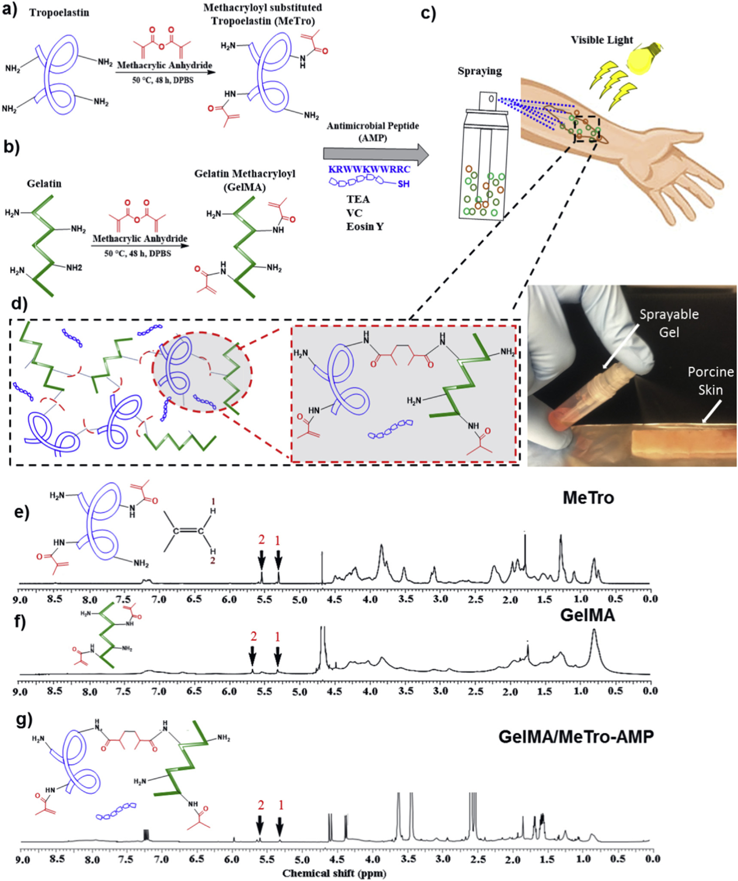
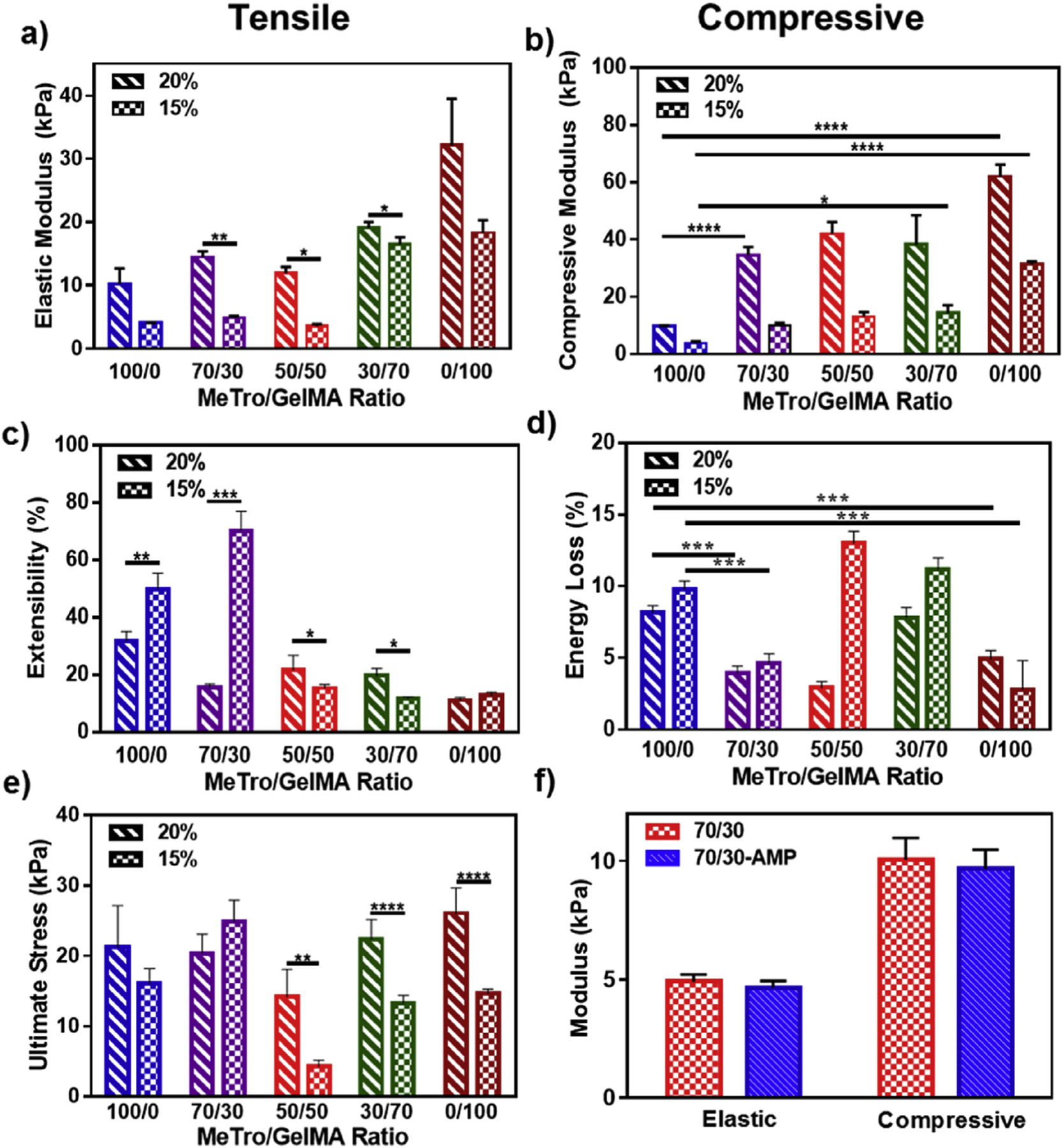
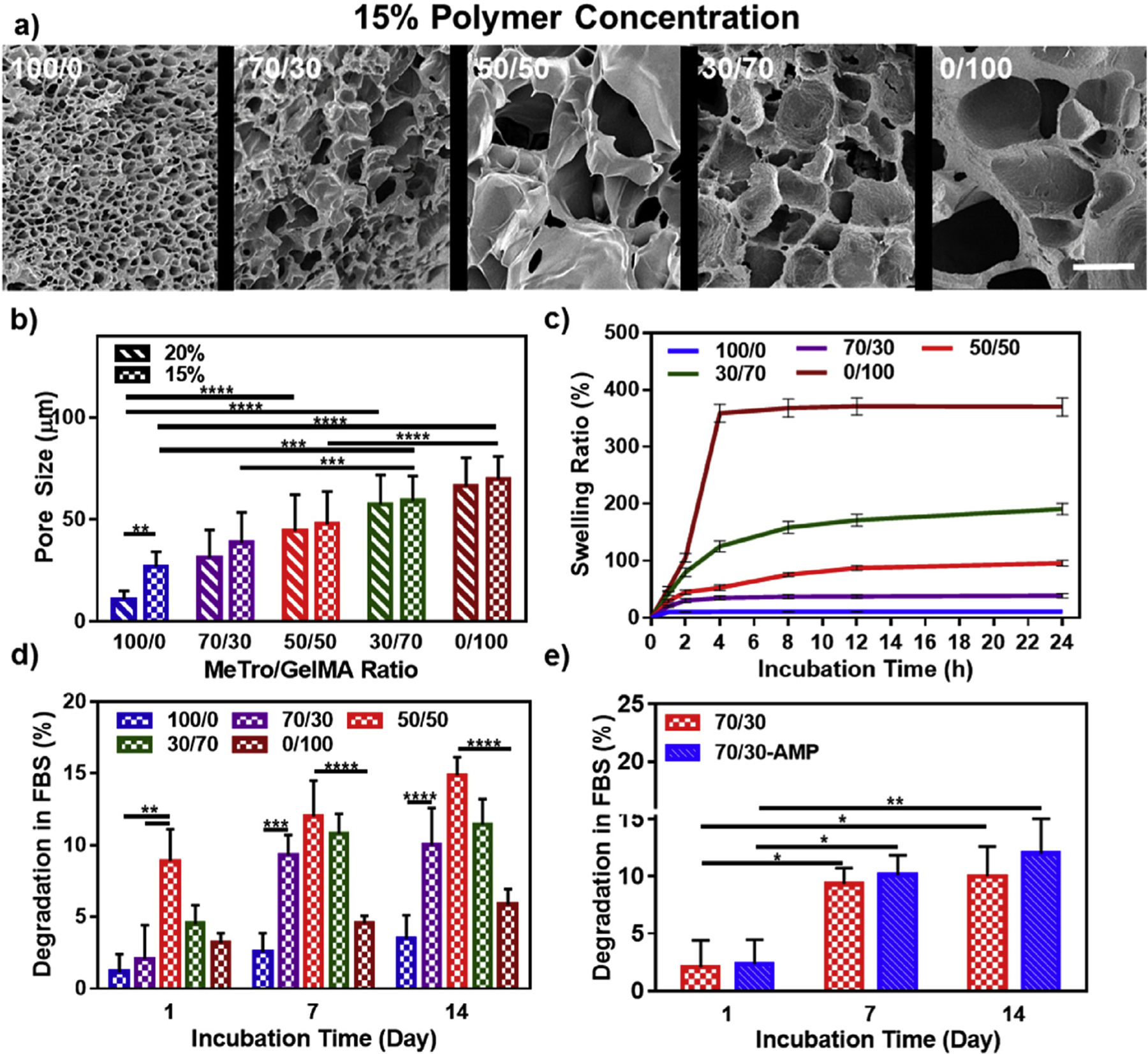
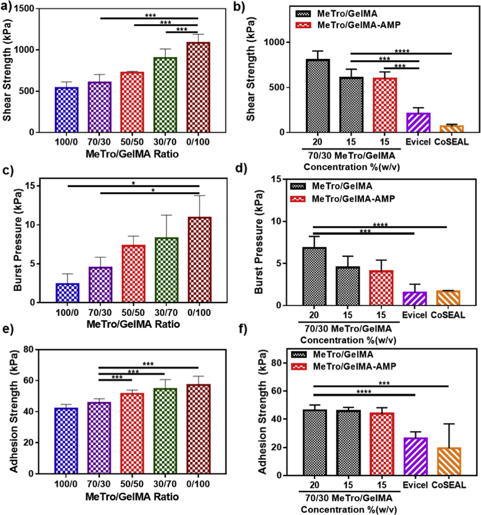
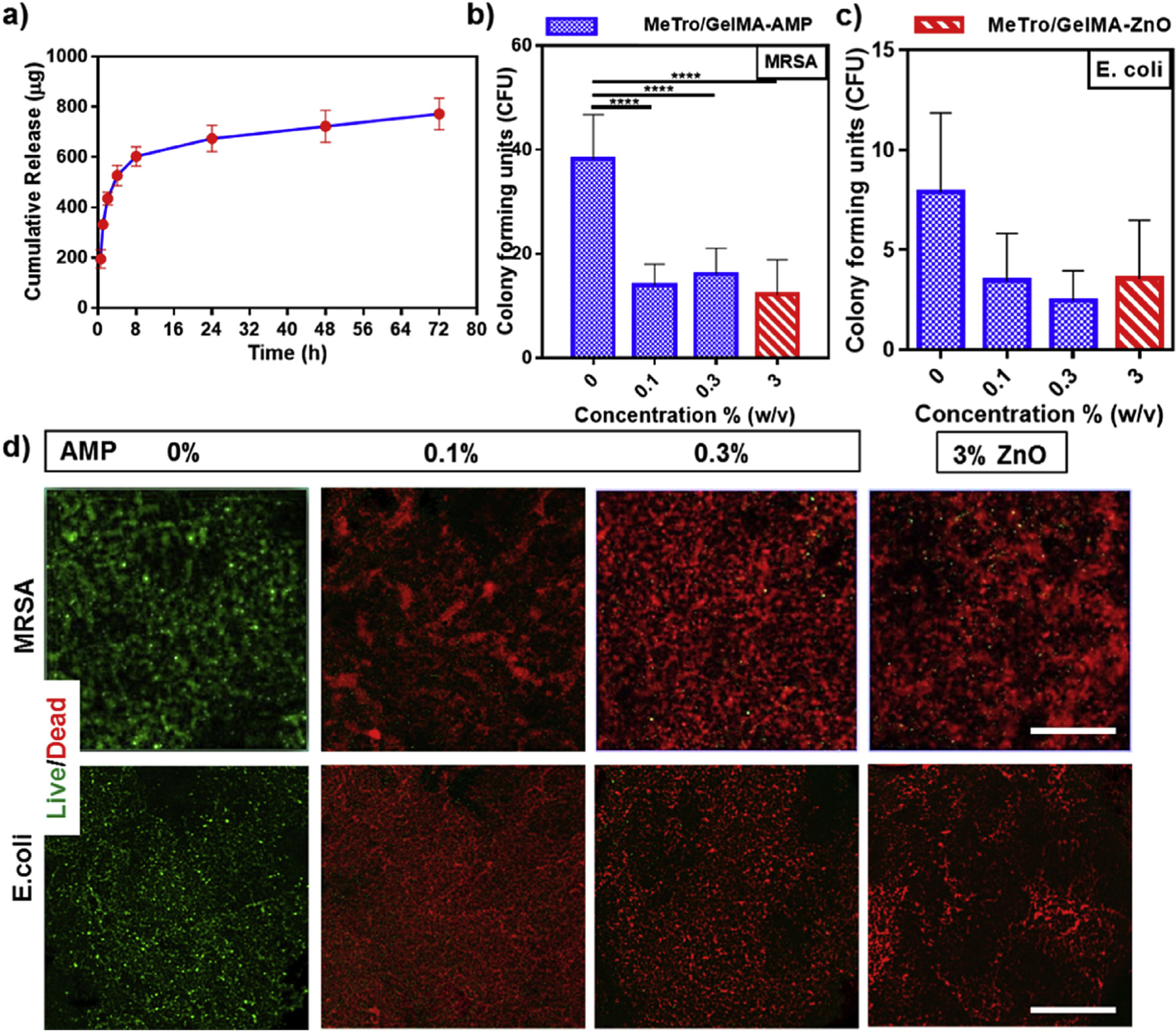
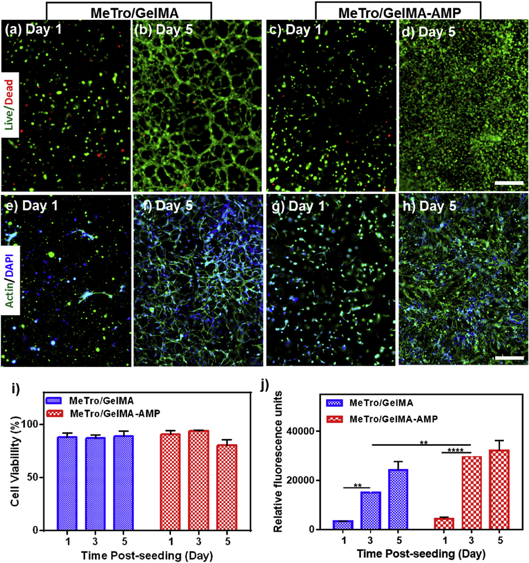
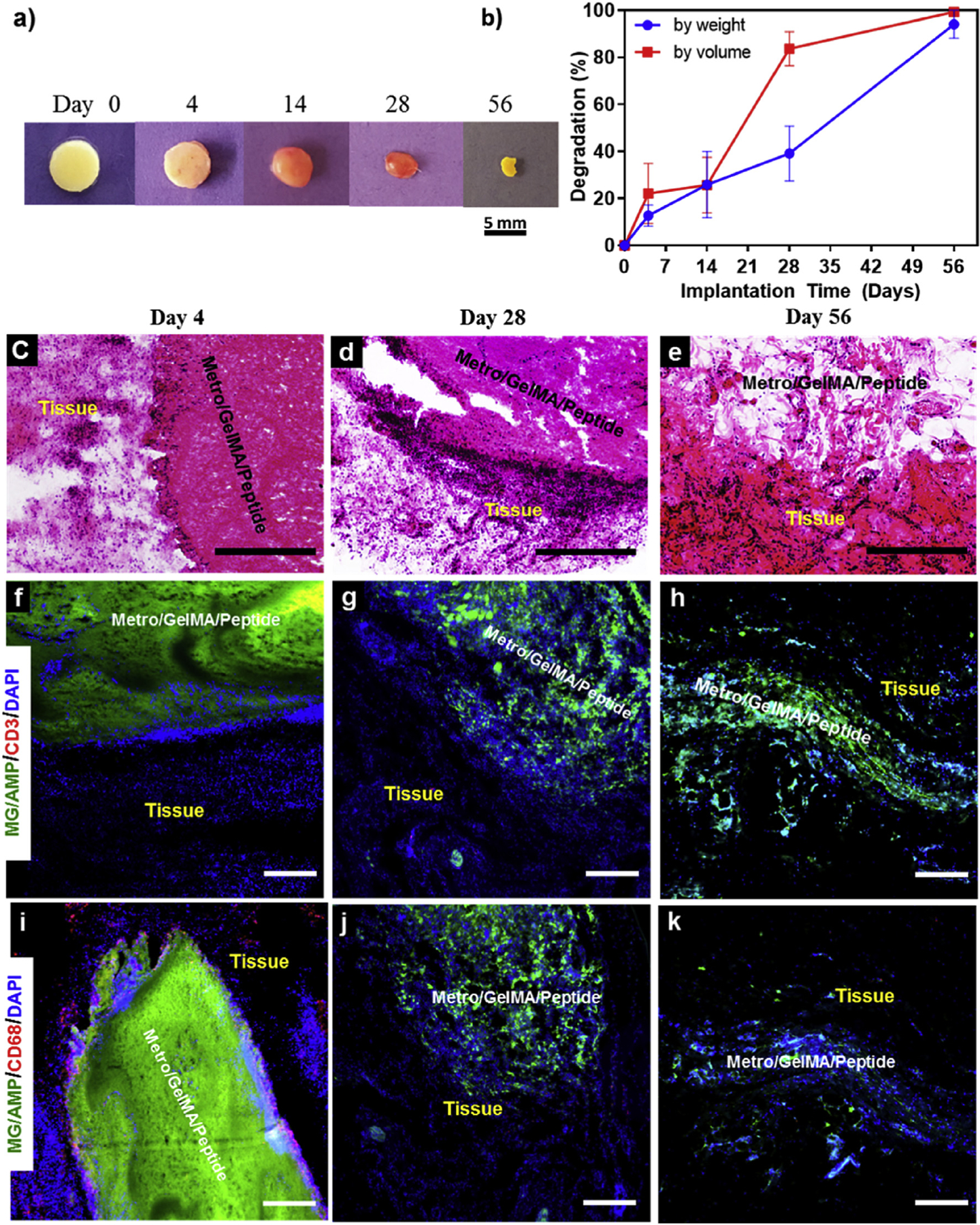
Hydrogel-based bioadhesives have emerged as alternatives for sutureless wound closure, since they can mimic the composition and physicochemical properties of the extracellular matrix. However, they are often associated with poor mechanical properties, low adhesion to native tissues, and lack of antimicrobial properties. Herein, a new sprayable, elastic, and biocompatible composite hydrogel, with broad-spectrum antimicrobial activity, for the treatment of chronic wounds is reported. The composite hydrogels were engineered using two ECM-derived biopolymers, gelatin methacryloyl (GelMA) and methacryloyl-substituted recombinant human tropoelastin (MeTro). MeTro/GelMA composite hydrogel adhesives were formed via visible light-induced crosslinking. Additionally, the antimicrobial peptide Tet213 was conjugated to the hydrogels, instilling antimicrobial activity against Gram (+) and (-) bacteria. The physical properties (e.g. porosity, degradability, swellability, mechanical, and adhesive properties) of the engineered hydrogel could be fine-tuned by varying the ratio of MeTro/GelMA and the final polymer concentration. The hydrogels supported in vitro mammalian cellular growth in both two-dimensional and three dimensional cultures. The subcutaneous implantation of the hydrogels in rats confirmed their biocompatibility and biodegradation in vivo. The engineered MeTro/GelMA-Tet213 hydrogels can be used for sutureless wound closure strategies to prevent infection and promote healing of chronic wounds.

Keywords: Antimicrobial hydrogels; GelMA; MeTro; Tissue adhesive; Wound healing.


Conflict of interest statement

Competing financial interests

The authors declare no competing financial interests.

Figures

Fig. 1.
Fig. 1.. Synthesis and 1H NMR analysis of MeTro/GelMA-AMP composite hydrogels.
Design and photocrosslinking of composite hydrogels. The panel shows a schematic of the proposed reaction for synthesis of (a) MeTro, and (b) GelMA, and (cd) MeTro-GelMA-AMP; AMP, GelMA, and MeTro were added in a TEA (co-initiator) and VC (co-initiator) solution. Immediately prior to photocrosslinking, Eosin Y (photoinitiator) was introduced into the solution. The solution could be sprayed onto a wound area and exposed to visible light to form an adhesive and elastic antimicrobial hydrogel layer. 1H NMR (500 MHz; D2O) spectra of (e) MeTro prepolymer, (f) GelMA prepolymer, and (g) MeTro/GelMA-AMP hydrogels.
Fig. 2.
Fig. 2.. Mechanical characterization of MeTro/GelMA and MeTro/GelMA-AMP composite hydrogels.
(a) Elastic modulus, (b) compressive modulus, (c) extensibility, (d) energy loss, and (e) ultimate stress of hydrogels produced by using 15% and 20% (w/v) total polymer concentration, at varying ratios of MeTro to GelMA. (f) Elastic and compressive modulus of MeTro/GelMA and MeTro/GelMA-AMP (containing 0.1% (w/v) AMP) hydrogels produced by using 15% (w/v) total polymer concentration; the results show no significant difference in mechanical properties of hydrogels with and without AMP. Data is represented as mean ± SD (*p < 0.05, **p < 0.01, ***p < 0.001, ****p < 0.0001 and n ≥ 5).
Fig. 3.
Fig. 3.. Pore characteristics, swelling ratios and in vitro degradation properties of MeTro/GelMA composite hydrogels.
(a) Representative SEM images and (b) pore size characterization of MeTro/GelMA composite hydrogels at varying MeTro/GelMA ratios and 15% final polymer concentration (scale bar = 100 μm). (c) Swelling ratios (in DPBS) and (d) degradation properties (in DPBS+10% FBS solution) of 15% composite hydrogels at varying ratios of MeTro/GelMA at 37 °C. (e) In vitro degradation of MeTro/GelMA and MeTro/GelMA-AMP (containing 0.1% (w/v) AMP) hydrogels at 15% (w/v) total polymer concentration and 70/30 MeTro/GelMA ratio; the results show no significant difference in degradation properties of hydrogels with and without AMP. Data is represented as mean ± SD (*p < 0.05, **p < 0.01, ***p < 0.001, ****p < 0.0001 and n ≥ 5).
Fig. 4.
Fig. 4.. In vitro adhesion properties of MeTro/GelMA composite hydrogels.
The in vitro shear strength of (a) composite hydrogels at varying MeTro/GelMA ratios and 15% polymer concentration, and (b) 70/30 MeTro/GelMA hydrogels at 20% and 15% polymer concentrations with and without AMP and different commercially available adhesives (Evicel and Coseal). The in vitro burst pressure of composite hydrogels at (c) varying MeTro/GelMA ratios and 15% polymer concentration, and (d) 70/30 MeTro/GelMA hydrogels at 20% and 15% polymer concentrations with and without AMP and different commercially available adhesives (Evicel and Coseal). The in vitro adhesion strength of composite hydrogels at (e) varying MeTro/GelMA ratios and 15% polymer concentration, and (f) 70/30 MeTro/GelMA hydrogels at 20% and 15% polymer concentrations with and without AMP and different commercially available adhesives (Evicel and Coseal). Data is represented as mean ± SD (*p < 0.05, **p < 0.01, ***p < 0.001, ****p < 0.0001, n ≥ 5).
Fig. 5.
Fig. 5.. In vitro AMP release profile and antibacterial properties of Metro/GelMA-AMP hydrogels as compared to MeTro/GelMA (control) and Metro/GelMA-ZnO hydrogels.
(a) In vitro release profile of AMP from 70/30 MeTro/GelMA-AMP hydrogels at 15% (w/v) total polymer concentration. Colony forming units test for MeTro/GelMA-AMP hydrogels with different AMP content (0% as a control, 0.1% and 0.3% (w/v)) and 3% (w/v) ZnO nanoparticles seeded with (b) methicillin-resistant S aureus (MRSA) and (c) E. coli. (d) Representative live/dead images from MRSA and E. coli seeded on MeTro/GelMA-AMP hydrogels with different AMP content (0% as a control, 0.1% and 0.3% (w/v)) and 3% (w/v) ZnO nanoparticles. (Scale bar = 200 μm). Data is represented as mean ± SD (*p < 0.05, **p < 0.01, ***p < 0.001, ****p < 0.0001, n ≥ 3).
Fig. 6.
Fig. 6.. In vitro 3D cell encapsulation in MeTro/GelMA and MeTro/GelMA-AMP (0.1% (w/v) AMP) hydrogels using 3T3 cells.
Representative live/dead images from 3T3 encapsulated within the (ab) MeTro/GelMA (cd) and MeTro/GelMA-AMP hydrogels on days 1 and 5. Representative Actin/DAPI stained images for 3T3 cells encapsulated within (ef) MeTro/GelMA (g, h) and MeTro/GelMA-AMP hydrogels on days 1 and 5 (scale bar = 200 μm). (i) Quantification of cell viability encapsulated in MeTro/GelMA and MeTro/GelMA-AMP hydrogels after 1, 3, and 5 days of encapsulation. (j) Quantification of metabolic activity of 3T3 cells encapsulated in MeTro/GelMA and MeTro/GelMA-AMP hydrogels after 1, 3, and 5 days. 30/70 MeTro/GelMA hydrogels at 10% (w/v) total polymer concentration were used for 3D cell encapsulation. Data is represented as mean ± SD (*p < 0.05, **p < 0.01, ***p < 0.001 and ****p < 0.0001, n ≥ 3).
Fig. 7.
Fig. 7.. In vivo biocompatibility and biodegradation of MeTro/GelMA-AMP composite hydrogel using a rat subcutaneous model.
(a) Representative images of the MeTro/GelMA-AMP hydrogels before implantation (Day 0) and on days 4, 14, 28, 56 post-implantation. (b) In vivo biodegradation of MeTro/GelMA-AMP hydrogels on days 0, 4, 14, 28 and 56 of implantation (n = 4). Hematoxylin and eosin (H&E) staining of MeTro/GelMA-AMP sections (hydrogels with the surrounding tissue) after (c) 4 days, (d) 28 days, and (e) 56 days of implantation (scale bars = 500 μm). (c) Fluorescent immunohistochemical analysis of subcutaneously implanted MeTro/GelMA-AMP hydrogels showing no significant local lymphocyte infiltration (CD3) at days (f) 4, (g) 28 and (h) 56 (scale bars = 200 μm), and exhibiting macrophages (CD68) at (i) day 4 but not at days (j) 28 and (k) 56 (scale bars = 200 μm). Green, red and blue colors in (f–k) represent the MeTro/GelMA-AMP hydrogels, the immune cells, and the cell nuclei (DAPI) respectively. 50/50 MeTro/GelMA hydrogels at 15% (w/v) total polymer concentration were used for the in vivo test.

References

    1. Frykberg RG, Banks J, Challenges in the treatment of chronic wounds, Adv. Wound Care (New Rochelle) 4 (9) (2015) 560–582. - PMC - PubMed
    1. Humphreys G, Lee GL, Percival SL, McBain AJ, Combinatorial activities of ionic silver and sodium hexametaphosphate against microorganisms associated with chronic wounds, J. Antimicrob. Chemother 66 (11) (2011) 255–2561. - PubMed
    1. Werdin F, Tenenhaus M, Rennekampff HO, Chronic wound care, Lancet 372 (9653) (2008) 1860–1862. - PubMed
    1. Ammons MCB, Ward LS, James GA, Anti-biofilm efficacy of a lactoferrin/xylitol wound hydrogel used in combination with silver wound dressings, Int. Wound J 8 (3) (2011) 268–273. - PMC - PubMed
    1. Ng VW, Chan JM, Sardon H, Ono RJ, Garcia JM, Yang YY, Hedrick JL, Antimicrobial hydrogels: a new weapon in the arsenal against multidrug-resistant infections, Adv. Drug Deliv. Rev 78 (2014) 46–62. - PubMed

MeSH terms