Review
. 2017 Sep:69:9-17.
doi: 10.1016/j.semcdb.2017.05.020. Epub 2017 Jun 1.

The role of MACF1 in nervous system development and maintenance

Affiliations
Review

The role of MACF1 in nervous system development and maintenance

Jeffrey J Moffat et al. Semin Cell Dev Biol. 2017 Sep.

Abstract

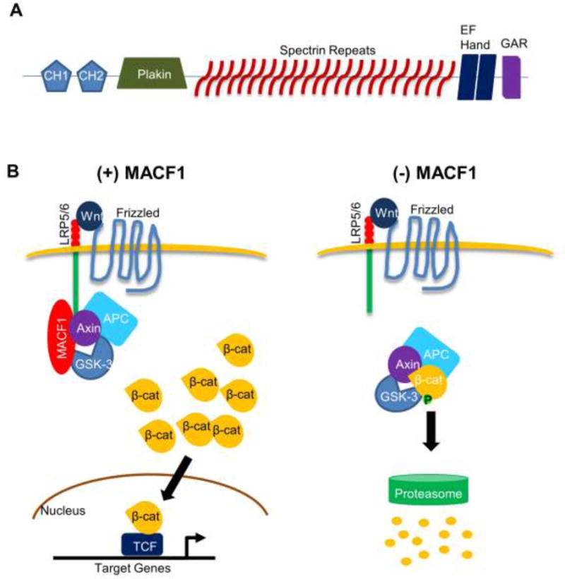
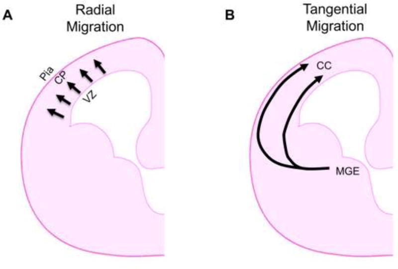
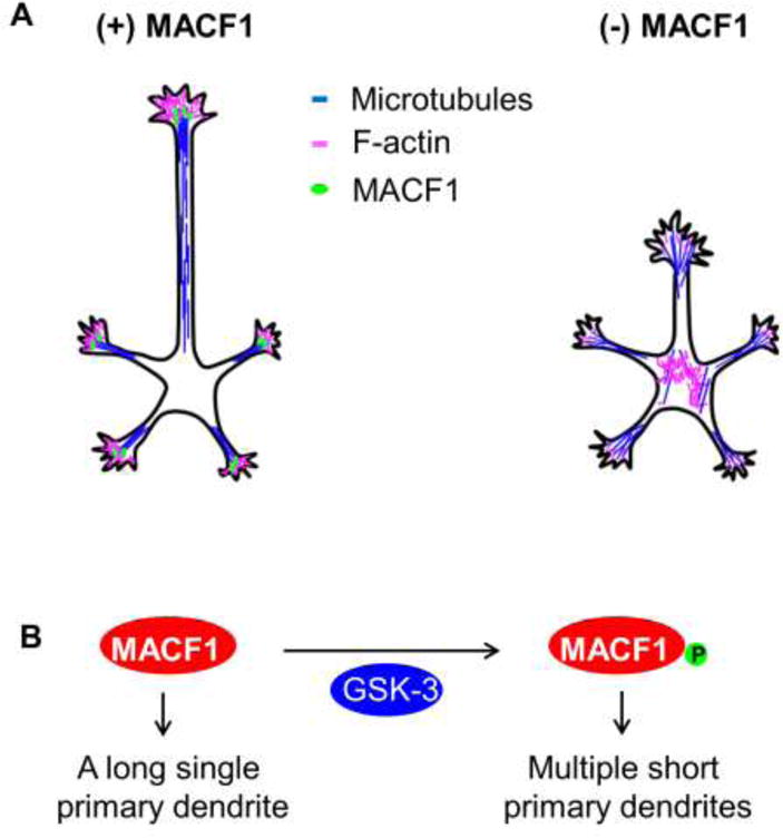
Microtubule-actin crosslinking factor 1 (MACF1), also known as actin crosslinking factor 7 (ACF7), is essential for proper modulation of actin and microtubule cytoskeletal networks. Most MACF1 isoforms are expressed broadly in the body, but some are exclusively found in the nervous system. Consequentially, MACF1 is integrally involved in multiple neural processes during development and in adulthood, including neurite outgrowth and neuronal migration. Furthermore, MACF1 participates in several signaling pathways, including the Wnt/β-catenin and GSK-3 signaling pathways, which regulate key cellular processes, such as proliferation and cell migration. Genetic mutation or dysregulation of the MACF1 gene has been associated with neurodevelopmental and neurodegenerative diseases, specifically schizophrenia and Parkinson's disease. MACF1 may also play a part in neuromuscular disorders and have a neuroprotective role in the optic nerve. In this review, the authors seek to synthesize recent findings relating to the roles of MACF1 within the nervous system and explore potential novel functions of MACF1 not yet examined.

Keywords: Development; MACF1; Nervous system; Neuron migration; Proliferation.


Conflict of interest statement

Competing interests

None

Figures

Figure 1
Figure 1. MACF1 structure and role in the Wnt/β-catenin signaling
(A) General protein structure of MACF1. The five functional domains found in most MACF1 isotypes are shown: the actin-binding domain (ABD) comprised of CH1 and CH2 fragments, a plakin domain, 23 α-helical spectrin repeats, two EF hand motifs, and a GAR domain at the C-terminus. CH1: calponin homology domain 1. CH2: calponin homology domain 2. GAR: Gas2-related domain. (B) MACF1 knockdown inhibits Wnt/β-catenin signaling. Upon Wnt binding to the receptor, MACF1 translocates axin and associated molecules to the cell membrane, allowing accumulation of β-catenin in the cytosol. Some β-catenin proteins enter the nucleus to turn on target gene expression. In the absence of MACF1, Axin is unable to translocate to the cell membrane and facilitate formation of the destruction complex containing β-catenin, resulting in proteasome-mediated β-catenin degradation. LRP5/6: low-density lipoprotein receptor-related protein 5/6. GSK-3: glycogen synthase kinase-3. APC: adenomatous polyposis coli.
Figure 2
Figure 2. Modes of neuronal migration in the developing brain
(A) Radial migration. Excitatory pyramidal projection neurons migrate outward from the ventricular zone of the cerebral cortex toward the cortical plate during brain development. CP: cortical plate. VZ: ventricular zone. (B) Tangential migration. Inhibitory interneurons migrate tangentially from the medial ganglionic eminence (MGE) in the ventral brain to the cerebral cortex, where they undergo further movements until they reach their final cortical destinations. CC: cerebral cortex.
Figure 3
Figure 3. MACF1 in neurite outgrowth
(A) MACF1 regulation of neurite outgrowth. MACF1 localizes near the distal ends of growing axons and dendrites, where it stabilizes microtubule bundles and leads to the assembly of polymerized actin. In the absence of MACF1, microtubules and actin are disorganized and unstable at the neurite tip and scattered in the cytosol. (B) GSK-3 regulates the activity of MACF1 in dendrite outgrowth and arborization. When MACF1 is phosphorylated by GSK-3, it results in multiple short primary dendrites. When MACF1 remains unphosphorylated, neurons extend a single long primary dendrite.

References

    1. Suozzi KC, Wu X, Fuchs E. Spectraplakins: master orchestrators of cytoskeletal dynamics. J Cell Biol. 2012;197(4):465–75. - PMC - PubMed
    1. Byers TJ, Beggs AH, McNally EM, Kunkel LM. Novel actin crosslinker superfamily member identified by a two step degenerate PCR procedure. FEBS Lett. 1995;368(3):500–4. - PubMed
    1. Gupta T, Marlow FL, Ferriola D, Mackiewicz K, Dapprich J, Monos D, Mullins MC. Microtubule actin crosslinking factor 1 regulates the Balbiani body and animal-vegetal polarity of the zebrafish oocyte. PLoS Genet. 2010;6(8):e1001073. - PMC - PubMed
    1. Leung CL, Sun D, Zheng M, Knowles DR, Liem RK. Microtubule actin cross-linking factor (MACF): a hybrid of dystonin and dystrophin that can interact with the actin and microtubule cytoskeletons. J Cell Biol. 1999;147(6):1275–86. - PMC - PubMed
    1. Goode BL, Drubin DG, Barnes G. Functional cooperation between the microtubule and actin cytoskeletons. Curr Opin Cell Biol. 2000;12(1):63–71. - PubMed

Publication types

Substances

LinkOut - more resources