Review
. 2017 Jun;34(2):145-166.
doi: 10.1055/s-0037-1602712. Epub 2017 Jun 1.

Metastatic Liver Disease: Indications for Locoregional Therapy and Supporting Data

Affiliations
Review

Metastatic Liver Disease: Indications for Locoregional Therapy and Supporting Data

Susan Shamimi-Noori et al. Semin Intervent Radiol. 2017 Jun.

Abstract

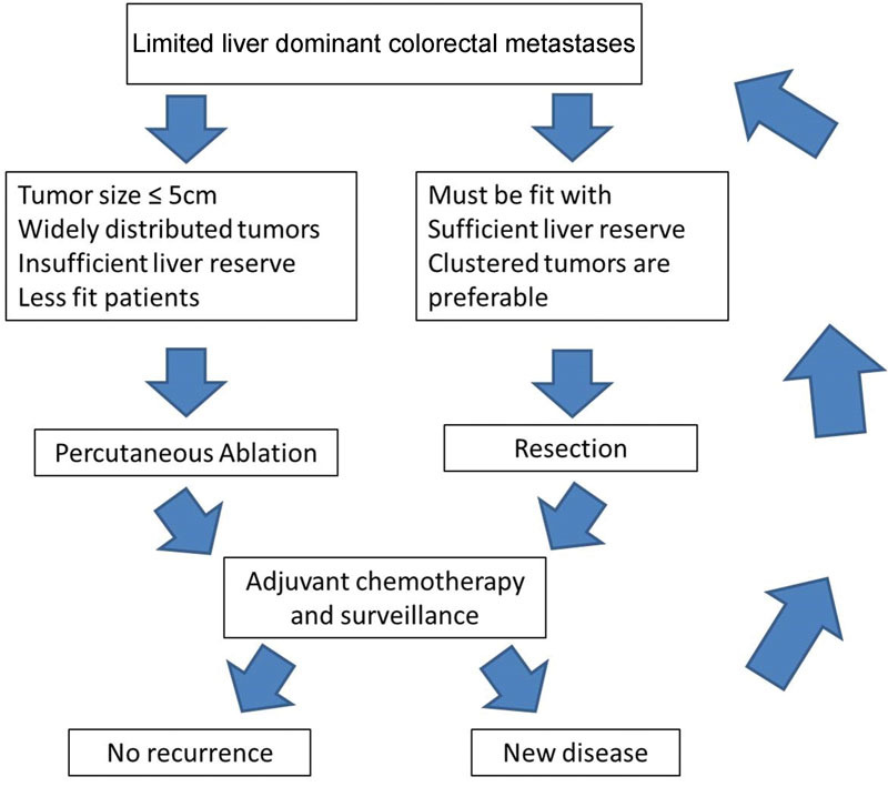
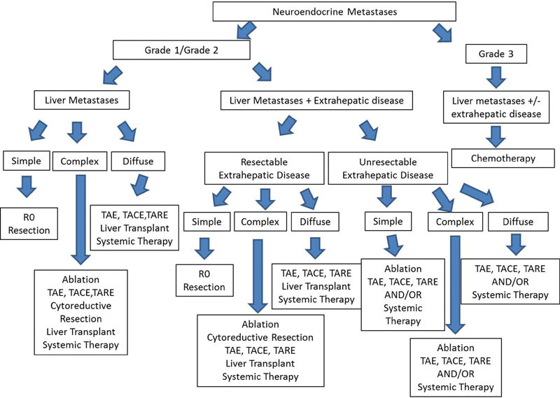
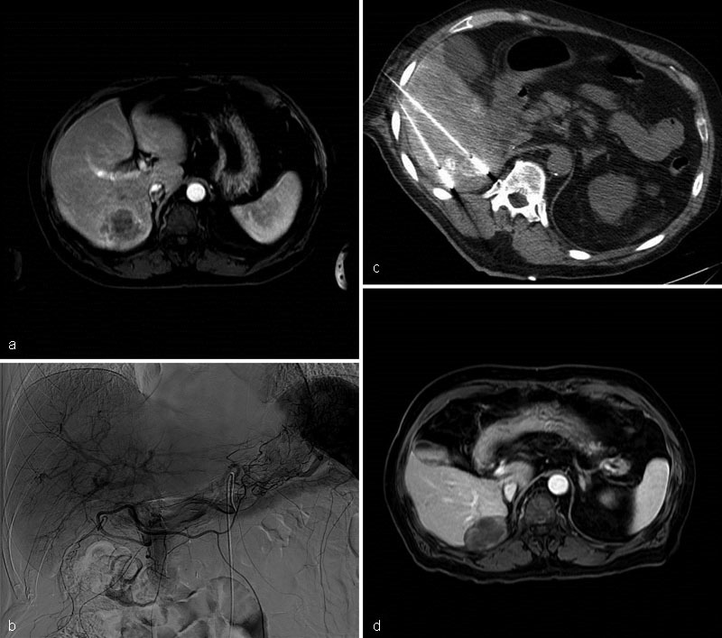
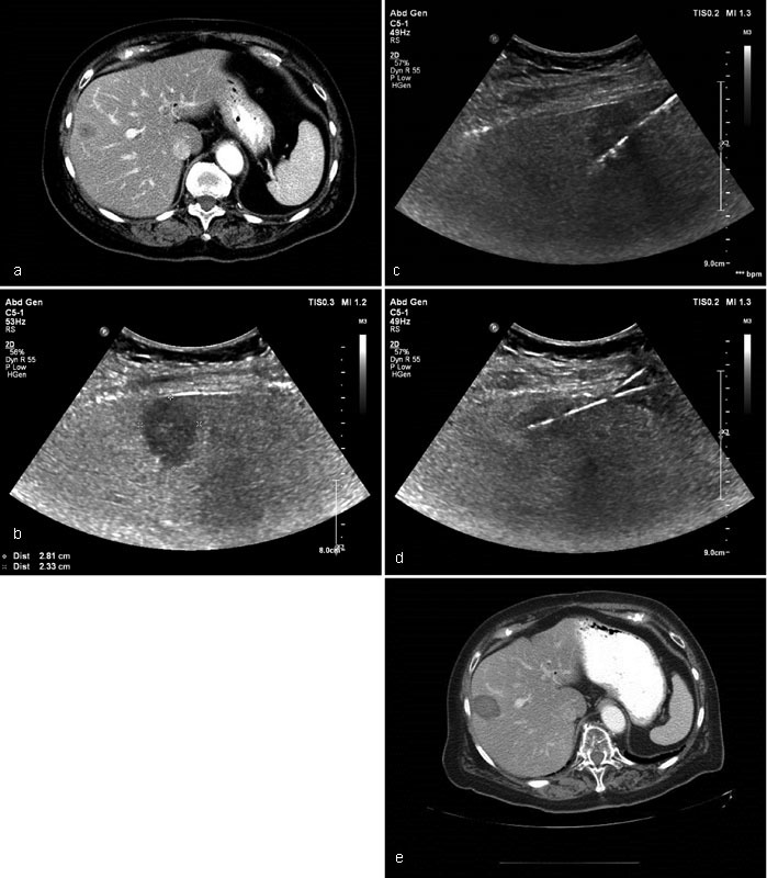
Metastatic liver disease is a major cause of cancer-related morbidity and mortality. Surgical resection is considered the only curative treatment, yet only a minority is eligible. Patients who present with unresectable disease are treated with systemic agents and/or locoregional therapies. The latter include thermal ablation and catheter-based transarterial interventions. Thermal ablation is reserved for those with limited tumor burden. It is used to downstage the disease to enable curative surgical resection, as an adjunct to surgery, or in select patients it is potentially curative. Transarterial therapies are indicated in those with more diffuse disease. The goals of care are to palliate symptoms and prolong survival. The indications and supporting data for thermal ablation and transarterial interventions are reviewed, technical and tumor factors that need to be considered prior to intervention are outlined, and finally several cases are presented.

Keywords: chemoembolization; interventional radiology; liver metastases; locoregional therapy; radioembolization; thermal ablation.


Figures

Fig. 1
Fig. 1
Management algorithm for patients presenting with limited liver-dominant colorectal metastases (image courtesy: Gillams et al 32 ).
Fig. 2
Fig. 2
Management algorithm for neuroendocrine liver metastases (image courtesy: Frilling and Clift 157 ).
Fig. 3
Fig. 3
Case 1: MRI of the abdomen postcontrast T1-weighted sequence demonstrates an enhancing 4.6 cm × 4 cm mass in segments 6/7 ( a ). Celiac arteriogram: a large hypervascular mass in the right lobe of liver is identified. cTACE with Doxorubicin and Mitomycin was performed ( b ). A week later, the patient underwent MWA using 3 antennae ( c ). MRI of the abdomen postcontrast T1-weighted sequence 18 months posttreatment shows no evidence of recurrent disease. No extrahepatic metastases were identified ( d ).
Fig. 4
Fig. 4
Case 2: Contrast-enhanced CT demonstrates a hypoattenuating mass in segment 8 of the liver consistent with metastatic disease. Air in the biliary tree status post bilioenteric anastomosis ( a ). Ultrasound confirmed a 2.8 × 2.3 cm hypoechoic mass at the periphery of segment 8 ( b ). Two antennae were placed with ultrasound guidance. Microwave ablation was performed ( c and d ). Contrast-enhanced CT abdomen 6 months posttreatment shows no evidence of recurrent disease. No new metastases were identified ( e ).
Fig. 5
Fig. 5
Case 3: MRI of the liver postcontrast T1-weighted sequence shows multiple bilobar hypervascular liver masses ( a ). Octreotide SPECT CT coronal reconstruction demonstrates significant metabolic activity within the known neuroendocrine liver metastases ( b ). MRI of the liver T1-weighted sequence postcontrast performed 6 months following the initial bland embolization shows significant decrease in size of the liver metastases. No new lesions are seen ( c ).
Fig. 6
Fig. 6
Case 4: CT of the abdomen portal venous phase shows bilobar liver metastases ( a ). Celiac arteriography shows multiple hypervascular masses within both lobes of the liver ( b ). Fluoroscopic spot image shows Ethiodol staining within the tumors following cTACE with Doxorubicin and Mitomycin ( c ). CT of the abdomen portal venous phase one year later shows decreased vascularity and Ethiodol staining within multiple liver masses. New left lateral segment bile duct dilation secondary to mass effect by the metastases ( d ).
Fig. 7
Fig. 7
Case 5: MRI of the abdomen postcontrast fat-saturated T1-weighted sequence demonstrates a heterogeneously enhancing 5 × 5 × 3.9 cm mass in segment 7 consistent with metastases ( a ). Celiac arteriogram shows a large enhancing mass in the posterior right lobe ( b and c ). MRI of the abdomen postcontrast fat-saturated T1-weighted sequence 6 months post-radioembolization shows gross necrosis and reduction in size of the segment 7 mass. It now measures 3.6 × 2.6 × 2.7 cm ( d) .

Similar articles

Cited by

References

    1. Namasivayam S, Martin D R, Saini S. Imaging of liver metastases: MRI. Cancer Imaging. 2007;7:2–9. - PMC - PubMed
    1. Robinson P J. Imaging liver metastases: current limitations and future prospects. Br J Radiol. 2000;73(867):234–241. - PubMed
    1. Jemal A, Murray T, Ward E et al.Cancer statistics, 2005. CA Cancer J Clin. 2005;55(01):10–30. - PubMed
    1. Forner A, Llovet J M, Bruix J.Hepatocellular carcinoma Lancet 2012379(9822):1245–1255. - PubMed
    1. Brace C L. Microwave tissue ablation: biophysics, technology, and applications. Crit Rev Biomed Eng. 2010;38(01):65–78. - PMC - PubMed