

Persistent proatlas with additional segmentation of the craniovertebral junction - The Tsuang-Goehmann-Malformation
- PMID: 28580053
- PMCID: PMC5443582
- DOI: 10.3941/jrcr.v10i10.2890
Persistent proatlas with additional segmentation of the craniovertebral junction - The Tsuang-Goehmann-Malformation
Abstract
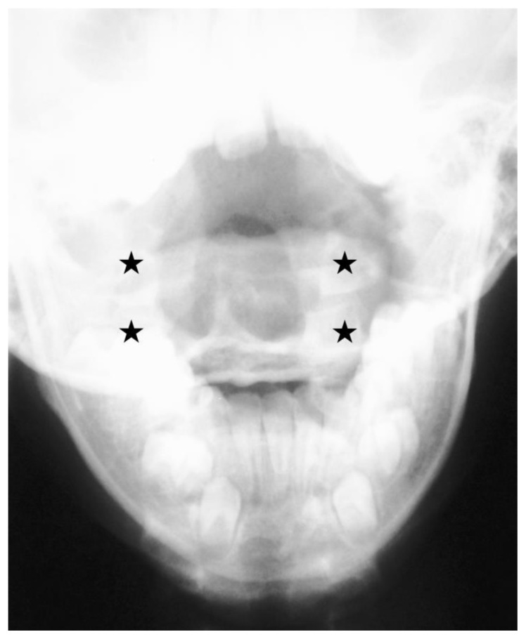
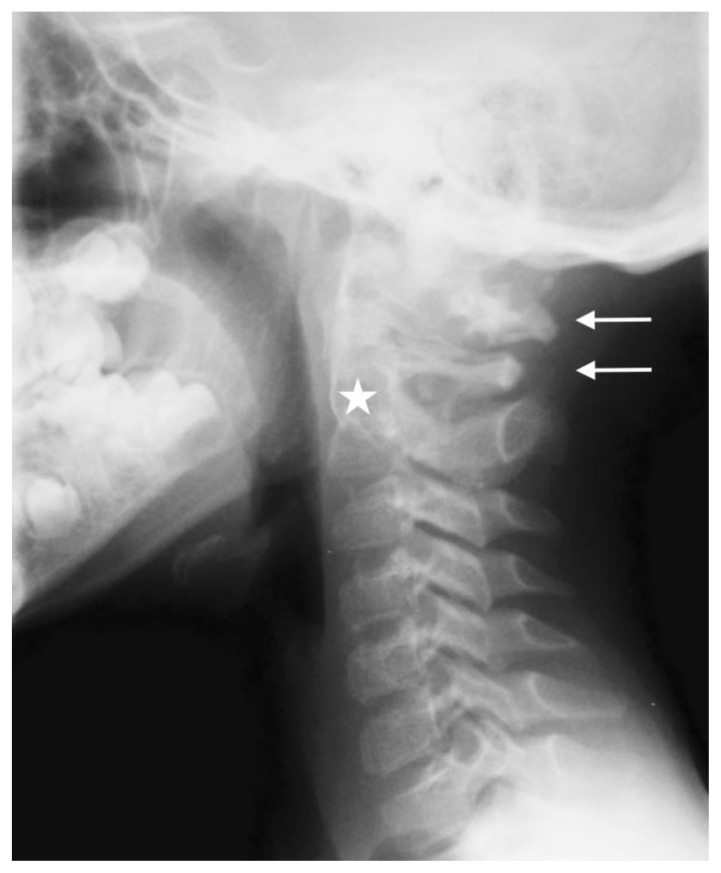
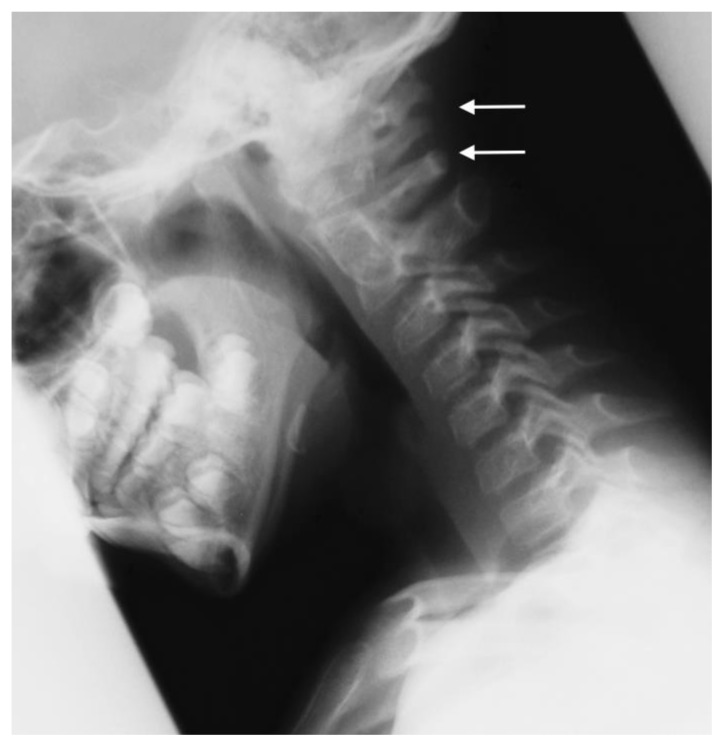
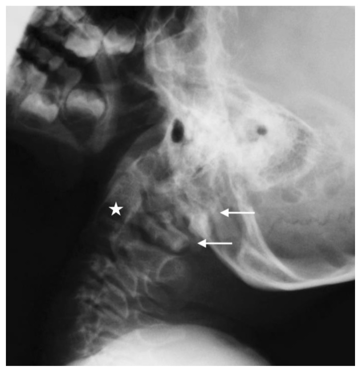
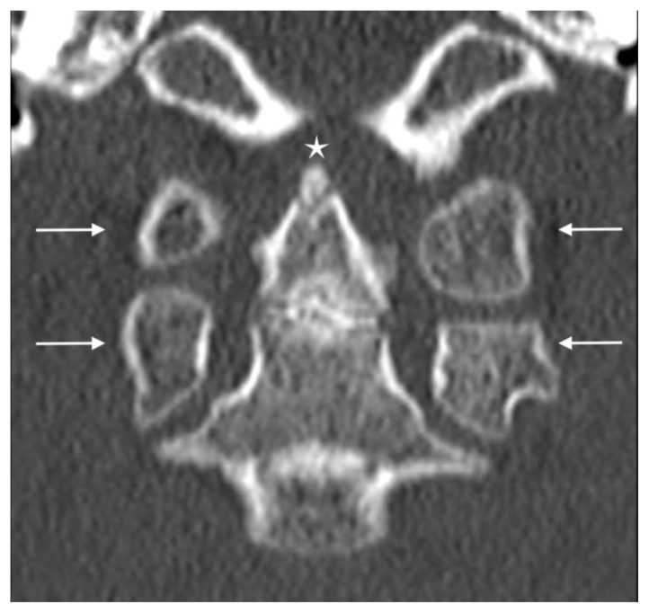
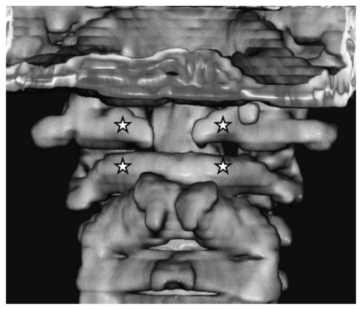
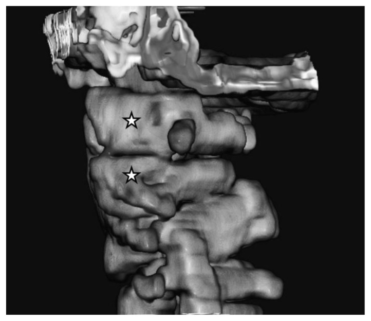
Case study description and analysis of a complex craniovertebral dysplasia in an 8-year-old male patient, in which conventional cervical spine radiographs demonstrated a regularly differentiated occipital base, as well as the presence of two lateral masses of the proatlas vertebra and two lateral masses of the atlas vertebra. Further assessment included computed tomography of the occipital base and the upper cervical spine as well as three-dimensional reconstruction. Malsegmentation of the fourth occipital vertebra can result in various anomalies that are known as 'manifestation of the proatlas'. The occurrence of a persistent proatlas with additional segmentation of the craniovertebral junction represents an extremely rare dysplasia. To our knowledge, it is the second report concerning the persistence of a complete human proatlas vertebra. We consider the biomechanical and embryological particularities of this complex dysplasia to represent sufficient basis for future differentiation from other malformations of the fourth occipital vertebra. Comprehensive literature review and discussion about the entity will be provided.
Keywords: CT scan; Sandberg-Gutmann; X-ray; additional segmentation; atlas vertebra; cervical spine; computed tomography; craniovertebral junction; malformation; manifestation of occipital vertebra; manifestation of proatlas; occipital vertebra; persistent proatlas; plain radiographs.
Figures


Similar articles
-
The proatlas: a comprehensive review with clinical implications.Childs Nerv Syst. 2012 Mar;28(3):349-56. doi: 10.1007/s00381-012-1698-8. Epub 2012 Jan 27. Childs Nerv Syst. 2012. PMID: 22282080 Review.
-
Elongated Clivus with Deficient Anterior Atlantal Arch and Congenital Posterior Atlantooccipital Dislocation: Pathoembryology and Management Nuances of a Rare Form of Proatlas Segmentation Anomaly.World Neurosurg. 2019 Jun;126:286-290. doi: 10.1016/j.wneu.2019.03.091. Epub 2019 Mar 18. World Neurosurg. 2019. PMID: 30898752
-
Proatlas segmentation anomalies: Surgical management of five cases and review of the literature.J Pediatr Neurosci. 2016 Jan-Mar;11(1):14-9. doi: 10.4103/1817-1745.181246. J Pediatr Neurosci. 2016. PMID: 27195027 Free PMC article.
-
Unusual case of occipital vertebra in a medieval skeleton.Anat Sci Int. 2008 Dec;83(4):286-90. doi: 10.1111/j.1447-073X.2007.00213.x. Anat Sci Int. 2008. PMID: 19159361
-
Bony anomalies of the craniocervical junction.Cent Afr J Med. 2002 Jan-Feb;48(1-2):17-23. doi: 10.4314/cajm.v48i1.8419. Cent Afr J Med. 2002. PMID: 12808784 Review.
Cited by
-
Proposing novel dorsal Proatlas-manifestations: Description and classification of three rare phenomena in humans.J Anat. 2023 Jul;243(1):138-147. doi: 10.1111/joa.13851. Epub 2023 Mar 2. J Anat. 2023. PMID: 36863846 Free PMC article.
-
Osseous bridging of the condylar fossa: report of a rare anatomical variation at the outer skull base.Surg Radiol Anat. 2024 Aug;46(8):1231-1235. doi: 10.1007/s00276-024-03422-w. Epub 2024 Jun 26. Surg Radiol Anat. 2024. PMID: 38926224 Free PMC article.
-
IVD Development: Nucleus pulposus development and sclerotome specification.Curr Mol Biol Rep. 2018 Sep;4(3):132-141. doi: 10.1007/s40610-018-0100-3. Epub 2018 Jul 13. Curr Mol Biol Rep. 2018. PMID: 30505649 Free PMC article.
-
Development of the axial skeleton and intervertebral disc.Curr Top Dev Biol. 2019;133:49-90. doi: 10.1016/bs.ctdb.2018.11.018. Epub 2019 Jan 3. Curr Top Dev Biol. 2019. PMID: 30902259 Free PMC article. Review.
-
The Ligamentum condylicum posterius as a precursor structure of the Processus condylicus posterior, another Proatlas-Manifestation of the human occipital bone.J Anat. 2021 Sep;239(3):611-621. doi: 10.1111/joa.13444. Epub 2021 Apr 12. J Anat. 2021. PMID: 33846976 Free PMC article.
References
-
- Kessel M, Balling R, Gruss P. Variations of the cervical vertebrae after expression of a Hox-1.1 transgene in mice. Cell. 1990;61:301–8. - PubMed
-
- Tsuang FY, Chen JY, Wang YH, et al. Neurological Picture. Occipitocervical malformation with atlas duplication. J Neurol Neurosurg Psychiatry. 2011;82:1101–2. - PubMed
-
- Went H. Zur Manifestation eines Proatlas. Fortschr Röntgenstr. 1961;95:370–4. - PubMed
-
- Goldfeld M, Loberant N, Herskovits M. Manifestations of persistent proatlas: radiographic and computed tomographic appearance. Eur Radiol. 1994;4:71–4.
Publication types
MeSH terms
LinkOut - more resources
Full Text Sources
Other Literature Sources