Case Reports
. 2017 Jan 31;11(1):1-6.
doi: 10.3941/jrcr.v11i1.2983. eCollection 2017 Jan.

Imaging Findings of Ulceroglandular Tularemia

Affiliations
Case Reports

Imaging Findings of Ulceroglandular Tularemia

Neil Anand et al. J Radiol Case Rep. .

Abstract

Francisella tularensis, the causative organism in Tularemia, is a relatively rare disease. There are a few radiological clues to elucidate its presence when suspicion arises. There should be strong consideration for Tularemia in the differential of any patient with its classic symptoms, diffuse cervical lymphadenopathy with evidence of necrosis, and enlarged adenoids. Ultrasound may demonstrate suppurative lymphadenopathy suggestive of infection, as in the case presented. CT often will demonstrate the extent of lymphadenopathy. On chest radiography, tularemia pneumonia is often the presenting finding, which may demonstrate bilateral or lobar infiltrates. Additionally, hilar lymphadenopathy and pleural effusions are often associated findings. Cavitary lesions may be present, which are better delineated on CT scan. We present a case of a 7-year-old male who presented with a painful right-sided palpable neck mass for 9 days, who was diagnosed with Tularemia after numerous admissions.

Keywords: Computer Tomography; Francisella; Lymphadenopathy; Rabbits; Suppurative; Tularemia; Tularensis; Ulceroglandular; Ultrasound.


Figures
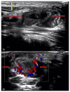
Figure 1
Figure 1
7 year old male patient: Diagnosis of Ulceroglandular Tularemia Findings: A) Real time sonography of the neck soft tissues demonstrate a right neck mass [between red arrows] measuring approximately 3.8 cm × 3.0 cm, corresponding to the area of palpable abnormality. A central area of decreased echogenicity within this nodal mass [asterisk] likely represents necrosis. Overlying subcutaneous edema [double ended yellow arrow] is also present. Numerous other enlarged adjacent lymph nodes were identified on the real-time exam [not displayed]. Findings were found to be consistent with suppurative nodal masses, seen in the setting of Tularemia. B) Color Doppler image of the neck soft tissues demonstrate a right neck mass [between red arrows], corresponding to the area of palpable abnormality. There is no flow within the center of this neck mass. Findings were found to be consistent with suppurative nodal masses, seen in the setting of Tularemia. Technique: Ultrasonography was performed using a linear transducer at 12 MHz. A) B-mode Transabdominal sonogram of the neck soft tissues. B) Power Doppler sonogram of the neck soft tissues. Ultrasonography was performed using a linear transducer at 12 MHz.
Figure 2
Figure 2
7 year old male patient: Diagnosis of Ulceroglandular Tularemia Findings: Contrast enhanced coronal CT image of the neck soft tissues, 24 days prior to the ultrasound images, demonstrate multiple enlarged, enhancing, right cervical lymph nodes [red circle]. Findings were found to be consistent with reactive lymphadenopathy, seen in the setting of Tularemia. Suppurative lymphadenopathy is not demonstrated at this time. Technique: mA 300, kvp 100, 2.0 mm slice thickness, with 100ml, Omnipaque 350.
Figure 3
Figure 3
7 year old male patient: Diagnosis of Ulceroglandular Tularemia Findings: PA view of the chest demonstrates clear lung fields. Technique: Postero-anterior view of the chest.

Similar articles

Cited by

References

    1. Baggett M, Gonzalez G, Bhattacharyya R, et al. Case 4-2016. New England Journal of Medicine. 2016;374(6):573–581. - PubMed
    1. Dogan S, Ekinci A, Demiraslan H, et al. Ultrasonography and contrast-enhanced CT findings of tularemia in the neck. Diagnostic and Interventional Radiology. 2016;22(5):430–435. - PMC - PubMed
    1. Treat J, Hess S, McGowan K, Yan A, Kovarik C. Ulceroglandular Tularemia. Pediatric Dermatology. 2010;28(3):318–320. - PubMed
    1. Umlas S, Jaramillo D. Massive adenopathy in oropharyngeal tularemia; C. T. demonstration. Pediatric Radiology. 1990;20(6):483–484. - PubMed
    1. Sjöstedt A. Special Topic on Francisella tularensis and Tularemia. Front Microbio. 2011;2 - PMC - PubMed

Publication types