. 2017 May 2;8(3):257-266.
doi: 10.14336/AD.2017.0112. eCollection 2017 May.

Cerebral Microvascular Accumulation of Tau Oligomers in Alzheimer's Disease and Related Tauopathies

Affiliations

Cerebral Microvascular Accumulation of Tau Oligomers in Alzheimer's Disease and Related Tauopathies

Diana L Castillo-Carranza et al. Aging Dis. .

Abstract

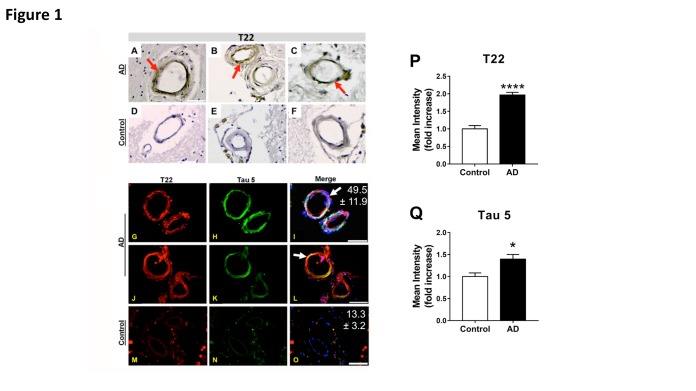
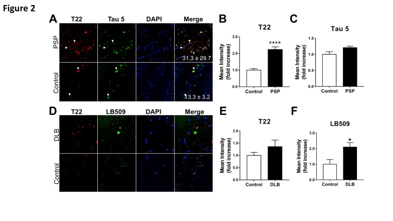
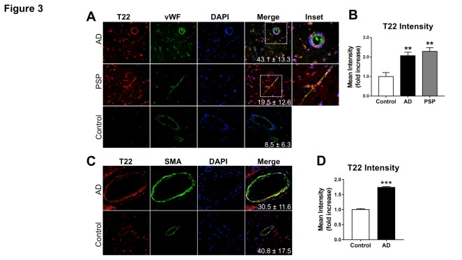
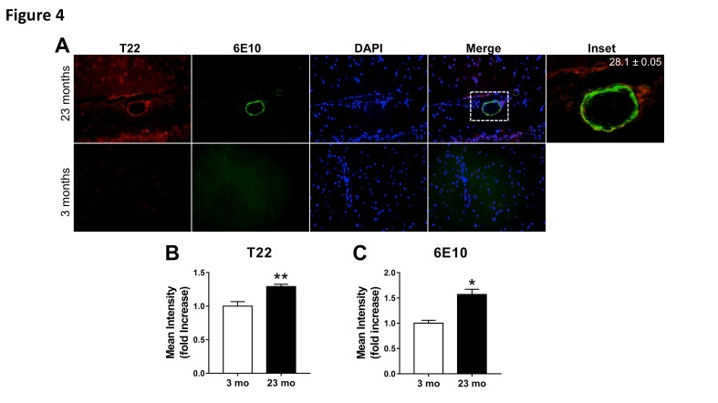
The importance of vascular contributions to cognitive impairment and dementia (VCID) associated with Alzheimer's disease (AD) and related neurodegenerative diseases is increasingly recognized, however, the underlying mechanisms remain obscure. There is growing evidence that in addition to Aβ deposition, accumulation of hyperphosphorylated oligomeric tau contributes significantly to AD etiology. Tau oligomers are toxic and it has been suggested that they propagate in a "prion-like" fashion, inducing endogenous tau misfolding in cells. Their role in VCID, however, is not yet understood. The present study was designed to determine the severity of vascular deposition of oligomeric tau in the brain in patients with AD and related tauopathies, including dementia with Lewy bodies (DLB) and progressive supranuclear palsy (PSP). Further, we examined a potential link between vascular deposition of fibrillar Aβ and that of tau oligomers in the Tg2576 mouse model. We found that tau oligomers accumulate in cerebral microvasculature of human patients with AD and PSP, in association with vascular endothelial and smooth muscle cells. Cerebrovascular deposition of tau oligomers was also found in DLB patients. We also show that tau oligomers accumulate in cerebral microvasculature of Tg2576 mice, partially in association with cerebrovascular Aβ deposits. Thus, our findings add to the growing evidence for multifaceted microvascular involvement in the pathogenesis of AD and other neurodegenerative diseases. Accumulation of tau oligomers may represent a potential novel mechanism by which functional and structural integrity of the cerebral microvessels is compromised.

Keywords: Alzheimer’s disease; brain vascular dysfunction; cerebrovascular dysfunction; cerebrovasculature; oligomers; tau; tauopathies.


Figures

Figure 1.
Figure 1.. Deposition of tau oligomers in cerebrovasculature of human Alzheimer’s Disease (AD) brains
(A-F) Representative images of immunohistochemistry studies using an oligomeric tau-specific antibody (T22) in cortical sections from AD (A-C) and age-matched controls (D-F). (G-O) Representative images of oligomeric tau (T22, red), and total tau (tau 5, green) cerebrovascular immunoreactivity in cortical sections of AD (G-L) and age-matched control (M-O) brains. Quantitative analyses of mean fluorescence intensity show increased tau oligomer- (T22, P) and total tau-specific (Tau5, Q) immunoreactivity in vasculature of AD brains compared to age-matched controls [****, t (28) = 8.12, p < 0.0001, and *, t(17) = 2.39, p = 0.029, for T22 and tau5 immunoreactivity respectively]. Our tau oligomer antibody T22 [18, 41] has been validated by immunoblot, ELISA, coimmunoprecipitation as well as rodent and human tissue staining, is produced endotoxin-free, and is commercially available (Millipore ABN454). For all studies, n=3 brains/group; 10-15 sections from each sample were analyzed for tau oligomers. All AD samples were tested and were positive for tau oligomers. Merged images are shown with DAPI (blue). In all panels, arrows indicate tau inclusions. Mean percent colocalization ± SEM of T22 with Tau 5 is reported in the figure. Scale bar 50 µm.
Figure 2.
Figure 2.. Increased deposition of tau oligomers in cerebrovasculature of patients with progressive supranuclear palsy (PSP) but not with dementia with Lewy bodies (DLB)
(A) Representative images of pons sections from PSP patients and age-matched controls immunostained with antibodies specific for tau oligomers (T22, red) and total tau (Tau 5, green). Quantitative analyses of mean fluorescent intensity shows (B) increased levels of tau oligomers [****, t (18) = 7.38, p<0.0001] but not of total tau [C, t(7) = 1.67, p = 0.138] in cerebrovasculature of patients with PSP compared to age-matched controls. Examples of cerebrovascular oligomeric tau deposits are indicated with white arrows. (D) Representative images of brain sections from frontal cortex of DLB patients and age-matched controls immunostained with antibodies specific for tau oligomers (T22, red) and alpha-synuclein (LB509, green). (E) Quantitative analysis of mean fluorescence intensity did not reveal differences in oligomeric tau immunoreactivity in DLB patients compared to age-matched controls (t(9) = 1.289, p = 0.23). (F) Quantitative analysis of mean fluorescence intensity demonstrates an increase in alpha-synuclein abundance in brains of DLB patients compared to controls (*, t(9)=2.486, p = 0.035). For all studies, n=2 brains/group; 10-15 sections from each sample were analyzed for tau oligomers. All PSP and DLB samples were tested and were positive for tau oligomers.
Figure 3.
Figure 3.. Association of tau oligomers with vascular endothelium and smooth muscle in Alzheimer’s disease brain
Representative images of brain sections from AD patients and age-matched controls reacted with (A) antibodies specific for tau oligomers (T22, red) and the endothelial cell marker Von Willebrand Factor (vWF, green). (B) Quantitative analysis shows increased tau oligomer immuonoreactivity in the cerebrovasculature of AD and PSP patients compared to age-matched controls [Control-AD **, q=5.24 and p=0.0078, Control-PSP **, q=6.728 and p=0.0013, AD-PSP, q=1.03 and p=0.7517 as a result of Tukey’s post hoc test applied to a significant effect of group in ANOVA, F(2,12)=13.03, p=0.001]. (C) Representative images of brain sections from AD patients and age-matched controls reacted with antibodies specific for tau oligomers (T22, red) and the vascular smooth muscle cell marker smooth muscle actin (SMA, green). (D) Quantitative analysis demonstrates increased tau oligomer immuonoreactivity associated with smooth muscle actin-positive cells [***, t(4)=15.35, p=0.0001). For all studies, n=2 brains/group; 10-15 sections from each sample were analyzed for tau oligomers. All AD and PSP samples were tested and were positive for tau oligomers. Mean percent colocalization values ± SEM are shown as insets. Data in panels B and D were also included in the analyses of tau oligomer abundance in Figure 1P.
Figure 4.
Figure 4.. Age-dependent increase of oligomeric tau and fibrillar amyloid pathology in cerebrovasculature of Tg2576 mice
(A) Representative images of brain sections from 23-month-old (top) and 3-month-old (bottom) transgenic Tg2576 mice reacted with antibodies specific for tau oligomers (T22, red) and amyloid-β (Aβ, 6E10, green). Mander’s colocalization coefficient suggests partial association of oligomeric tau and fibrillar Aβ in cerebrovasculature of 23-month-old mice. Because both T22 and Aβ immunoreactivity were absent in 3-month-old mice, a colocalization coefficient could not be computed for this group. Mean percent colocalization ± SEM are shown in the inset, which was merged without DAPI. (B) Quantitative analysis of T22-specific immunoreactivity shows increased cerebrovascular oligomeric tau deposition in 23 month-old compared to 3 month-old Tg2576 mice (**, t(19)=3.87, p=0.010). (C) Quantitative analysis of 6E10-specific immunoreactivity shows an age-dependent increase in cerebrovascular Aβ deposition in 23 month-old compared to 3 month-old mice (*, t(20)=2.62, p=0.016). n=3; 10-15 sections from each sample were analyzed for tau oligomers. All sections were positive for tau oligomers. All of the transgenic Tg2576 mice used were positive for both Abeta and tau oligomers.

References

    1. Ruitenberg A, den Heijer T, Bakker SL, van Swieten JC, Koudstaal PJ, Hofman A, et al. (2005). Cerebral hypoperfusion and clinical onset of dementia: the Rotterdam Study. Ann Neurol, 57: 789-794 - PubMed
    1. Cantin S, Villien M, Moreaud O, Tropres I, Keignart S, Chipon E, et al. (2011). Impaired cerebral vasoreactivity to CO2 in Alzheimer's disease using BOLD fMRI. NeuroImage, 58: 579-587 - PubMed
    1. Iadecola C (2004). Neurovascular regulation in the normal brain and in Alzheimer's disease. Nat Rev Neurosci, 5: 347-360 - PubMed
    1. Zlokovic BV (2011). Neurovascular pathways to neurodegeneration in Alzheimer's disease and other disorders. Nat Rev Neurosci, 12: 723-738 - PMC - PubMed
    1. Corriveau RA, Bosetti F, Emr M, Gladman JT, Koenig JI, Moy CS, et al. (2016). The Science of Vascular Contributions to Cognitive Impairment and Dementia (VCID): A Framework for Advancing Research Priorities in the Cerebrovascular Biology of Cognitive Decline. Cell Mol Neurobiol, 36: 281-288 - PMC - PubMed

LinkOut - more resources