. 2015 Oct 2;5(10):6153-6176.
doi: 10.1021/acscatal.5b01539. Epub 2015 Sep 9.

How the Same Core Catalytic Machinery Catalyzes 17 Different Reactions: the Serine-Histidine-Aspartate Catalytic Triad of α/β-Hydrolase Fold Enzymes

Affiliations

How the Same Core Catalytic Machinery Catalyzes 17 Different Reactions: the Serine-Histidine-Aspartate Catalytic Triad of α/β-Hydrolase Fold Enzymes

Alissa Rauwerdink et al. ACS Catal. .

Abstract

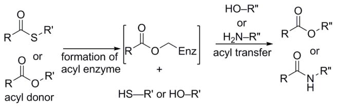
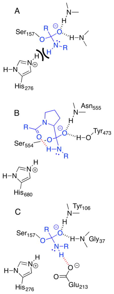
Enzymes within a family often catalyze different reactions. In some cases, this variety stems from different catalytic machinery, but in other cases the machinery is identical; nevertheless, the enzymes catalyze different reactions. In this review, we examine the subset of α/β-hydrolase fold enzymes that contain the serine-histidine-aspartate catalytic triad. In spite of having the same protein fold and the same core catalytic machinery, these enzymes catalyze seventeen different reaction mechanisms. The most common reactions are hydrolysis of C-O, C-N and C-C bonds (Enzyme Classification (EC) group 3), but other enzymes are oxidoreductases (EC group 1), acyl transferases (EC group 2), lyases (EC group 4) or isomerases (EC group 5). Hydrolysis reactions often follow the canonical esterase mechanism, but eight variations occur where either the formation or cleavage of the acyl enzyme intermediate differs. The remaining eight mechanisms are lyase-type elimination reactions, which do not have an acyl enzyme intermediate and, in four cases, do not even require the catalytic serine. This diversity of mechanisms from the same catalytic triad stems from the ability of the enzymes to bind different substrates, from the requirements for different chemical steps imposed by these new substrates and, only in about half of the cases, from additional hydrogen bond partners or additional general acids/bases in the active site. This detailed analysis shows that binding differences and non-catalytic residues create new mechanisms and are essential for understanding and designing efficient enzymes.

Keywords: X-ray structures; catalytic triad; divergent evolution; hydrolase; lyase; mechanism; oxyanion hole.


Figures

Figure 1
Figure 1
Schematic of the α/β-hydrolase fold showing sequence of α-helices (red rectangles) and β-sheets (blue arrows) and location of the catalytic triad residues and the oxyanion loop. The oxyanion loop positions one main chain N-H to donate a hydrogen bond to the oxyanion. The other N-H comes from the residue after the catalytic serine. These two residues form the oxyanion hole.
Figure 2
Figure 2
The substrate-binding site of α/β-hydrolases. A) Schematic showing how the active site accommodates different parts of a tetrahedral intermediate. The active site contains a region to stabilize the oxyanion as well as regions for the acyl and alcohol parts of an ester. B) Ribbon diagram of carboxylesterase Est30 from Geobacillus stearothermophilus (cap domain in gray, catalytic domain in green; pdb id: 1tqh) with tetrahedral intermediate for hydrolysis of propyl acetate and the catalytic serine and histidine in sticks representation. The active site lies between the two domains. The inset shows a close-up of the active site. The Ser-His-Asp triad (aspartate not shown) and the oxyanion hole residues are in the catalytic domain. The cap domain contributes substrate binding residues.
Figure 3
Figure 3
Venn diagrams of differences between the seventeen different reactions catalyzed by α/β-hydrolase fold enzymes with a Ser-His-Asp catalytic triad. Nine reactions follow a hydrolase-mechanism with an acyl enzyme intermediate. The first is the canonical esterase mechanism. Eight other mechanisms are similar, but differ in either the formation of the acyl enzyme (five examples) or the release of the acyl enzyme (three examples). Eight reactions follow a lyase-type mechanism where no acyl enzyme forms. Some of these reactions do not require active site serine, while the others do, but use it differently. The oxyanion hole is used by most reactions to bind the oxygen of a carbonyl, but it can also bind a nitrile nitrogen, a dioxygen molecule, or nothing at all. The decarboxylase mechanism does not use the catalytic serine, nor the oxyanion hole.
Figure 4
Figure 4
Crystal structure of BphD S112A with HOPDA (blue) in the nonplanar keto form (pdb id: 2PUH). Catalytic triad is shown in grey carbons. The C-C hydrolases contain conserved substrate-binding residues shown in magenta carbons (side chain interacts with substrate) or green carbons (backbone interacts with substrate). Residues from the catalytic domain include Gly42, Asn111, Ser112, Met113, Asp237, His265 and Trp266. Phe175 and Arg190 belong to the cap domain. Another conserved residue, Cys263, lies outside the active site and is not shown.
Scheme 1
Scheme 1
Canonical esterase mechanism for hydrolysis of methyl acetate, a typical ester. The top left structure shows the free enzyme with the catalytic triad (Asp-His-Ser) and the oxyanion hole (two main chain amide N-H’s). The first step is binding the substrate, methyl acetate, in the active site. Attack of serine on the ester carbonyl carbon yields the first tetrahedral intermediate, Td1. The catalytic histidine acts as a base to deprotonate the serine in this step. Reformation of the carbon-oxygen double bond leads to the release of methanol and the formation of the acyl enzyme intermediate. Histidine acts as an acid in this step enabling the leaving group to be methanol instead of methoxide. Next, water binds to the acyl enzyme intermediate. The active site histidine again acts as a base to deprotonate the water so that it can attack the carbonyl carbon of the acyl enzyme to form the second tetrahedral intermediate, Td2. Lastly, reformation of the carbon-oxygen double bond releases acetic acid and restores the free enzyme state. To draw the mechanistic steps clearly, only selected lone pairs of electrons are shown.
Scheme 2
Scheme 2
Direct epoxidation of α.β-unsaturated aldehydes with hydrogen peroxide catalyzed by CAL-B or the CAL-B S105A variant. A) The epoxidation occurs in buffer or organic solvent and yields racemic epoxide. The active site serine is not needed for catalysis so no acyl enzyme intermediate can form. B) The proposed mechanism supported by calculations involves deprotonation of H2O2 by the active site histidine and stabilization of the oxyanion intermediate by the oxyanion hole.
Scheme 3
Scheme 3
Enzyme-catalyzed perhydrolysis accounts for the bromoperoxidase activity in the α/β-hydrolase family. A) Bromoperoxidases catalyze the formation of hypobromite from bromide and peroxides like hydrogen peroxide. In a second step, hypobromite reacts spontaneously with organic compounds like monochlorodimedone to replace a hydrogen with bromine. B) Some α/β-hydrolases show bromoperoxidase activity in acetate buffer. The α/β-hydrolase catalyze the perhydrolysis of acetic acid to form peracetic acid. Next, the peracetic acid spontaneously reacts with bromide to form hypobromite. This hypobromite can spontaneously brominate organic compounds as in panel A.
Scheme 4
Scheme 4
Perhydrolysis of acetic acid yields peracetic acid, top equation. The reaction involves an acetyl enzyme intermediate, whose formation limits the reaction rate, bottom equation.
Scheme 5
Scheme 5
Bacterial oxidation of quinaldine (R = CH3) to catechol. The α/β-hydrolase 1H-3-hydroxy-4-oxoquinoline 2,4-dioxygenase (HOD) converts 1H-3-hydroxy-4-oxoquinaldine (QND) to carbon monoxide and N-acetylanthranilate by oxidative cleavage of the C2–C3 and C3–C4 bonds.
Scheme 6
Scheme 6
Proposed reaction mechanism proposed for co-factor free dioxygenase cleavage of QND with oxygen. The reaction does not involve an acyl enzyme intermediate and the Ser-His-Asp triad serves only as a general acid-base catalyst. The aspartate residue of the catalytic triad (Asp126) is not shown for clarity. Similarly only selected electrons are shown. Dioxygen is shown in green.
Scheme 7
Scheme 7
Like hydrolases, acyl transferases form an acyl-serine enzyme intermediate, but unlike hydrolases, acyl transferase transfer this group to an acceptor (usually an alcohol or amine), not to water. The acyl donor is usually an ester or thioester.
Scheme 8
Scheme 8
Reaction and key catalytic steps catalyzed by N-acyl homoserine lactone hydrolase. A) The lactonase catalyzes hydrolysis of the homoserine lactone ring. B) Proposed mechanism for formation of the acyl enzyme intermediate based on the X-ray structure with bound substrate (pdb id: 4g8b). The lone pairs on the lactone’s alcohol oxygen point away from the catalytic histidine and accept a hydrogen bond from Tyr160. We propose that this residue, which is essential for catalysis, acts as the proton donor in place of the catalytic histidine.
Scheme 9
Scheme 9
Palmitoyl-protein thioesterase (EC 3.2.1.22) catalyzes hydrolysis of palmitoyl cysteine residues on the surface of H-Ras protein.
Scheme 10
Scheme 10
Organophosphates inhibit esterases by forming stable phosphoryl enzyme intermediates, but a histidine in a new region of the active site can promote hydrolysis. A. Slow hydrolysis of the cholinesterase inhibitor echothiophate by a human butyrylcholinesterase variant. B. The Gly117His substitution allows positioning and activation of a water molecule to hydrolyze phosphorylserine intermediate.
Scheme 11
Scheme 11
Prolyl oligopeptidase catalyzed catalyzes the hydrolysis of amide links after proline. The natural substrates are various peptides; the substrate shown is a chromogenic substrate often used in assays.
Scheme 12
Scheme 12
Kynurenine formamidase catalyzes hydrolysis of an amide during tryptophan degradation. An internal hydrogen bond, shown in red dashes, may contribute to catalysis by preventing the N-H from disrupting the catalytic histidine.
Scheme 13
Scheme 13
Microbial oxidation of biphenyl proceeds via the biphenyl meta-cleavage product (MCP) 2-hydroxy-6-oxo-6-phenylhexa-2,4-dienoic acid (HOPDA). The MCP is the dienol tautomer of a vinylogous 1,5-diketone. The MCP hydrolase catalyzes the hydrolysis of the C5–C6 bond yielding benzoic acid and 2-hydroxypenta-2,4-dienoic acid.
Scheme 14
Scheme 14
The MCP-hydrolase substrate, HOPDA, deprotonates readily to the dianion where the negative charge delocalizes over many atoms; only two of the many possible resonance structures are shown. Binding to the active site twists this substrate to a non-planar conformation called the keto form due to keto resonance at C2. The twist and the H-bond donors from the oxyanion hole localize the charge to C5 and the oxyanion oxygen. The reaction starts from this keto form.
Scheme 15
Scheme 15
Proposed mechanism for MCP-hydrolases with residues numbered to match those in BphD from the biphenyl degradation pathway of B. xenovorans LB400. The dianion of HOPDA binds in a non-planar conformation, which localizes the negative charge to C5. This negative charge, not the histidine, deprotonates the serine to create the nucleophile. The rest of the reaction steps are similar to those in the canonical esterase mechanism.
Scheme 16
Scheme 16
Degradation of nicotine by the bacteria Arthrobacter nicotinovorans includes a hydrolysis of a C–C bond in DHPON catalyzed by DHPON hydrolase. If DHPON hydrolase is not present, DHPON irreversibly cyclizes to 2,6-dihydroxy-N-methyl-myosmine.
Scheme 17
Scheme 17
Proposed mechanism for hydrolysis of the C–C bond in DHPON involves an enol to keto tautomerization followed by steps similar to those for ester hydrolysis. Glu148 catalyzes the enol to keto tautomerization by deprotonation of the phenolic OH at C2 followed by protonation at C3. An acyl enzyme intermediate forms at Ser217 in this mechanism.
Scheme 18
Scheme 18
Decarboxylation of β-ketomyristic acid catalyzed by methyl ketone synthase from tomato (MKS1). A) Tomato leaves convert β-ketomyristic acid, an intermediate in fatty acid biosynthesis, into 2-tridecanone for defense against insects. B) The active site contains a threonine that blocks the oxyanion hole and a catalytic triad of Ala-His-Asn, although the Ser-His-Asp variant is only 0.6-fold slower. The oxyanion hole and catalytic serine are not shown because there is no evidence that they contribute to catalysis. The key step is the protonation of the carboxylate by the catalytic histidine to the acid form, which decarboxylates yielding the enol and carbon dioxide. The enol later tautomerizes to the keto form (not shown).
Scheme 19
Scheme 19
Linamarin is a cyanogenic glucoside in plants. Hydrolysis of the glucoside by glycosidases yields acetone cyanohydrin. Hydroxynitrile lyase from Hevea brasiliensis catalyzes the elimination of hydrogen cyanide from this cyanohydrin.
Scheme 20
Scheme 20
Mechanism for the cleavage of acetone cyanohydrin by (S)-HNL from Hevea brasiliensis,. Blocking of the oxyanion hole by threonine forces the acetone carbonyl group into a different orientation from that for the ester carbonyl group in esterases. In HNL, the serine Oγ interacts with the oxygen of the carbonyl group, while in esterases it interacts with the carbon.
Scheme 21
Scheme 21
Proposed mechanism for cleavage of mandelonitrile by (R)-HNL from A. thaliana based on docking calculations. AtEST5 binds the substrate in an orientation that differs from both esterases (carbonyl is not in oxyanion hole) and from HbHNL (nitrile is in oxyananion hole). X-ray structures have not confirmed this proposed binding. For clarity, the catalytic aspartate is not shown.
Scheme 22
Scheme 22
Lipase-catalyzed aldol addition involves general acid-base catalysis, but no acyl enzyme intermediate. A) Lipase B from C.antarctica catalyzes the addition of hexanal to a second molecule of hexanal to form a β-hydroxy aldehyde. This addition is not enantioselective, but lipase from pig pancreas shows low enantioselectivity (E ~2.5) in a similar reaction. B) Theoretical calculations indicate that lipase catalyzes both the enolate formation step and the enolate addition step. The active site serine is not shown because it is not essential for the reaction, while the active site aspartate is omitted for clarity.
Scheme 23
Scheme 23
Lipases catalyze the addition of nucleophiles to α,β-unsaturated carbonyl compounds. These additions can form carbon-carbon, carbon-oxygen, carbon-nitrogen or carbon-sulfur bonds. v = measured rate at one substrate concentration.
Scheme 24
Scheme 24
Menaquinone synthase, MenH, catalyzes a 1,4-elimination of pyruvate from 2-succinyl-5-enolpyruvyl-6-hydroxy-3-cyclohexene-1-carboxylate. A) The ring numbering makes this reaction a 2,5-elimination for this substrate. The kcat value refers to the enzyme from E. coli. B) The proposed mechanism starts with the formation of an enolate, whose oxyanion is stabilized by the oxyanion hole. Elimination of pyruvate from this enolate is aided by protonation of the leaving group by the catalytic histidine. The amino acid numbers correspond to the E. coli enzyme.
Scheme 25
Scheme 25
A) Lipase from B. cepacia (BCL) catalyzes the racemization of N-methyl α-aminonitriles. B) The proposed mechanism is the reversible elimination of cyanide to form an imine intermediate. The catalytic histidine serves as the base, while the oxyanion hole stabilizes the released cyanide.
Scheme 26
Scheme 26
Hydrolysis catalyzed by wt DLH and isomerization catalyzed by DLH-Cys123Ser. A) Loss of the best leaving group, thiol, from the second tetrahedral intermediate leads to hydrolysis of the dienelactone by wt DLH. Steps for formation of the acyl enzyme and second tetrahedral intermediate are omitted for clarity. B) DLH-Cys123Ser catalyzes not hydrolysis, but the isomerization of the (Z)- and (E)-lactones. C. Possible mechanism for isomerization beginning with second tetrahedral intermediate. Hydrolysis of the acyl enzyme is slow because the serine Oγ is a poorer leaving group than the cysteine Sγ thus favoring reformation of the acyl enzyme. Rotation about the C2–C3 bond followed by tautomerization to enol form allows the acyl enzyme to recyclize to form the isomeric lactone.
Chart 1
Chart 1
Phosphonates mimic the first tetrahedral intermediate in the hydrolysis of esters just before loss of the alcohol, while sulfonates mimic the second tetrahedral intermediate where water has added to the acyl enzyme intermediate.
Chart 2
Chart 2
Active site differences between esterase and variants with higher perhydrolase activity affect the rate of acetyl enzyme formation. A) In variant PFE-L29P, the main chain carbonyl of Trp28 accepts a hydrogen bond from the leaving water of the tetrahedral intermediate. This interaction speeds acetyl enzyme formation. B) In wild type PFE, this carbonyl group is farther from the active site. It can also accept a hydrogen bond via a water molecular bridge, but this the bridge can also donate a hydrogen bond, which would hinder water loss. C) In variant PFE-L29I, an acetate, held by the main chain amides of Ile29 and Leu30, accepts a hydrogen bond. A negatively charged acetate is a better hydrogen bond acceptor than water, which may account for the faster perhydrolysis catalyzed by PFE-L29I as compared to PFE-L29P. D) The general mechanism for a faster perhydrolase is to position a hydrogen bond acceptor for the leaving water in the tetrahedral intermediate.
Chart 3
Chart 3
A) The final step of cephalosporin C biosynthesis involves acetyl transfer from acetyl-CoA to deacetyl cephalosporin C (DAC). B) An X-ray structure shows many hydrogen bonds and ion pair interactions that bind the alcohol DAC (blue lines) in the alcohol site of DAC-acetyltransferase. These interactions favor binding of this alcohol instead of water and therefore favor acetyl transfer over acetyl hydrolysis. The structure shows the catalytic serine 149 in the acetylated form.
Chart 4
Chart 4
A different main chain conformation of the oxyanion loop in acyltransferases can lower the reactivity of water, but not alcohols. In esterases (top), the main chain carbonyl oxygen acts as a base via a bridging water molecule to activate the attacking water molecule. In acyltransferases, the N–H acts as an acid via a bridging water molecule to deactivate the attacking water molecule. For the acyl transfer reaction, the attacking nucleophile is an alcohol, which is larger than water. This alcohol displaces the bridging water molecule thereby eliminating any activating or deactivating effects. Acyltransferases may also favor binding of the incoming alcohol nucleophile.
Chart 5
Chart 5
The cyclic nature of a lactone creates a different shape in the alcohol part as compared to an ester. In esters, the alcohol carbon orients cis to the carbonyl oxygen to cancel the two dipoles shown. In lactones, rings smaller than eight force the alcohol carbon trans to the carbonyl oxygen. The different alcohol carbon location also changes the location of the lone pairs on the alcohol oxygen in esters versus lactones.
Chart 6
Chart 6
The N–H on an amide may interfere with catalysis, but peptidases avoid this interference with hydrogen bond acceptors that speed inversion at the nitrogen. A) Breakdown of the first tetrahedral intermediate in the hydrolysis of an amide requires protonation of the nitrogen. If the N-H of the amide, not the lone pair of the amide, points toward the catalytic histidine, it prevents protonation. B) Prolyl endopeptidases use an intramolecular hydrogen bond promote inversion of the nitrogen. The prolyl carbonyl oxygen of the prolyl peptide substrate accepts a hydrogen bond from the N-H of the leaving amide. This drawing is based on an X-ray structure of the catalytically inactive Ser554Ala variant complexed with an octapeptide (pdb id:1e8m). C) Glutamate 213 is a hydrogen bond acceptor in the active site of tricorn-interacting aminopeptidase F1, which may similarly promote inversion of the nitrogen.

References

    1. Albery W, Knowles J. Biochemistry. 1976;15:5631–5640. - PubMed
    1. Wolfenden R. Chem Rev. 2006;106:3379–3396. - PubMed
    1. Siegel JB, Gallaher JL, Zanghellini A, Lovick HM, Kiss G, Lambert AR, St Clair JL, Hilvert D, Gelb MH, Stoddard BL, Houk KN, Michael FE, Baker D. Science. 2010;329:309–313. - PMC - PubMed
    1. Kiss G, Çelebi-Ölçüm N, Moretti R, Baker D, Houk KN. Angew Chem Int Ed. 2013;52:5700–5725. - PubMed
    1. For example, Savile CK, Janey JM, Mundorff EC, Moore JC, Tam S, Jarvis WR, Colbeck JC, Krebber A, Fleitz FJ, Brands J, Devine PN, Huisman GW, Hughes GJ. Science. 2010;329:305–309.

LinkOut - more resources