Review
. 2017 Apr 19;7(3):1-18.
doi: 10.1080/2159256X.2017.1318201. eCollection 2017.

Transposable elements in Drosophila

Affiliations
Review

Transposable elements in Drosophila

Tabitha J McCullers et al. Mob Genet Elements. .

Abstract

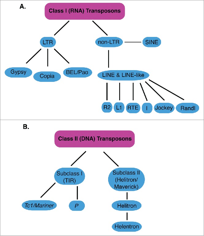
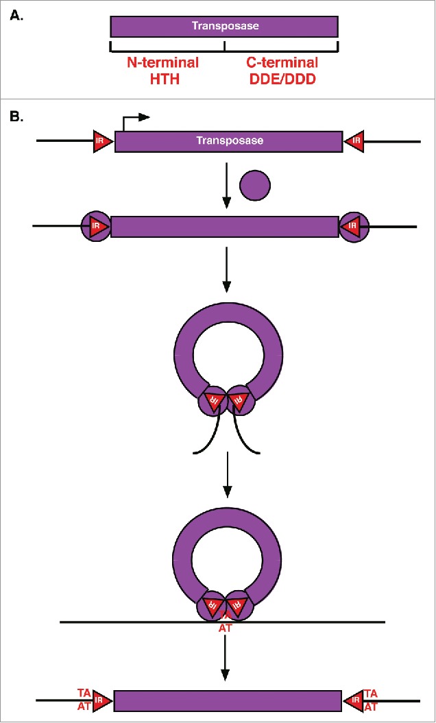
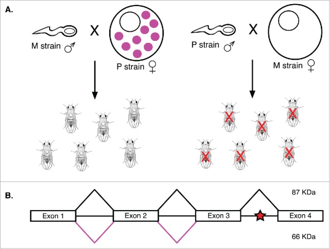
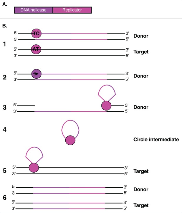
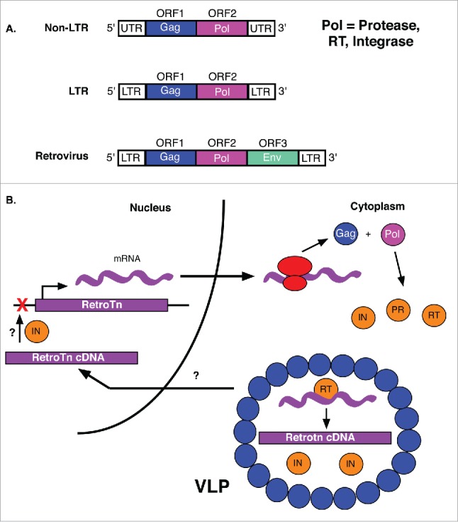
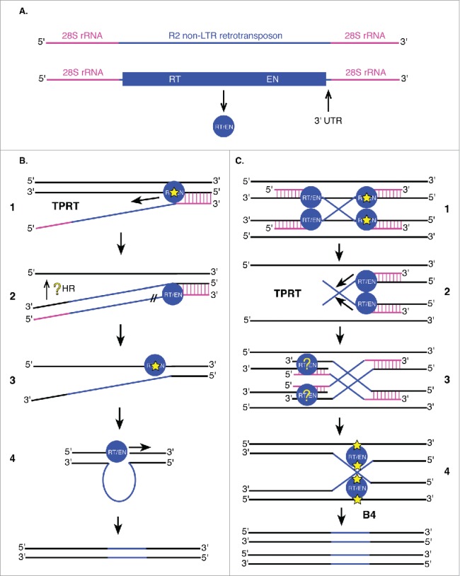
Transposable elements (TEs) are mobile genetic elements that can mobilize within host genomes. As TEs comprise more than 40% of the human genome and are linked to numerous diseases, understanding their mechanisms of mobilization and regulation is important. Drosophila melanogaster is an ideal model organism for the study of eukaryotic TEs as its genome contains a diverse array of active TEs. TEs universally impact host genome size via transposition and deletion events, but may also adopt unique functional roles in host organisms. There are 2 main classes of TEs: DNA transposons and retrotransposons. These classes are further divided into subgroups of TEs with unique structural and functional characteristics, demonstrating the significant variability among these elements. Despite this variability, D. melanogaster and other eukaryotic organisms utilize conserved mechanisms to regulate TEs. This review focuses on the transposition mechanisms and regulatory pathways of TEs, and their functional roles in D. melanogaster.

Keywords: LTR retrotransposons; P elements; TEs; TIR transposons; helitrons; non-LTR retrotransposons; retrovirus; transposons.


Figures

Figure 1.
Figure 1.
classes of Transposons. Shows classes, subclasses and groups of TEs described in this review. (A) classes, subclasses and groups of class I (RNA) transposons are shown in light blue. LTR is Long Terminal Repeat, SINE is Short Interspersed Nuclear Elements, LINE is Long Interspersed Nuclear Elements (B) classes, subclasses and families of class II (DNA) transposons are shown in light blue. Only the Tc1/mariner and P families are shown for simplicity. TIR is Terminal Inverted Repeats.
Figure 2.
Figure 2.
TIR transposase and transposition mechanism. (A) TIR Transposases have an N-terminal DNA binding domain with HTH motifs and a C-terminal DDE or DDD catalytic domain. (B) For transposition, TIR transposases (purple circles) first bind to inverted repeats (red triangles, IR) flanking the element. Bound transposases then dimerize followed by cleavage of the element from surrounding sequences (black lines) and integration into a new target site (AT) resulting in target site duplication.
Figure 3.
Figure 3.
P element splicing and hybrid dysgenesis. (A) Hybrid dysgenesis results when M strain females are crossed with P strain males. Because the P element repressor (pink circles) is only transmitted by P cytotype females, progeny of the P strain male-M strain female cross have many mutations caused by germline P element transposition. These mutations often result in sterility (red X). (B) Exons 1-4 of P element transcripts are spliced to form a functional 87 kDa transposase (black lines). When intron 3 is not properly spliced, a stop codon (red star) generates a 66 kDa truncated repressor of P element transposition (pink lines).
Figure 4.
Figure 4.
Helitron enzymes and transposition mechanism. (A) Helitron transposons encode a protein with both DNA helicase and Replicator functions. (B) The Helitron is represented with purple and pink lines. The Replicator domain (pink circle) first binds to both donor (TC) and target (AT) creating nicks in both. The DNA helicase domain (purple circle) then displaces the donor strand. The Replicator domain cleaves the 3′ end of the element, promoting formation of a circular single-stranded DNA intermediate. Rep cleaves the circular single-stranded intermediate and promotes covalent bond formation between the 5′ and 3′ ends of the donor strand and target site. Host DNA replication generates a second DNA strand at both the donor and target sites. While the Replicator nicks the other end of the donor and facilitates attachment to the target site. The second strand of the element is generated at both the donor and target sites upon host DNA replication.
Figure 5.
Figure 5.
Retrotransposons and the LTR retrotransposition mechanism. (A) Non-LTR transposons encode both Gag and Pol, but are not flanked by LTRs. LTR retrotransposons contain gag and pol genes surrounded by LTRs. In addition to gag and pol genes, retroviruses encode an env gene. (B) Gag and pol of retrotransposon mRNAs are first translated into a polyprotein. The protease (PR) of the Pol cleaves the peptide into integrase (IN) and reverse transcriptase (RT) enzymes. The RT, retrotransposon and IN are then packaged into virus-like particles (VLPs) for import into the nucleus where retrotransposon cDNA is integrated into the genome (red X). The mechanisms by which VLP contents are localized to the nucleus and retrotransposon cDNA is integrated into the target site are unknown (?).
Figure 6.
Figure 6.
Non-LTR retrotransposons utilize target primed reverse transcription (TPRT) for integration. (A) R2 non-LTR retrotransposons (blue) are flanked by 28S rRNA genomic sequences (pink). The single R2 ORF encodes an enzyme with reverse transcriptase (RT) and endonuclease (EN) activities (blue circle). Other non-LTR retrotransposons may encode these enzymes as 2 separate proteins (RT and integrase with EN activity). The 3′ UTR is important for integration of R2 retrotransposons into 28S rRNA genes. (B and C) Proposed models of non-LTR retrotransposon (B) and R2 (C) insertion. DNA is shown in black (including reverse transcribed flanking sequences), 28S rRNA sequences in pink and retrotransposon sequences (mRNA and DNA) in blue, following the color scheme in (A). (B1) Non-LTR retrotransposon transcripts first hybridize to 28S rRNA sequences (vertical pink lines) followed by initiation of TPRT by single-stranded nicking of the target DNA (yellow star) by the element's encoded endonuclease. (B2) Following reverse transcription of the element, element mRNA is degraded by R2 RT/EN (//). Integration of the 5′ end of the element is not well understood (yellow ?). (B3) Cleavage of the second strand (yellow star) may occur at the same location as the first strand, or 2 base pairs upstream or downstream of this site. (B4) R2 RT/EN also generates the complementary R2 strand at the target site to fully transpose the element. (C1, C2, C3) The initial steps of this alternative mechanism are identical to those described in B1 and B2 except they take place on 2 homologous targets simultaneously resulting in a Holliday junction intermediate (C4). The Holliday junction intermediate is resolved by R2 RT/EN (C4) followed by second strand synthesis resulting in fully-integrated R2 non-LTR retrotransposons in 2 new locations.

Similar articles

Cited by

References

    1. Pagel M, Johnstone RA. Variation across species in the size of the nuclear genome supports the junk-DNA explanation for the C-value paradox. Proc Biol Sci 1992; 249:119-24; PMID:1360673; https://doi.org/10.1098/rspb.1992.0093 - DOI - PubMed
    1. Vieira C, Nardon C, Arpin C, Lepetit D, Biemont C. Evolution of genome size in Drosophila. Is the invader's genome being invaded by transposable elements? Mol Biol Evol 2002; 19:1154-161; PMID:12082134 - PubMed
    1. Bosco G, Campbell P, Leiva-Neto JT, Markow TA. Analysis of Drosophila species genome size and satellite DNA content reveals significant differences among strains as well as between species. Genetics 2007; 177:1277-90; PMID:18039867; https://doi.org/10.1534/genetics.107.075069 - DOI - PMC - PubMed
    1. Gregory TR, Johnston JS. Genome size diversity in the family Drosophilidae. Heredity (Edinb) 2008; 101:228-38; PMID:18523443; https://doi.org/10.1038/hdy.2008.49 - DOI - PubMed
    1. SanMiguel P, Tikhonov A, Jin YK, Motchoulskaia N, Zakharov D, Melake-Berhan A, Springer PS, Edwards KJ, Lee M, Avramova Z, et al.. Nested retrotransposons in the intergenic regions of the maize genome. Science 1996; 274:765-8; PMID:8864112 - PubMed

LinkOut - more resources