. 2017 Apr 8;6(6):459-470.
doi: 10.1016/j.molmet.2017.03.011. eCollection 2017 Jun.

Expression and functional assessment of candidate type 2 diabetes susceptibility genes identify four new genes contributing to human insulin secretion

Affiliations

Expression and functional assessment of candidate type 2 diabetes susceptibility genes identify four new genes contributing to human insulin secretion

Fatou K Ndiaye et al. Mol Metab. .

Abstract

Objectives: Genome-wide association studies (GWAS) have identified >100 loci independently contributing to type 2 diabetes (T2D) risk. However, translational implications for precision medicine and for the development of novel treatments have been disappointing, due to poor knowledge of how these loci impact T2D pathophysiology. Here, we aimed to measure the expression of genes located nearby T2D associated signals and to assess their effect on insulin secretion from pancreatic beta cells.

Methods: The expression of 104 candidate T2D susceptibility genes was measured in a human multi-tissue panel, through PCR-free expression assay. The effects of the knockdown of beta-cell enriched genes were next investigated on insulin secretion from the human EndoC-βH1 beta-cell line. Finally, we performed RNA-sequencing (RNA-seq) so as to assess the pathways affected by the knockdown of the new genes impacting insulin secretion from EndoC-βH1, and we analyzed the expression of the new genes in mouse models with altered pancreatic beta-cell function.

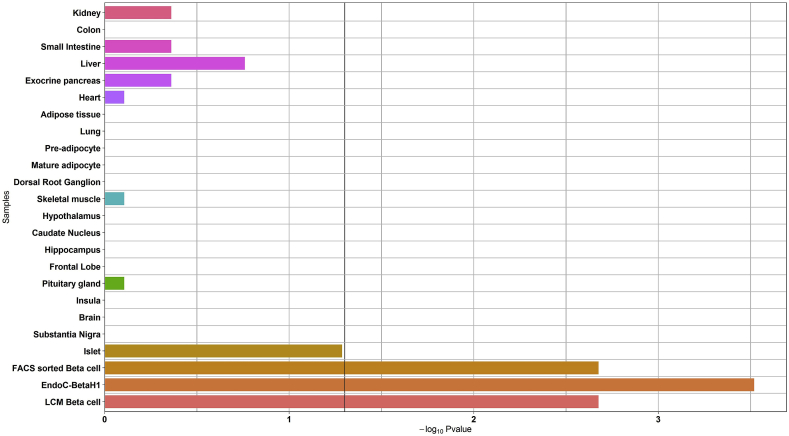
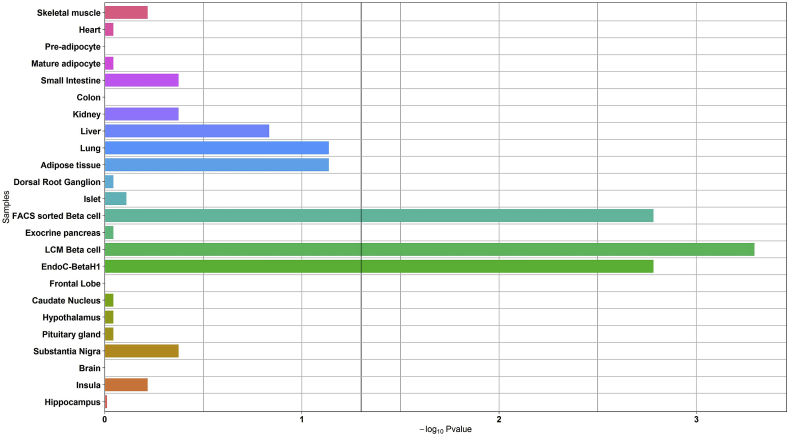
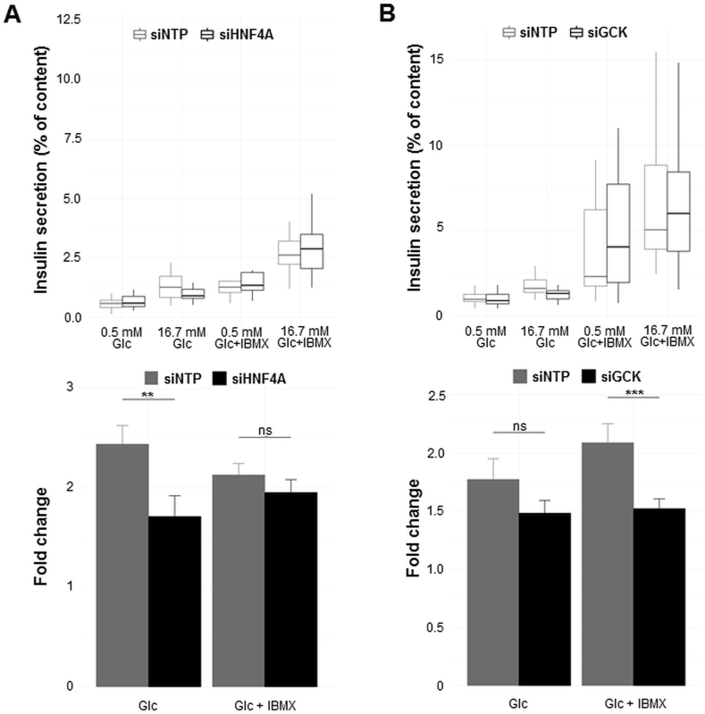
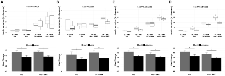
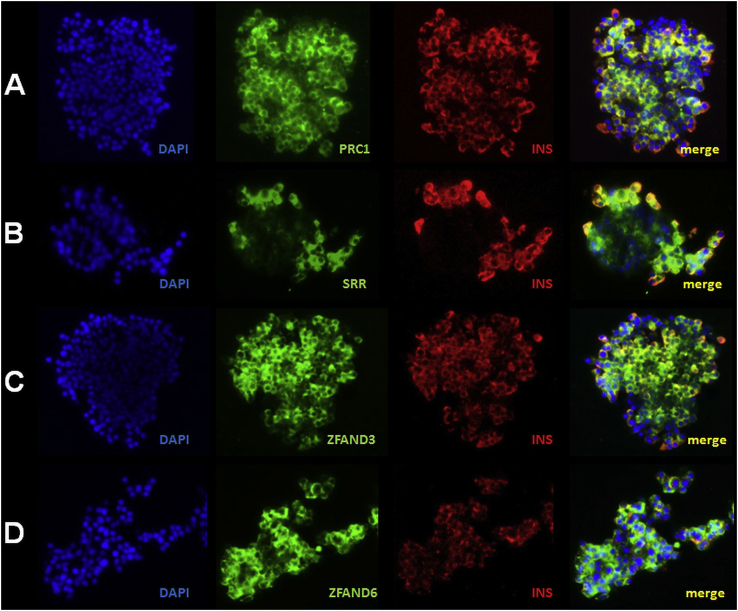
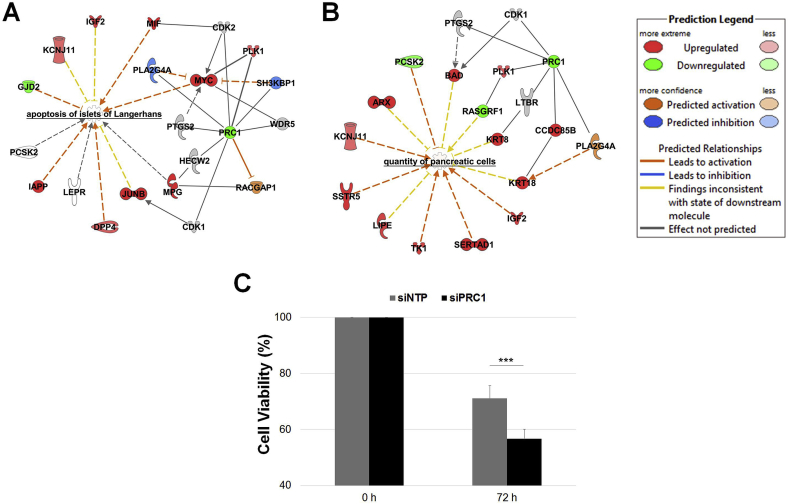
Results: We found that the candidate T2D susceptibility genes' expression is significantly enriched in pancreatic beta cells obtained by laser capture microdissection or sorted by flow cytometry and in EndoC-βH1 cells, but not in insulin sensitive tissues. Furthermore, the knockdown of seven T2D-susceptibility genes (CDKN2A, GCK, HNF4A, KCNK16, SLC30A8, TBC1D4, and TCF19) with already known expression and/or function in beta cells changed insulin secretion, supporting our functional approach. We showed first evidence for a role in insulin secretion of four candidate T2D-susceptibility genes (PRC1, SRR, ZFAND3, and ZFAND6) with no previous knowledge of presence and function in beta cells. RNA-seq in EndoC-βH1 cells with decreased expression of PRC1, SRR, ZFAND6, or ZFAND3 identified specific gene networks related to T2D pathophysiology. Finally, a positive correlation between the expression of Ins2 and the expression of Prc1, Srr, Zfand6, and Zfand3 was found in mouse pancreatic islets with altered beta-cell function.

Conclusions: This study showed the ability of post-GWAS functional studies to identify new genes and pathways involved in human pancreatic beta-cell function and in T2D pathophysiology.

Keywords: EndoC-βH1; Expression analysis; Genome-wide association study; Insulin secretion; RNAi screening; Type 2 diabetes.


Figures

Figure 1
Figure 1
Enrichment analysis of the expression of the genes causing monogenic diabetes in the panel of human organs, tissues, and cells. The black vertical line denotes a p-value of 0.05.
Figure 2
Figure 2
Enrichment analysis of the expression of candidate T2D-susceptibility genes in the panel of human organs, tissues, and cells. The black vertical line denotes a p-value of 0.05.
Figure 3
Figure 3
Decreased expression of HNF4A (A) or GCK (B), causing monogenic diabetes, leads to impaired insulin secretion from EndoC-βH1 cells. EndoC-βH1 cells were transfected with control non-targeting pool siRNA (siNTP) or target gene siRNA and were analyzed 72 h post-transfection. Insulin secretion (percentage of secretion of the total insulin content) was analyzed in response to 60 min incubation with 0.5 mM glucose (±0.5 mM IBMX), followed by 60 min incubation with 16.7 mM glucose (±0.5 mM IBMX). Data represent mean values ± SEM of at least three independent experiments. **p < 0.01; ***p < 0.001; ns, not significant. Glc, glucose; IBMX, phosphodiesterase inhibitor 3-isobutyl-1-methylxanthine.
Figure 4
Figure 4
Decreased expression of TCF19 (A), SLC30A8 (B), TBC1D4 (C), CDKN2A (D), or KCNK16 (E), already suggested to be expressed in beta cell and/or to contribute to beta-cell function, significantly modifies insulin secretion from EndoC-βH1 cells. EndoC-βH1 cells were transfected with control non-targeting pool siRNA (siNTP) or target gene siRNA and were analyzed 72 h post-transfection. Insulin secretion (percentage of secretion of the total insulin content) was analyzed in response to 60 min incubation with 0.5 mM glucose (±0.5 mM IBMX), followed by 60 min incubation with 16.7 mM glucose (±0.5 mM IBMX). Data represent mean values ± SEM of at least three independent experiments. *p < 0.05; **p < 0.01; ns, not significant. Glc, glucose; IBMX, phosphodiesterase inhibitor 3-isobutyl-1-methylxanthine.
Figure 5
Figure 5
Decreased expression of PRC1 (A), SRR (B), ZFAND3 (C), or ZFAND6 (D), with no evidence of expression and role in pancreatic beta cells, leads to impaired insulin secretion from EndoC-βH1 cells. EndoC-βH1 cells were transfected with control non-targeting pool siRNA (siNTP) or target gene siRNA and were analyzed 72 h post-transfection. Insulin secretion (percentage of secretion of the total insulin content) was analyzed in response to 60 min incubation with 0.5 mM glucose (±0.5 mM IBMX), followed by 60 min incubation with 16.7 mM glucose (±0.5 mM IBMX). Data represent mean values ± SEM of at least three independent experiments. *p < 0.05; **p < 0.01; ns, not significant. Glc, glucose; IBMX, phosphodiesterase inhibitor 3-isobutyl-1-methylxanthine.
Figure 6
Figure 6
PRC1 (A), SRR (B), ZFAND3 (C), and ZFAND6 (D) are expressed in human pancreatic islets and beta cells. Representative images of immunofluorescence staining for PRC1 (green; A), SRR (green; B), ZFAND3 (green; C), ZFAND6 (green, D), and insulin (red) performed on fixed isolated human islet clusters. Yellow (merged images) indicates co-localization of PRC1, SRR, ZFAND3, or ZFAND6 with insulin. Blue, DAPI.
Figure 7
Figure 7
Decreased PRC1 expression in EndoC-βH1 cells leads to increased apoptosis and decreased cell viability. List of deregulated genes emphasized by the decreased expression of PRC1 in EndoC-βH1 cells within the network related to apoptosis of islets of Langerhans (A) and the quantity of pancreatic cells (B) through IPA. Cell viability measurement (C) performed at baseline (100%) and 72 h post transfection of siRNA (% compared to baseline). Data at 72 h represent mean values ± SEM of at least five independent experiments. ***p < 0.001.

References

    1. Bonnefond A., Froguel P. Rare and common genetic events in type 2 diabetes: what should biologists know? Cell Metabolism. 2015;21(3):357–368. - PubMed
    1. Marullo L., El-Sayed Moustafa J.S., Prokopenko I. Insights into the genetic susceptibility to type 2 diabetes from genome-wide association studies of glycaemic traits. Current Diabetes Reports. 2014;14(11):551. - PubMed
    1. Pasquali L., Gaulton K.J., Rodríguez-Seguí S.A., Mularoni L., Miguel-Escalada I., Akerman I. Pancreatic islet enhancer clusters enriched in type 2 diabetes risk-associated variants. Nature Genetics. 2014;46(2):136–143. - PMC - PubMed
    1. Warren H.R., Evangelou E., Cabrera C.P., Gao H., Ren M., Mifsud B. Genome-wide association analysis identifies novel blood pressure loci and offers biological insights into cardiovascular risk. Nature Genetics Advance Online Publication. 2017 - PMC - PubMed
    1. Bonnefond A., Lomberk G., Buttar N., Busiah K., Vaillant E., Lobbens S. Disruption of a novel Kruppel-like transcription factor p300-regulated pathway for insulin biosynthesis revealed by studies of the c.-331 INS mutation found in neonatal diabetes mellitus. The Journal of Biological Chemistry. 2011;286(32):28414–28424. - PMC - PubMed

Publication types

MeSH terms