

Case Reports
doi: 10.1016/j.jdcr.2017.02.020.
eCollection 2017 May.
Recurrent halo nevus: Dermoscopy and confocal microscopy features
Affiliations
- PMID: 28580411
- PMCID: PMC5447382
- DOI: 10.1016/j.jdcr.2017.02.020
Item in Clipboard
Case Reports
Recurrent halo nevus: Dermoscopy and confocal microscopy features
JAAD Case Rep.
.
No abstract available
Keywords: HN, halo nevi; RCM, reflectance confocal microscopy; RN, recurrent nevi; confocal microscopy; dermoscopy; halo nevus; melanoma; nevus; skin cancer.
Figures
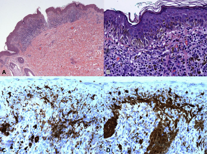
Histopathologic analysis and immunohistochemical stainings of the recurrent halo nevus. A, Panoramic view of the lesion. Lesion is characterized by a dense inflammatory infiltrate obscuring the dermal-epidermal junction of the skin. B, Moderate atypical melanocytic proliferation in the dermal-epidermal junction with nest formation and without pagetoid migration; in the dermis, an intense lymphocytic infiltration with melanocytes nests and numerous melanophages. C, Immunohistochemistry study revealing the intraepidermal Langerhans cells, confirming that some of the dendritic cells seen on confocal microscopy are Langerhans cells. D, S100 staining highlighting the melanocytic nests in the dermis confirming the compound nature of the lesion, and an increased number of Langerhans cells in the epidermis. (A and B, Hematoxylin-eosin stain; C, CD1a staining; D, S100 stain; original magnifications: A, ×40; B-D, ×400.)
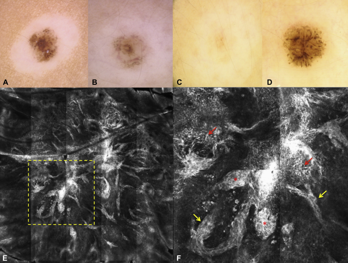
Dermoscopic images of recurrent halo nevus. A-C, Gradual regression of the halo nevus. A, Dermoscopy shows a regular reticular and globular nevus surrounded by a symmetric halo. B, After 6 months, a partial disappearance of the nevus was observed. C, After 1 year, an almost complete regression of the nevus occurred. D and E, recurrent halo nevus. D, After a 2-year follow-up, repigmentation in the center of the lesion occurred. Dermoscopy of the recurrent halo nevus showed an atypical network in the center and asymmetric and irregular globules in the periphery. E, Reflectance confocal microscopy (RCM) mosaic image (2.5 × 2.5 mm) of the recurrent nevus shows an atypical meshwork pattern at the level of the dermo-epidermal junction. F, RCM mosaic image (zoom in of the dashed square in E, 1 × 1 mm) shows homogeneous dense nests (red asterisks), atypical meshwork patterns (yellow arrows), and dendritic cells (red arrows).
Similar articles
-
Sequential Dermoscopy and Reflectance Confocal Microscopy Paired With Pigmented Lesion Assay Gene Expression Profiling for In Vivo Monitoring of Multiple Halo Nevi on an Adult Patient.Cureus. 2023 Nov 26;15(11):e49465. doi: 10.7759/cureus.49465. eCollection 2023 Nov. Cureus. 2023. PMID: 38152824 Free PMC article.
-
In vivo reflectance confocal microscopy of halo nevi.Skin Res Technol. 2021 Sep;27(5):841-845. doi: 10.1111/srt.13029. Epub 2021 Mar 10. Skin Res Technol. 2021. PMID: 33751665
-
In vivo reflectance confocal microscopy of halo nevus.J Cutan Med Surg. 2013 Jan-Feb;17(1):33-8. doi: 10.2310/7750.2012.12019. J Cutan Med Surg. 2013. PMID: 23364148
-
Melanocytic nevi with special features: clinical-dermoscopic and reflectance confocal microscopic-findings.J Eur Acad Dermatol Venereol. 2014 Jul;28(7):833-45. doi: 10.1111/jdv.12291. Epub 2013 Oct 31. J Eur Acad Dermatol Venereol. 2014. PMID: 24171788 Review.
-
Discriminating Nevi from Melanomas: Clues and Pitfalls.Dermatol Clin. 2016 Oct;34(4):395-409. doi: 10.1016/j.det.2016.05.003. Dermatol Clin. 2016. PMID: 27692446 Free PMC article. Review.
Cited by
-
Sequential Dermoscopy and Reflectance Confocal Microscopy Paired With Pigmented Lesion Assay Gene Expression Profiling for In Vivo Monitoring of Multiple Halo Nevi on an Adult Patient.Cureus. 2023 Nov 26;15(11):e49465. doi: 10.7759/cureus.49465. eCollection 2023 Nov. Cureus. 2023. PMID: 38152824 Free PMC article.
-
Predictive value of global dermoscopic pattern in patients diagnosed with cutaneous melanoma.Postepy Dermatol Alergol. 2021 Aug;38(4):572-577. doi: 10.5114/ada.2020.94593. Epub 2020 Apr 22. Postepy Dermatol Alergol. 2021. PMID: 34658696 Free PMC article.
References
-
- Vyas R., Rademaker M., Oakley A. An observational case series on dermatoscopic patterns of fading melanocytic naevi. Australas J Dermatol. 2016 published online March 1, 2016. - PubMed
-
- Terushkin V., Scope A., Halpern A.C., Marghoob A.A. Pathways to involution of nevi: insights from dermoscopic follow-up. Arch Dermatol. 2010;146(4):459–460. - PubMed
-
- Aouthmany M., Weinstein M., Zirwas M.J., Brodell R.T. The natural history of halo nevi: a retrospective case series. J Am Acad Dermatol. 2012;67(4):582–586. - PubMed
-
- Mooney M.A., Barr R.J., Buxton M.G. Halo nevus or halo phenomenon? A study of 142 cases. J Cutan Pathol. 1995;22(4):342–348. - PubMed
-
- Kolm I., Di Stefani A., Hofmann-Wellenhof R. Dermoscopy patterns of halo nevi. Arch Dermatol. 2006;142(12):1627–1632. - PubMed
Publication types
LinkOut - more resources
Full Text Sources
Other Literature Sources