. 2017 Jun 1;73(Pt 6):522-533.
doi: 10.1107/S2059798317007021. Epub 2017 May 31.

High-resolution cryo-EM proteasome structures in drug development

Affiliations

High-resolution cryo-EM proteasome structures in drug development

Edward P Morris et al. Acta Crystallogr D Struct Biol. .

Abstract

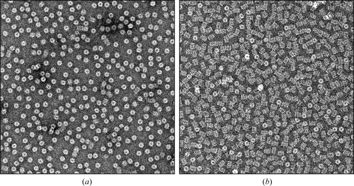
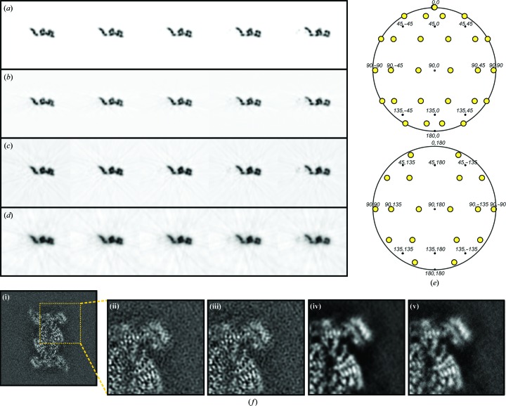
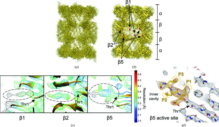
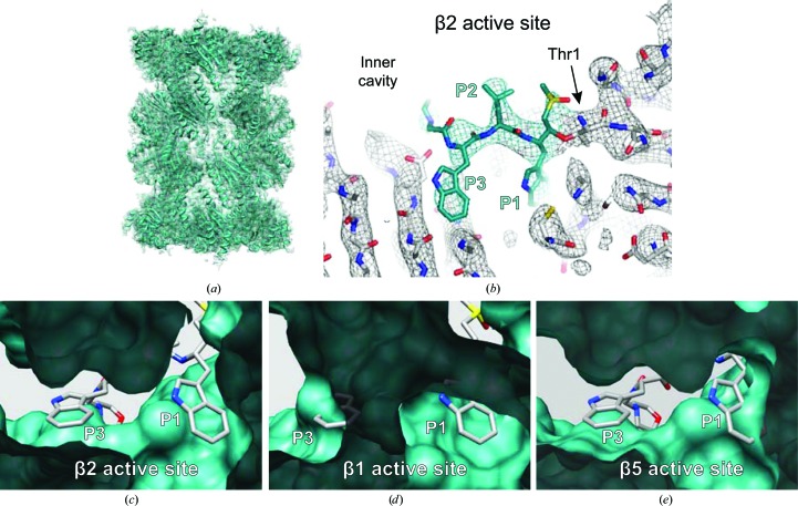
With the recent advances in biological structural electron microscopy (EM), protein structures can now be obtained by cryo-EM and single-particle analysis at resolutions that used to be achievable only by crystallographic or NMR methods. We have explored their application to study protein-ligand interactions using the human 20S proteasome, a well established target for cancer therapy that is also being investigated as a target for an increasing range of other medical conditions. The map of a ligand-bound human 20S proteasome served as a proof of principle that cryo-EM is emerging as a realistic approach for more general structural studies of protein-ligand interactions, with the potential benefits of extending such studies to complexes that are unfavourable to other methods and allowing structure determination under conditions that are closer to physiological, preserving ligand specificity towards closely related binding sites. Subsequently, the cryo-EM structure of the Plasmodium falciparum 20S proteasome, with a new prototype specific inhibitor bound, revealed the molecular basis for the ligand specificity towards the parasite complex, which provides a framework to guide the development of highly needed new-generation antimalarials. Here, the cryo-EM analysis of the ligand-bound human and P. falciparum 20S proteasomes is reviewed, and a complete description of the methods used for structure determination is provided, including the strategy to overcome the bias orientation of the human 20S proteasome on electron-microscope grids and details of the icr3d software used for three-dimensional reconstruction.

Keywords: Plasmodium falciparum; cryo-EM; drug design; electron microscopy; human; icr3d; icr3dpro; inhibitors; malaria; proteasome; single particle.


Figures

Figure 1
Figure 1
EM images of negatively stained fields of 20S proteasome complexes showing the effect of altering the surface charge of the carbon support film on the orientation of the human 20S proteasome. (a) Glow discharge of the EM grid in a partial vacuum of atmospheric air results in a strongly biased proteasome orientation with predominant top views. (b) Glow discharge in a partial vacuum containing pentylamine vapour results in a predominance of side views.
Figure 2
Figure 2
Scheme for the Fourier-space reconstruction used in the program icr3d. (a) Three-dimensional reconstruction in Fourier space by the summation of central sections (two are shown, one in blue and one in red), which derive from the projection angles of the two-dimensional images (indicated by red and blue arrows). The depth of each central section depends on the reciprocal dimension of the reconstructed object (1/D) measured in the projection direction. (b) Contributions from the input Fourier components of two individual particle images (red and blue points) to the output Fourier components of the three-dimensional reconstruction (black points) are confined to the central section (red and blue dashed lines) as in (a). These are added to neighbouring three-dimensional Fourier components within ellipsoidal contributing envelopes (red and blue ellipses), the dimensions of which are reciprocally related to the maximum dimensions of the reconstructed object in the relevant directions. (c) Maximum dimensions of a reconstructed object expressed as fractions of the cubic reconstructed volume shown in the xy and xz planes. (d) Corresponding ellipsoidal Fourier-space contribution envelopes (red ellipses), which can be compared with the spherical contribution envelopes (black circles) that relate to a reconstructed object with maximum dimensions equal to the cubic reconstructed volume. (e) The fractional distance (d Frac) used to determine sinc-weighted contributions to the output Fourier components is calculated from the distances (B and B′) between the input Fourier components (red dots) and the output Fourier components (black dots) as a fraction of the distance to the edge of the ellipsoidal contributing envelope (A and A′).
Figure 3
Figure 3
Effect of the different parameters used in icr3d reconstructions. (ae) Tests with the model density illustrated in Fig. 2 ▸(c). (a) Sections of model density. (bcd) Sections from reconstructed density produced from input projection images sampled at 30° intervals. (b) Sections from reconstruction with the contribution envelope set to match the dimensions of the reconstructed object as illustrated in Fig. 2 ▸(c) (i.e. the standard mode of usage for icr3d). (c) Sections from reconstruction with the contribution envelope set to match the dimensions of the reconstructed volume. (d) Sections from reconstruction calculated with the contribution envelope set to match the dimensions of a reconstructed volume of twice the size used in (c). (e) Distribution of Euler angles of the input projection images with 30° sampling, used to calculate the maps in (bd). (f) Reconstruction of the human 20S proteasome from cryo-EM data (da Fonseca & Morris, 2015 ▸) illustrating the effect of different parameters on a single section of reconstructed density. (i) Reconstruction using CTF weighting, CTF amplitude correction and set to match the dimensions of the 20S proteasome; (ii) magnified region of image (i); (iii) as (ii) but with the contribution envelope set to match the reconstructed volume; (iv) as (ii) but with no CTF amplitude correction; (v) as (iv) but with no CTF weighting.
Figure 4
Figure 4
Cryo-EM structure of the human 20S proteasome with an inhibitor bound, showing the map and coordinates deposited in the Electron Microscopy Data Bank and Protein Data Bank with accession codes EMD-2981 and 5a0q, respectively (da Fonseca & Morris, 2015 ▸). (a) Overall view of the density map (mesh) and protein coordinates (cartoon). (b) The map shown in (a) cut to reveal the location in the proteasome inner cavity of the Thr1 residues (shown as spheres) at the active sites of subunits β1, β2 and β5. (c) Close-up views of the three active sites, showing the map densities (mesh representation) colour-coded according to the local resolution estimated with ResMap (Kucukelbir et al., 2014 ▸) with protein coordinates represented as a cartoon. Densities for the ligand (encircled by dashed lines) are clearly seen extending from the Thr1 residues of subunits β1, β2 and β5 (indicated by arrows). (d) Close-up view of the cryo-EM map (mesh representation) and model coordinates (shown as sticks) for the β5 subunit of the human 20S proteasome, with the L3VS moiety of the inhibitor (coloured yellow) extending from the Thr1 residue (indicated by an arrow). The ligand LLL tripeptide mimics proteasome substrate positions P1–P3, as labelled, where the side chains at positions P1 and P3 are oriented towards the ligand-binding pocket, while that at position P2 is oriented towards the inner cavity of the proteasome. (a), (b) and (c) were created using UCSF Chimera (Pettersen et al., 2004 ▸) and (d) was created using PyMOL (Schrödinger).
Figure 5
Figure 5
Cryo-EM structure of the P. falciparum 20S proteasome with an inhibitor bound, showing the map and coordinates deposited in the Electron Microscopy Data Bank and Protein Data Bank with accession codes EMD-3231 and 5fmg, respectively (Li, O’Donoghue et al., 2016 ▸). (a) Overall view of the density map (mesh) and protein coordinates (cartoon). (b) Close-up view of the density map (mesh) and model coordinates (sticks) showing the WLW-vs inhibitor (teal) bound to Thr1 of the β2 subunit. The ligand WLW tripeptide mimics proteasome substrate positions P1–P3, as labelled. As for other tripeptide proteasome inhibitors, the side chains at positions P1 and P3 are oriented towards the ligand-binding pocket, while that at position P2 is oriented towards the inner cavity of the proteasome. (c) Model of the P. falciparum β2 active site with bound ligand, as determined by cryo-EM, viewed towards the inner cavity of the proteasome. (d) P. falciparum β1 active site with superimposed inhibitor coordinates, showing that the tryptophan side chains of the ligand, at positions P1 and P3, cannot be accommodated at this active site owing to steric constraints. (e) P. falciparum β5 active site with superimposed inhibitor coordinates, showing that the tryptophan side chain of the ligand at position P1 cannot be accommodated in this active site owing to steric constraints. In (c), (d) and (e) the protein model is represented as van der Waals surfaces and the ligand as sticks. (a), (c), (d) and (e) were created using UCSF Chimera (Pettersen et al., 2004 ▸) and (b) was created using PyMOL (Schrödinger).

References

    1. Aebi, U. & Pollard, T. D. (1987). J. Electron Microsc. Tech. 7, 29–33. - PubMed
    1. Amos, L. A., Henderson, R. & Unwin, P. N. (1982). Prog. Biophys. Mol. Biol. 39, 183–231. - PubMed
    1. Ashley, E. A. et al. (2014). N. Engl. J. Med. 371, 411–423.
    1. Banerjee, S. et al. (2016). Science, 351, 871–875. - PMC - PubMed
    1. Basler, M., Mundt, S., Bitzer, A., Schmidt, C. & Groettrup, M. (2015). Clin. Exp. Rheumatol. 33, S74–S79. - PubMed

MeSH terms