. 2017 Jun 5:8:15632.
doi: 10.1038/ncomms15632.

IL-17-producing γδ T cells switch migratory patterns between resting and activated states

Affiliations

IL-17-producing γδ T cells switch migratory patterns between resting and activated states

Duncan R McKenzie et al. Nat Commun. .

Abstract

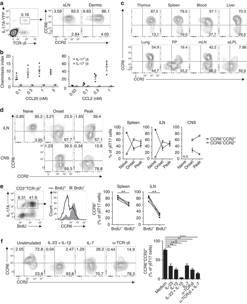
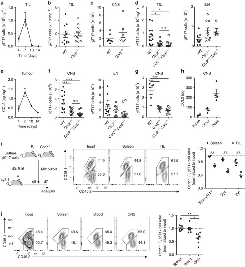
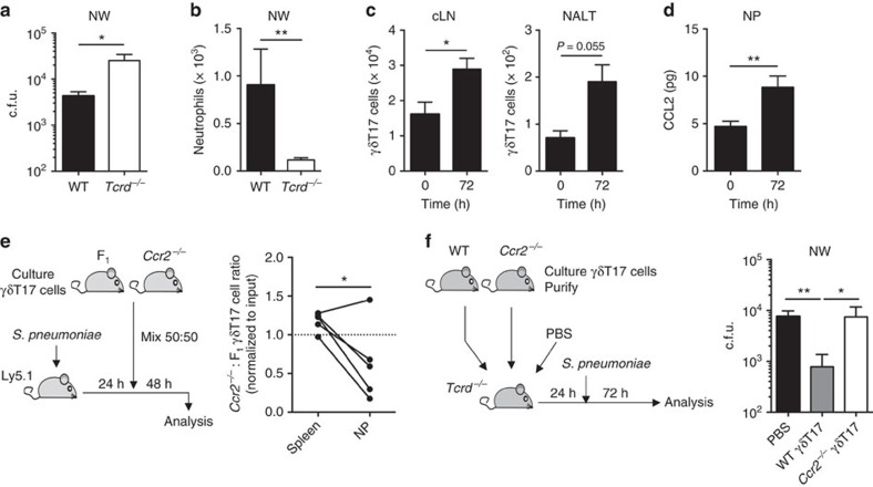
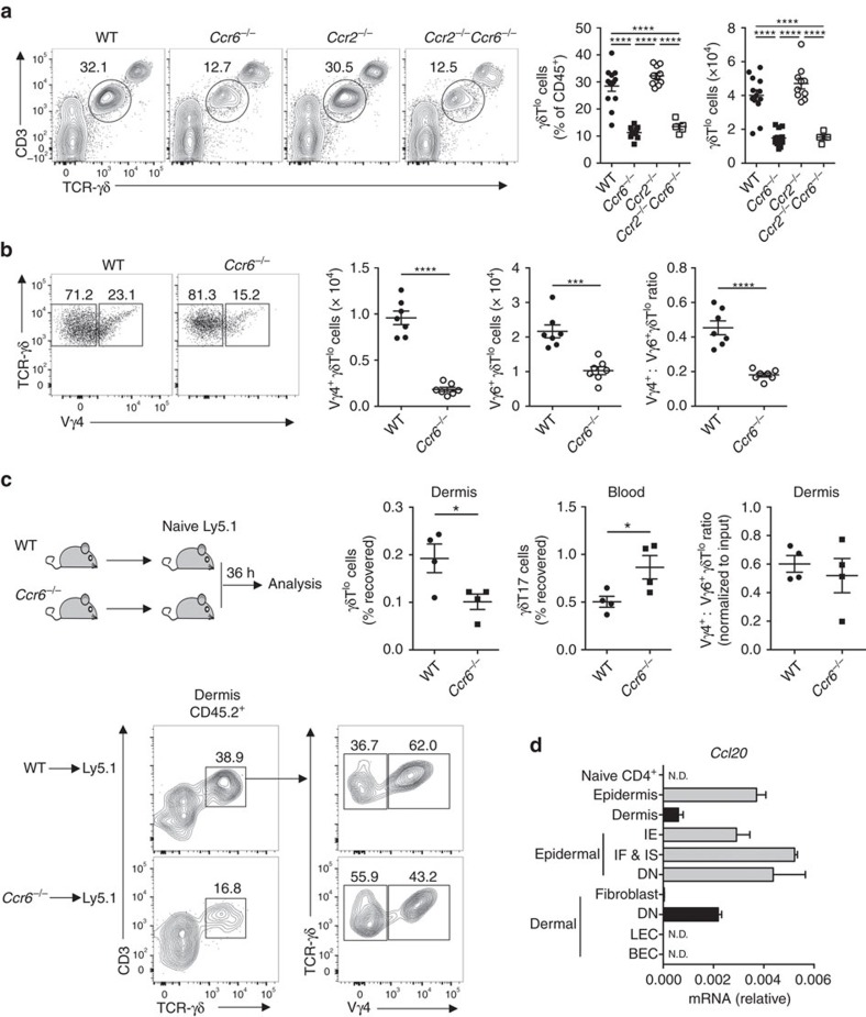
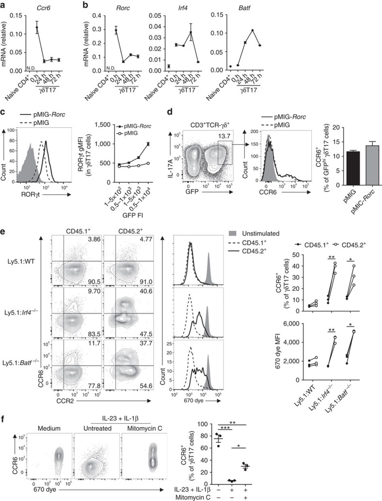
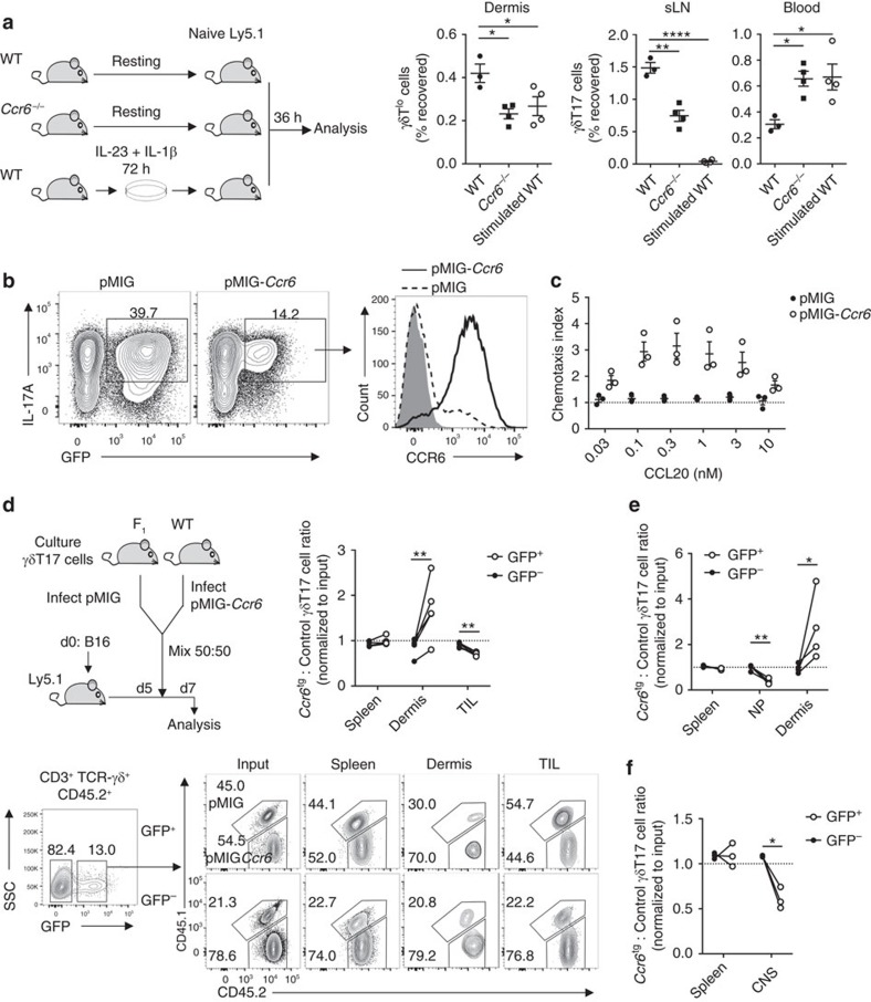
Interleukin 17-producing γδ T (γδT17) cells have unconventional trafficking characteristics, residing in mucocutaneous tissues but also homing into inflamed tissues via circulation. Despite being fundamental to γδT17-driven early protective immunity and exacerbation of autoimmunity and cancer, migratory cues controlling γδT17 cell positioning in barrier tissues and recruitment to inflammatory sites are still unclear. Here we show that γδT17 cells constitutively express chemokine receptors CCR6 and CCR2. While CCR6 recruits resting γδT17 cells to the dermis, CCR2 drives rapid γδT17 cell recruitment to inflamed tissues during autoimmunity, cancer and infection. Downregulation of CCR6 by IRF4 and BATF upon γδT17 activation is required for optimal recruitment of γδT17 cells to inflamed tissue by preventing their sequestration into uninflamed dermis. These findings establish a lymphocyte trafficking model whereby a hierarchy of homing signals is prioritized by dynamic receptor expression to drive both tissue surveillance and rapid recruitment of γδT17 cells to inflammatory lesions.


Conflict of interest statement

The authors declare no competing financial interests.

Figures

Figure 1
Figure 1. γδT17 cells downregulate CCR6 upon activation.
(a) Representative flow cytometry of CCR6 and CCR2 expression in skin-draining lymph nodes (sLN) and dermal CD3+TCR-γδ+IL-17A-YFP+ γδT17 cells from Il17aCre × Rosa26eYFP mice (n=3). (b) Ex vivo transwell chemotaxis of Il17aCre × Rosa26eYFPsplenic IL-17A+/− γδ T cells to CCL20 and CCL2 (n=3). (c) Representative flow cytometry of CD45+ γδT17 cells from organs of naïve Il17aCre × Rosa26eYFP mice (n=3). mLN, mesenteric lymph node; PP, Peyer's patches; siLPL, small intestinal lamina propria lymphocytes. (d) Representative flow cytometry and quantitation of CCR6 and CCR2 expression by γδT17 cells from organs of Il17aCre × Rosa26eYFP mice either naïve (n=6) or at experimental autoimmune encephalomyelitis (EAE) onset (n=7) or peak (n=5). CNS, central nervous system; iLN, inguinal lymph node; ND, not detected. (e) Representative flow cytometry and quantitation of CCR6 expression by γδT17 cells from wild type (WT) mice given BrdU at d3 post-immunization for EAE, and analysed at d8 (n=4). (f) Representative flow cytometry and frequency of CCR6 and CCR2 expression by γδT17 cells from Il17aCre × Rosa26eYFP lymphocytes cultured with indicated stimuli for 72 h (n=5). See also Supplementary Figs 1 and 2. Mean±s.e.m. (ac) Representative of two experiments. (d,f) Pooled from two experiments. (e) Paired two-tailed Student's t-test, (f) one-way paired ANOVA with Dunnett's multiple comparisons test relative to unstimulated control. *P<0.05, **P<0.01, ***P<0.001, ****P<0.0001.
Figure 2
Figure 2. CCR2 recruits γδT17 cells to inflammatory sites.
(a) CD45+CD3+TCRγδ+IL-17A+ γδT17 cell numbers in tumour-infiltrating lymphocytes (TIL) following B16 melanoma challenge (n=5/time point). (b) γδT17 cell numbers in TIL d7 post-challenge with B16 melanoma in wild type (WT) (n=12) and Ccr6−/− mice (n=13). (c) γδT17 cell numbers in central nervous system (CNS) at experimental autoimmune encephalomyelitis (EAE) onset in WT (n=7) and Ccr6−/− mice (n=6). (d) γδT17 cell numbers in TIL and inguinal lymph nodes (iLN) d7 post-challenge with B16 melanoma in WT (n=15 (TIL), 9 (iLN)), Ccr2−/− (n=13 (TIL), 10 (iLN)) and Ccr2−/−Ccr6−/− mice (n=9 (TIL), 5 (iLN)). (e) ELISA for CCL2 in tumour supernatant from WT mice challenged with B16 melanoma (n=5/time point). (f) γδT17 cell numbers in CNS and iLN at EAE onset in WT (n=14), Ccr2−/− (n=13) and Ccr2−/−Ccr6−/− mice (n=12). (g) γδT17 cell numbers in CNS at peak disease in WT (n=6), Ccr2−/− (n=5) and Ccr2−/−Ccr6−/− mice (n=6). (h) ELISA for CCL2 in CNS of WT mice with EAE (n=4/time point). (i) Ly5.1 mice (n=4) at d5 post-challenge with B16 melanoma were transferred i.v. with in vitro-expanded γδT17 cells from Ccr2−/− (CD45.2+) and F1 (CD45.1+CD45.2+) mice. Ccr2−/−:F1 total, Vγ4 and Vγ6 γδT17 cell ratios in spleen and tumours were normalized to input ratio. Vγ4 and Vγ6 γδT17 cells were determined by CD3bright gating. Representative flow cytometry of CD45.2+ γδT17 cells at d7 or input. (j) Ly5.1 mice (n=7) at EAE onset were transferred with F1 and Ccr2−/− γδT17 cells as in (i). Twenty-four hours later, ratios of Ccr2−/−:F1 γδT17 cells in spleen, blood and CNS were normalized to input. Representative flow cytometry of CD45.2+ γδT17 cells 24 h later or input. See also Supplementary Figs 3 and 4. Mean±s.e.m. (a,c,e,i) Representative of two experiments. (b,d,f,j) Pooled from two experiments. (b,c) Unpaired two-tailed Student's t-test, (d,fg,j) one-way ANOVA with Bonferroni's multiple comparisons test (paired in j)), (i) paired two-tailed Student's t-test. *P<0.05, **P<0.01, ***P<0.001, ****P<0.0001.
Figure 3
Figure 3. CCR2 drives protective γδT17 cell responses.
(a) Colony-forming units (c.f.u.) and (b) CD45+CD11b+Ly6G+ neutrophils recovered from nasal wash (NW) of wild type (WT) (n=9) and Tcrd−/− mice (n=10) 72 h post-infection with S. pneumoniae. (c) γδT17 cell numbers in cervical lymph node (cLN) and nasal-associated lymphoid tissue (NALT) and (d) ELISA for CCL2 in digested nasal passage (NP) supernatant in unimmunized mice (n=7) and at 72 h post-S. pneumoniae infection (n=13). (e) Ly5.1 mice (n=5) 24 h post-S. pneumoniae infection were transferred i.v. with expanded γδT17 cells from Ccr2−/− (CD45.2+) and F1 (CD45.1+CD45.2+) mice. The Ccr2−/−:F1 γδT17 cell ratio in spleen and NP was normalized to input ratio. (f) Twenty-four hours prior to S. pneumoniae infection, Tcrd−/− hosts received PBS (n=8) or expanded and purified γδT17 cells from WT (n=9) or Ccr2−/− (n=7) mice. c.f.u. recovered from NW 72 h post-infection. Mean±s.e.m. (ad) Pooled from two experiments. (a,b) Mann–Whitney test, (c,d) unpaired two-tailed Student's t-test, (e) paired two-tailed Student's t-test, (f) Kruskal–Wallis test with Dunn's multiple comparisons test. *P<0.05, **P<0.01.
Figure 4
Figure 4. CCR6 regulates homeostatic γδT17 cell recruitment to dermis.
(a) Representative flow cytometry and quantitation of CD45+CD3loTCR-γδlo (γδTlo) cells from ear skin dermis of naïve wild type (WT) (n=13), Ccr6−/− (n=11), Ccr2−/− (n=10) and Ccr2−/−Ccr6−/− mice (n=5). (b) Representative flow cytometry of Vγ4 expression by dermal γδTlo cells and quantitation of Vγ4+ and Vγ4 γδTlo cells in dermis of WT and Ccr6−/− mice (n=7/group). (c) WT or Ccr6−/− lymphocytes were transferred i.v. into naïve Ly5.1 mice (n=4/group). After 36 h, number of CD45.2+ γδTlo/γδT17 cells recovered was expressed as % of number transferred. Representative flow cytometry of dermal CD45.2+ cells and quantitation of γδT17 cell recovery and Vγ4+:Vγ4 ratio (normalized to input). (d) Ccl20 mRNA from whole tissues or sorted CD45 epidermal keratinoctyes (Sca-1+Ep-CAMlo interfollicular epidermis (IE), Sca-1lo/+Ep-CAM+ infundibulum and isthmus (IF & IS), Sca-1loEp-CAMlo double negative (DN)) or CD45 dermal populations (CD31CD90+CD140α+ fibroblast, gp38+CD31+ lymphatic endothelial cells (LEC), gp38loCD31+ blood endothelial cells (BEC), CD31CD90CD140α double negative (DN)) from naïve WT mice (pooled from 5 mice/experiment). ND, not detected. See also Supplementary Fig. 5. Mean±s.e.m. (a,d) Pooled from three experiments, (c) representative of two similar experiments. (a) One-way ANOVA with Bonferroni's multiple comparisons test, (b,c) unpaired two-tailed Student's t-test. *P<0.05, **P<0.01, ***P<0.001, ****P<0.0001.
Figure 5
Figure 5. IRF4 and BATF promote CCR6 downregulation in γδT17 cells.
(a) Ccr6 and (b) transcription factor mRNA in sorted γδT17 cells from Il17aCre × Rosa26eYFP lymphocytes ex vivo or cultured with IL-23/IL-1β for indicated times (pooled from 5 to 7 mice). ND, not detected. (c,d) Expanded γδT17 cells (n=3) were transduced with empty pMIG or pMIG-Rorc retrovirus. (c) Representative flow cytometry of RORγt expression in GFPhi γδT17 cells (gated as in d), relative to isotype (grey) and geometric mean fluorescence intensity (gMFI) relative to GFP fluorescence intensity (FI). (d) Representative flow cytometry of CCR6 expression and quantitation in GFPhi γδT17 cells. (e) Splenocytes from Ly5.1 and either wild type (WT), Irf4−/− or Batf−/− mice were 670 dye-labelled, mixed 50:50 and stimulated with IL-23/IL-1β for 72 h. Representative flow cytometry and quantitation of CCR6 expression and proliferation in CD45.1+ or CD45.2+ γδT17 cells (n=3/group). (f) Representative flow cytometry and quantitation of CCR6 expression by 670 dye-labelled γδT17 cells from WT splenocytes cultured with IL-23/IL-1β for 72 h with/without mitomycin C pre-treatment (n=3). See also Supplementary Figs 6 and 7. (a,b) Mean±s.d., (cf) Mean±s.e.m. (af) Representative of two similar experiments. (d,e) Paired two-tailed Student's t-test, (f) one-way paired ANOVA with Bonferroni's multiple comparisons test. *P<0.05, **P<0.01, ***P<0.001.
Figure 6
Figure 6. CCR6 downregulation by γδT17 cells enhances migration to inflamed tissue.
(a) Resting lymphocytes from wild type (WT) (n=3) or Ccr6−/− (n=4) mice, or WT lymphocytes stimulated with IL-23/IL-1β for 72 h (n=4) were transferred i.v. into separate naïve Ly5.1 hosts. After 36 h, number of CD45.2+ γδTlo/γδT17 cells recovered was expressed as % of number transferred. sLN, skin-draining lymph node. (b) Representative flow cytometry for CCR6 expression by GFP+ in vitro-expanded γδT17 cells transduced with empty pMIG or pMIG-Ccr6, relative to isotype (grey) (n=3). (c) Chemotaxis of GFP+ γδT17 cells transduced as in (b) to CCL20 (n=3). (d) In vitro-expanded γδT17 cells from F1 (CD45.1+CD45.2+) or WT (CD45.2+) mice were transduced with empty pMIG or pMIG-Ccr6, respectively. Equal numbers of mixed GFP+ cells were transferred i.v. into Ly5.1 mice challenged with B16 melanoma 5 days prior and analysed at d7 (n=5). Representative flow cytometry and ratio of recovered F1 to WT γδT17 cells within transduced (GFP+) and untransduced (GFP) populations. Recovered values were normalized to input values. TIL, tumour-infiltrating lymphocytes. (e,f) In vitro-expanded γδT17 cells from WT or F1 mice were transduced with empty pMIG or pMIG-Ccr6, respectively. Equal numbers of mixed GFP+ cells were transferred i.v. into Ly5.1 mice either (e) 24 h post-infection with S. pneumoniae (n=4) or (f) at experimental autoimmune encephalomyelitis (EAE) onset (n=3) and organs were analysed 48 h later. Ratio of recovered WT to F1 γδT17 cells within transduced (GFP+) and untransduced (GFP) populations, normalized to input values. CNS, central nervous system; NP, nasal passage. Mean±s.e.m. (a) Representative of three similar experiments, (b,d) representative of two experiments. (a) One-way ANOVA with Dunnett's multiple comparisons test relative to resting WT γδT17 cells, (df) paired two-tailed Student's t-test. *P<0.05, **P<0.01, ***P<0.001, ****P<0.0001.

References

    1. Sutton C. E. et al.. Interleukin-1 and IL-23 induce innate IL-17 production from gammadelta T cells, amplifying Th17 responses and autoimmunity. Immunity 31, 331–341 (2009). - PubMed
    1. Jensen K. D. et al.. Thymic selection determines gammadelta T cell effector fate: antigen-naive cells make interleukin-17 and antigen-experienced cells make interferon gamma. Immunity 29, 90–100 (2008). - PMC - PubMed
    1. Kashem S. W. et al.. Nociceptive sensory fibers drive interleukin-23 production from CD301b+ dermal dendritic cells and drive protective cutaneous immunity. Immunity 43, 515–526 (2015). - PMC - PubMed
    1. Cao J. et al.. Activation of IL-27 signalling promotes development of postinfluenza pneumococcal pneumonia. EMBO Mol. Med. 6, 120–140 (2014). - PMC - PubMed
    1. Cho J. S. et al.. IL-17 is essential for host defense against cutaneous Staphylococcus aureus infection in mice. J. Clin. Invest. 120, 1762–1773 (2010). - PMC - PubMed

Publication types