

Clinical Implication of Optical Coherence Tomography-Based Neoatherosclerosis
- PMID: 28581259
- PMCID: PMC5461306
- DOI: 10.3346/jkms.2017.32.7.1056
Clinical Implication of Optical Coherence Tomography-Based Neoatherosclerosis
Abstract
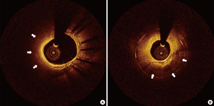
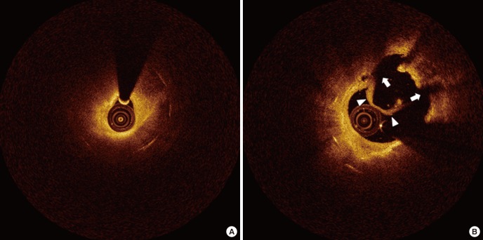
Recent research has indicated neoatherosclerosis (NA), the de novo development of atherosclerosis within the neointimal region of the stented segment after coronary stent implantation, as a mechanism of late/very late stent thrombosis (VLST) and restenosis. This research is based on histologic and intravascular imaging studies. Optical coherence tomography (OCT) is an imaging tool that is superior with regard to resolution capacity, and can be used to visualize detailed information about distinct morphological characteristics of the restenotic tissue. Thus, OCT is a valuable imaging tool for examining NA, such as macrophage infiltration, lipid accumulation, in-stent calcification, or neointimal rupture. This article discusses the prevalence, predictors, and clinical implications of NA that can be observed by OCT.
Keywords: Atherosclerosis; Drug-Eluting Stent; Optical Coherence Tomography.
© 2017 The Korean Academy of Medical Sciences.
Conflict of interest statement
The authors have no potential conflicts of interest to disclose.
Figures
Similar articles
-
In-stent neoatherosclerosis and tissue characteristics of restenotic lesions following implantation of second generation drug-eluting stents in unrestricted coronary lesions: Optical frequency domain imaging study.J Interv Cardiol. 2017 Jun;30(3):195-203. doi: 10.1111/joic.12375. Epub 2017 Mar 10. J Interv Cardiol. 2017. PMID: 28295660
-
Neoatherosclerosis in Patients With Coronary Stent Thrombosis: Findings From Optical Coherence Tomography Imaging (A Report of the PRESTIGE Consortium).JACC Cardiovasc Interv. 2018 Jul 23;11(14):1340-1350. doi: 10.1016/j.jcin.2018.02.029. JACC Cardiovasc Interv. 2018. PMID: 30025727
-
Ex vivo assessment of neointimal characteristics after drug-eluting stent implantation: Optical coherence tomography and histopathology validation study.Int J Cardiol. 2016 Oct 15;221:1043-7. doi: 10.1016/j.ijcard.2016.07.110. Epub 2016 Jul 9. Int J Cardiol. 2016. PMID: 27447812
-
OCT demonstrating neoatherosclerosis as part of the continuous process of coronary artery disease.Herz. 2015 Sep;40(6):845-54. doi: 10.1007/s00059-015-4343-y. Herz. 2015. PMID: 26259732 Free PMC article. Review.
-
Neoatherosclerosis: from basic principles to intravascular imaging.Minerva Cardioangiol. 2018 Jun;66(3):292-300. doi: 10.23736/S0026-4725.17.04573-X. Epub 2017 Nov 20. Minerva Cardioangiol. 2018. PMID: 29160047 Review.
Cited by
-
In Stent Neo-Atherosclerosis: Pathophysiology, Clinical Implications, Prevention, and Therapeutic Approaches.Life (Basel). 2022 Mar 8;12(3):393. doi: 10.3390/life12030393. Life (Basel). 2022. PMID: 35330144 Free PMC article. Review.
-
Neoatherosclerosis prediction using plaque markers in intravascular optical coherence tomography images.Front Cardiovasc Med. 2022 Dec 14;9:1079046. doi: 10.3389/fcvm.2022.1079046. eCollection 2022. Front Cardiovasc Med. 2022. PMID: 36588557 Free PMC article.
-
Comparison of neoatherosclerosis and a clinical outcomes between bioabsorbable versus durable polymer drug-eluting stent: Verification by optical coherence tomography analysis.Cardiol J. 2022 Apr 26;30(6):911-20. doi: 10.5603/CJ.a2022.0025. Online ahead of print. Cardiol J. 2022. PMID: 35470415 Free PMC article.
-
Shedding light on stent thrombosis.J Thorac Dis. 2017 Dec;9(12):4903-4907. doi: 10.21037/jtd.2017.11.47. J Thorac Dis. 2017. PMID: 29312688 Free PMC article. No abstract available.
-
Noninvasive monitoring of vascular alterations in mice with acute lower limb ischemia using multimodal photoacoustic imaging.Bioeng Transl Med. 2025 Feb 17;10(4):e70005. doi: 10.1002/btm2.70005. eCollection 2025 Jul. Bioeng Transl Med. 2025. PMID: 40708973 Free PMC article.
References
-
- Stone GW, Ellis SG, Cannon L, Mann JT, Greenberg JD, Spriggs D, O’Shaughnessy CD, DeMaio S, Hall P, Popma JJ, et al. Comparison of a polymer-based paclitaxel-eluting stent with a bare metal stent in patients with complex coronary artery disease: a randomized controlled trial. JAMA. 2005;294:1215–1223. - PubMed
-
- Stone GW, Ellis SG, Cox DA, Hermiller J, O’Shaughnessy C, Mann JT, Turco M, Caputo R, Bergin P, Greenberg J, et al. A polymer-based, paclitaxel-eluting stent in patients with coronary artery disease. N Engl J Med. 2004;350:221–231. - PubMed
-
- Räber L, Magro M, Stefanini GG, Kalesan B, van Domburg RT, Onuma Y, Wenaweser P, Daemen J, Meier B, Jüni P, et al. Very late coronary stent thrombosis of a newer-generation everolimus-eluting stent compared with early-generation drug-eluting stents: a prospective cohort study. Circulation. 2012;125:1110–1121. - PubMed
-
- Yamaji K, Kimura T, Morimoto T, Nakagawa Y, Inoue K, Soga Y, Arita T, Shirai S, Ando K, Kondo K, et al. Very long-term (15 to 20 years) clinical and angiographic outcome after coronary bare metal stent implantation. Circ Cardiovasc Interv. 2010;3:468–475. - PubMed
Publication types
MeSH terms
LinkOut - more resources
Full Text Sources
Other Literature Sources
Medical
