. 2017 Jun 30;127(7):2777-2788.
doi: 10.1172/JCI92958. Epub 2017 Jun 5.

NK cell heparanase controls tumor invasion and immune surveillance

Affiliations

NK cell heparanase controls tumor invasion and immune surveillance

Eva M Putz et al. J Clin Invest. .

Retraction in

  • NK cell heparanase controls tumor invasion and immune surveillance.
    Putz EM, Mayfosh AJ, Kos K, Barkauskas DS, Nakamura K, Town L, Goodall KJ, Yee DY, Poon IK, Baschuk N, Souza-Fonseca-Guimaraes F, Hulett MD, Smyth MJ. Putz EM, et al. J Clin Invest. 2024 Jul 1;134(13):e183295. doi: 10.1172/JCI183295. J Clin Invest. 2024. PMID: 38949030 Free PMC article. No abstract available.

Abstract

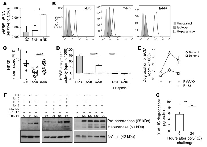
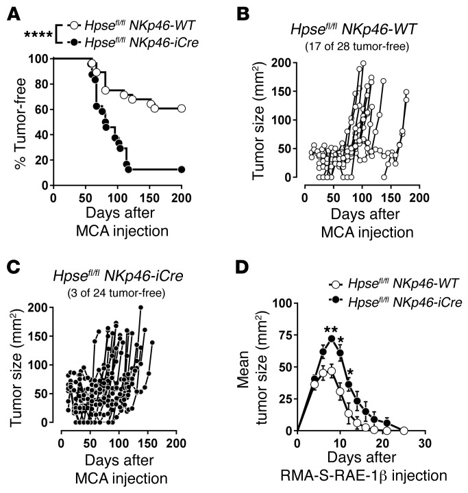
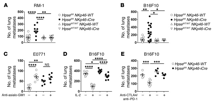
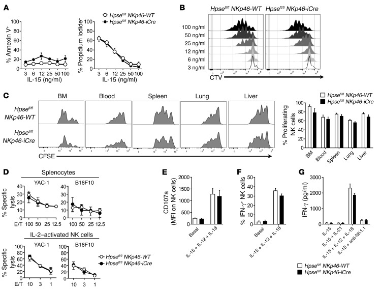
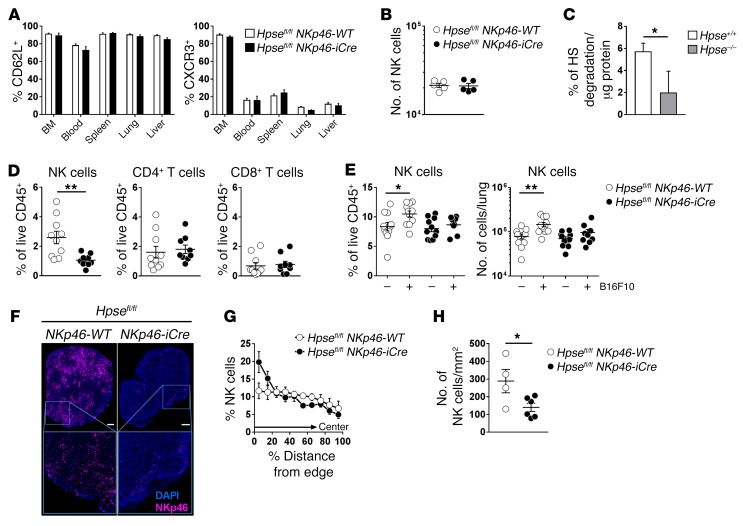
NK cells are highly efficient at preventing cancer metastasis but are infrequently found in the core of primary tumors. Here, have we demonstrated that freshly isolated mouse and human NK cells express low levels of the endo-β-D-glucuronidase heparanase that increase upon NK cell activation. Heparanase deficiency did not affect development, differentiation, or tissue localization of NK cells under steady-state conditions. However, mice lacking heparanase specifically in NK cells (Hpsefl/fl NKp46-iCre mice) were highly tumor prone when challenged with the carcinogen methylcholanthrene (MCA). Hpsefl/fl NKp46-iCre mice were also more susceptible to tumor growth than were their littermate controls when challenged with the established mouse lymphoma cell line RMA-S-RAE-1β, which overexpresses the NK cell group 2D (NKG2D) ligand RAE-1β, or when inoculated with metastatic melanoma, prostate carcinoma, or mammary carcinoma cell lines. NK cell invasion of primary tumors and recruitment to the site of metastasis were strictly dependent on the presence of heparanase. Cytokine and immune checkpoint blockade immunotherapy for metastases was compromised when NK cells lacked heparanase. Our data suggest that heparanase plays a critical role in NK cell invasion into tumors and thereby tumor progression and metastases. This should be considered when systemically treating cancer patients with heparanase inhibitors, since the potential adverse effect on NK cell infiltration might limit the antitumor activity of the inhibitors.


Conflict of interest statement

Conflict of interest: M.J. Smyth has research agreements with Bristol-Myers Squibb, Aduro Biotech, and Corvus Pharmaceuticals.

Figures

Figure 1
Figure 1. Activated NK cells express enzymatically active heparanase.
(AE) NK cells isolated from human donors were assayed as f-NK or a-NK cells. i-DCs were included as a control. (A) mRNA expression of HPSE relative to UBC was assessed by quantitative PCR (qPCR) (mean ± SD; n = 3 individual donors; 1 representative experiment of 2 experiments). (B and C) Heparanase protein expression was determined by intracellular staining and flow cytometry (mean ± SEM; n = 5–13 donors per group). MFI, mean fluorescence intensity. (D) HPSE enzymatic activity was determined by incubating 2 × 10⁵ f-NK or a-NK cells with 3H-HS for 16 hours ± 1 U heparin. Human platelet heparanase (2.5 ng) was included as a control (mean ± SEM; n = 4–11 per group; data were pooled from 2 independent experiments). (E) a-NK cells (2 × 10⁶) from 2 individual donors were cultured on 35S-ECM plates ± 2 ng/ml PMA/0.1 μM ionomycin (IO) ± 200 μg/ml PI-88. ECM degradation was measured after 20 hours (mean ± SD; n = 3 technical replicates; data are representative of 5 individual donors). (F) Heparanase expression was analyzed by Western blotting. FACS-purified mouse TCRβNK1.1+NKp46+DX5+ NK cells were analyzed ex vivo or after stimulation for the indicated durations by cytokines (500 U/ml IL-2, 1 ng/ml IL-12, 10 ng/ml IL-15, and 10 ng/ml IL-18) or by NK cell receptor cross-linking (α-Ly49D or α-NK1.1). (G) The enzymatic activity of heparanase was determined by a TR-FRET–based HS degradation assay. Splenic NK cells were isolated by negative depletion from WT mice that had been injected with 250 μg poly(I:C) 24 hours prior to the analysis or were left untreated (mean ± SD; n = 3). Statistically significant differences between the groups were determined by 1-way ANOVA with Tukey’s post test (A, C, and D) or unpaired Student’s t test (G). *P < 0.05, **P < 0.01, ***P < 0.001, and ****P < 0.0001.
Figure 2
Figure 2. NK cell–intrinsic heparanase is indispensable for efficient surveillance of MCA-induced fibrosarcoma and RAE-1–expressing lymphoma.
(AC) Hpsefl/fl NKp46-WT and Hpsefl/fl NKp46-iCre mice were inoculated s.c. in the hind flank with 100 μg MCA in 0.1 ml corn oil. Mice were then monitored over a 200-day period for fibrosarcoma development. Tumors were measured every week with a caliper (n = 24–28 per group; data were pooled from 2 independent experiments). (A) Data were recorded as the percentage of tumor-free mice (tumors were defined as measuring >3 mm in diameter and consistently growing). Tumor growth curves of individual (B) Hpsefl/fl NKp46-WT and (C) Hpsefl/fl NKp46-iCre mice. (D) Hpsefl/fl NKp46-WT and Hpsefl/fl NKp46-iCre mice were injected s.c. with 5 × 10⁶ RMA-S-RAE-1β cells. Tumor growth was measured every 2 to 3 days with a caliper (mean ± SEM; n = 7 per group; 1 representative experiment of 2 experiments). Statistically significant differences were determined by log-rank Mantel-Cox test (A) and Mann-Whitney U test (D). *P < 0.05, **P < 0.01, and ****P < 0.0001.
Figure 3
Figure 3. Heparanase-deficient NK cells display impaired control of lung metastases.
(A) Hpsefl/fl NKp46-WT, Hpsefl/fl NKp46-iCre, HpseWT/WT NKp46-WT (B6.WT), and HpseWT/WT NKp46-iCre mice were injected i.v. with 1 × 10⁵ RM-1 prostate carcinoma cells. Lungs were harvested on day 14 and macrometastases counted (mean ± SEM; n = 4–16 mice per group; data were pooled from 2 independent experiments). (B) Hpsefl/fl NKp46-WT, Hpsefl/fl NKp46-iCre, HpseWT/WT NKp46-WT (B6.WT), and HpseWT/WT NKp46-iCre mice were injected i.v. with 2 × 10⁵ B16F10 melanoma cells. Lungs were harvested on day 14 and macrometastases counted (mean ± SEM; n = 6–22 mice per group; data were pooled from 3 independent experiments). (C) Hpsefl/fl NKp46-WT and Hpsefl/fl NKp46-iCre mice were injected with 2 × 10⁴ E0771 cells into the mammary fat pad and treated with either 50 μg control Ig (–) or anti–asialo-GM1 (+) (NK cell depletion) on days –1, 0, 7, 14, and 23 after tumor transplantation. Tumors were removed surgically on day 12. Lungs were harvested on day 35 and macrometastases counted (mean ± SEM; n = 6–8 mice per group). (D) Hpsefl/fl NKp46-WT and Hpsefl/fl NKp46-iCre mice were injected i.v. with 5 × 10⁵ B16F10 melanoma cells and treated i.p. with either PBS or 100,000 IU IL-2 on days 0, 1, 2, 3, and 4. Lungs were harvested on day 14 and macrometastases counted (mean ± SEM; n = 10–11 mice per group; data were pooled from 2 independent experiments). (E) Hpsefl/fl NKp46-WT and Hpsefl/fl NKp46-iCre mice were injected i.v. with 5 × 10⁵ B16F10 melanoma cells and treated with either 500 μg control Ig (–) or 250 μg each of anti-CTLA4 and anti–PD-1 (+) on days 0 and 3 after injection, respectively. Lungs were harvested on day 14 and macrometastases counted (mean ± SEM; n = 5–7 mice per group). (AE) Statistically significant differences between the groups were determined by 1-way ANOVA with Tukey’s post test (*P < 0.05, **P < 0.01, ***P < 0.001, and ****P < 0.0001).
Figure 4
Figure 4. NK cell proliferation and function are unchanged by loss of heparanase.
(A and B) Purified BM NK cells from Hpsefl/fl NKp46-WT or Hpsefl/fl NKp46-iCre mice were labeled with CTV and cultured for 3 days in IL-15 as indicated (mean ± SD; n = 2 biological replicates). (A) Apoptosis was determined by annexin V and propidium iodide staining. (B) Proliferation was assessed by CTV dilution. (C) Purified splenic CFSE-labeled NK cells (2 × 10⁵) were injected i.v. into B6.Rag2–/– Il2rg–/– mice. After 3 days, the proliferation of CD45+TCRβNK1.1+DX5+ NK cells in the indicated organs was determined by flow cytometry. Flow cytometric plot shows a representative proliferation profile. Data in the bar graph were pooled from 2 independent experiments (mean ± SEM; n = 8 per group). (D) The cytotoxicity of freshly isolated splenocytes or IL-2–activated NK cells (1,000 U/ml for 5 days) against YAC-1 and B16F10 target cells was tested at the indicated E/T ratios after 4 hours (mean ± SD; n = 3 biological replicates; 1 representative experiment of 2 experiments). (E) Splenocytes (5 × 10⁶) were stimulated for 4 hours with 1 ng/ml IL-12, 100 ng/ml IL-15, and 10 ng/ml IL-18, and the expression of CD107a was assessed on TCRβNK1.1+DX5+ NK cells (mean ± SD; n = 4 mice per group). (F) Lung cells were stimulated for 4 hours in 1 ng/ml IL-12, 100 ng/ml IL-15, and 10 ng/ml IL-18, and the production of IFN-γ was measured by intracellular staining (mean ± SEM; n = 10; data were pooled from 3 independent experiments). (G) Purified splenic NK cells were stimulated in 50 ng/ml IL-15, 100 ng/ml IL-21, 1 ng/ml IL-12, 10 ng/ml IL-18, or anti-NK1.1 precoated wells. The release of IFN-γ was measured after 24 hours by CBA (mean ± SD; n = 2 biological replicates; 1 representative experiment of 2 experiments).
Figure 5
Figure 5. NK cell invasion is impaired in the absence of heparanase.
(A) The expression of CD62L and CXCR3 on TCRβNK1.1+DX5+ NK cells was analyzed by flow cytometry in the indicated organs of Hpsefl/fl NKp46-WT and Hpsefl/fl NKp46-iCre mice (mean ± SEM; n = 3–10). (B) Preactivated splenic NK cells (7.5 × 10⁴) were seeded in the upper chamber of a Transwell insert. The number of migrating cells in response to 10% FBS and 20 ng/ml CXCL10 was assessed after 17 hours (mean ± SEM; n = 5; data were pooled from 2 independent experiments). (C) Heparanase enzymatic activity of isolated Hpse+/+ and Hpse–/– splenic NK cells was determined by a TR-FRET–based HS degradation assay (mean ± SD; n = 3). (D) Mice were injected s.c. with 100 μl Matrigel. Leukocyte infiltration into the Matrigel plugs was determined by flow cytometry after 3 days. Depicted are TCRβNK1.1+NKp46+ NK, TCRβ+CD4+, and TCRβ+CD8+ T cells, respectively (mean ± SEM; n = 9–10 mice per group; data were pooled from 3 independent experiments). (E) Mice were injected i.v. with 5 × 10⁵ B16F10 melanoma cells. Lungs were harvested after 24 hours, and NK cell proportions and numbers were determined by flow cytometry (mean ± SEM; n = 9–12 mice per group; data were pooled from 3 independent experiments; direct comparison of NK cell proportions between B16F10 challenged NKp46-WT and NKp46-iCre mice P = 0.0118, Mann-Whitney U test, and NK cell numbers between B16F10 challenged NKp46-WT and NKp46-iCre mice: P = 0.0278, Mann-Whitney U test). (FH) Mice were injected s.c. with 5 × 10⁶ RMA-S-RAE-1β cells. Tumors were harvested on day 5 and analyzed by immunofluorescence (mean ± SEM; n = 4–6 per group). (F) Representative images of sections stained for NKp46+ NK cells (magenta) and DAPI (blue). Scale bars: 500 μm. Original magnification: ×20, tiled scan of whole tumor. (G) The distance of individual NK cells from the edge was calculated. (H) The number of NKp46+ cells per section was quantified by Imaris. Statistically significant differences were determined by Student’s t test (C), Mann-Whitney U test (D and H), or 1-way ANOVA with Tukey’s post test (E). *P < 0.05 and **P < 0.01.

References

    1. Halama N, et al. Natural killer cells are scarce in colorectal carcinoma tissue despite high levels of chemokines and cytokines. Clin Cancer Res. 2011;17(4):678–689. doi: 10.1158/1078-0432.CCR-10-2173. - DOI - PubMed
    1. Cantoni C, et al. NK cells, tumor cell transition, and tumor progression in solid malignancies: New hints for NK-based immunotherapy? J Immunol Res. 2016;2016:4684268. - PMC - PubMed
    1. Fridman WH, Pagès F, Sautès-Fridman C, Galon J. The immune contexture in human tumours: impact on clinical outcome. Nat Rev Cancer. 2012;12(4):298–306. doi: 10.1038/nrc3245. - DOI - PubMed
    1. Sarrazin S, Lamanna WC, Esko JD. Heparan sulfate proteoglycans. Cold Spring Harb Perspect Biol. 2011;3(7):a004952. - PMC - PubMed
    1. Zcharia E, et al. Newly generated heparanase knock-out mice unravel co-regulation of heparanase and matrix metalloproteinases. PLoS ONE. 2009;4(4):e5181. doi: 10.1371/journal.pone.0005181. - DOI - PMC - PubMed

Publication types

MeSH terms