. 2017 Jul;97(7):843-853.
doi: 10.1038/labinvest.2017.29. Epub 2017 Jun 5.

The Hippo signaling functions through the Notch signaling to regulate intrahepatic bile duct development in mammals

Affiliations

The Hippo signaling functions through the Notch signaling to regulate intrahepatic bile duct development in mammals

Nan Wu et al. Lab Invest. 2017 Jul.

Abstract

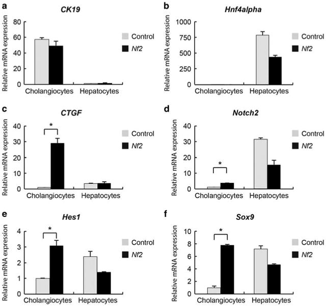
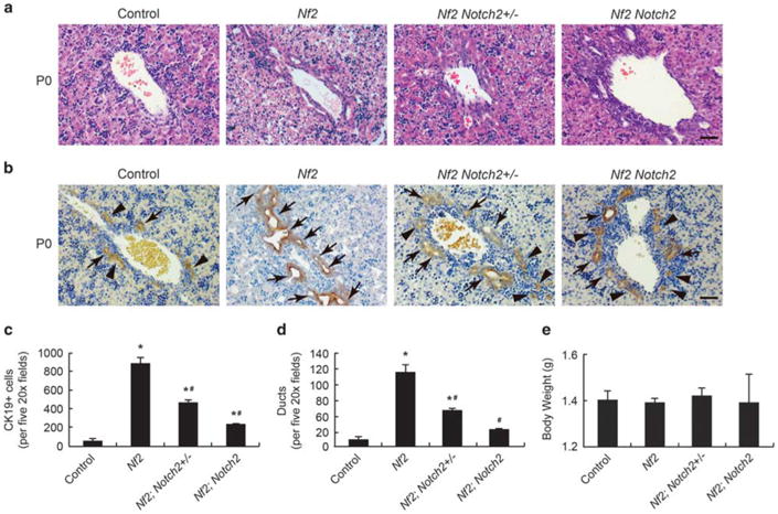
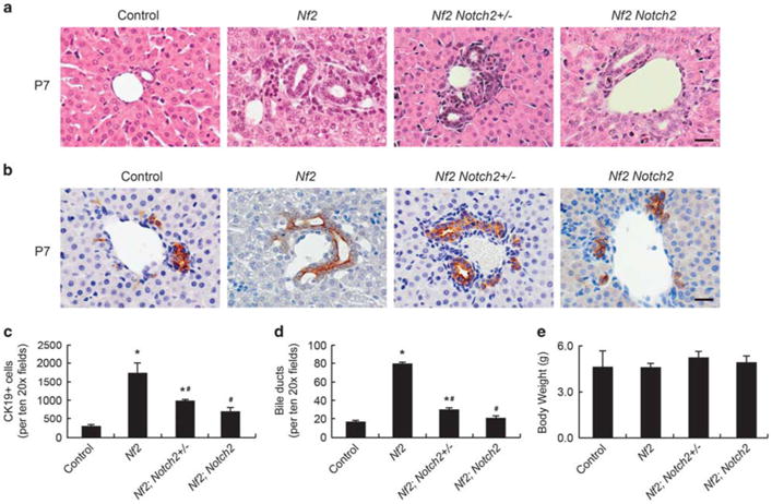
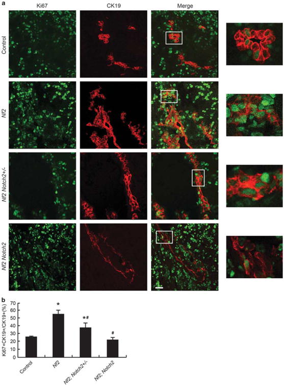
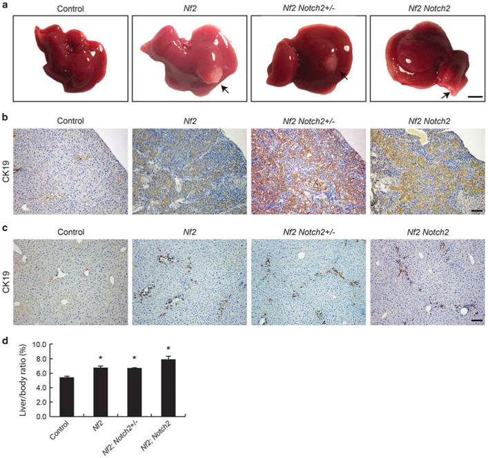
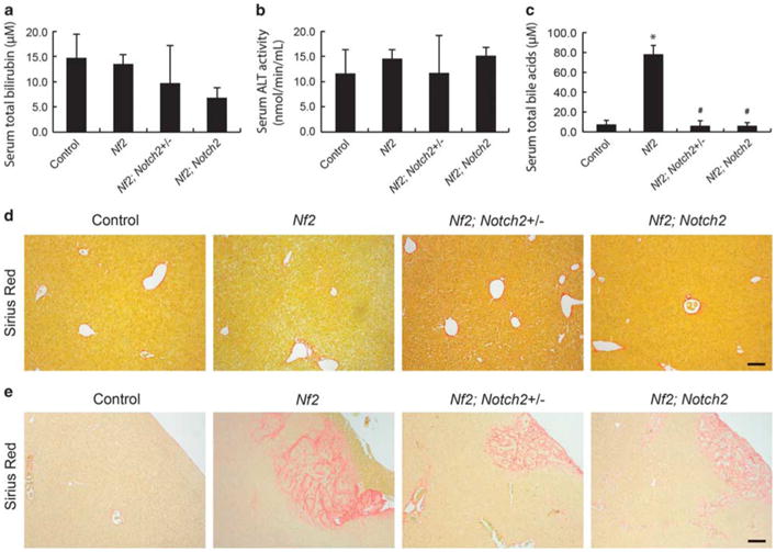
The Hippo signaling pathway and the Notch signaling pathway are evolutionary conserved signaling cascades that have important roles in embryonic development of many organs. In murine liver, disruption of either pathway impairs intrahepatic bile duct development. Recent studies suggested that the Notch signaling receptor Notch2 is a direct transcriptional target of the Hippo signaling pathway effector YAP, and the Notch signaling is a major mediator of the Hippo signaling in maintaining biliary cell characteristics in adult mice. However, it remains to be determined whether the Hippo signaling pathway functions through the Notch signaling in intrahepatic bile duct development. We found that loss of the Hippo signaling pathway tumor suppressor Nf2 resulted in increased expression levels of the Notch signaling pathway receptor Notch2 in cholangiocytes but not in hepatocytes. When knocking down Notch2 on the background of Nf2 deficiency in mouse livers, the excessive bile duct development induced by Nf2 deficiency was suppressed by heterozygous and homozygous deletion of Notch2 in a dose-dependent manner. These results implicated that Notch signaling is one of the downstream effectors of the Hippo signaling pathway in regulating intrahepatic bile duct development.


Conflict of interest statement

Disclosure/Conflict of Interest: The authors declare no conflict of interest.

Figures

Figure 1
Figure 1
Ablating Nf2 increases the expression levels of Notch2 and Notch2 targets in cholangiocytes. (a–f) Quantitative real-time PCR analysis of selective genes from fresh isolated cholangiocytes and hepatocytes from control and Nf2 mice at the age of 8 weeks. Bars represent mean±s.e.m. (n = 3 mice from each genotype). *P<0.05, compared with control, unpaired t-test.
Figure 2
Figure 2
Suppression of Nf2 mutant intrahepatic bile duct development phenotypes by loss of Notch2 (P0). (a and b) H&E staining (a) and CK19 staining (b) of large portal vein area in P0 Control, Nf2, Nf2;Notch2+/ and Nf2;Notch2 livers. Arrows, primitive ducts. Arrowheads, nontubular ductal plate structures. Scale bars =50 μm. (c) Quantification of CK19-positive cholangiocytes for P0 livers. (d) Quantification of primitive ducts for P0 livers. (e) Comparison of body weights for P0 mice. Bars represent mean ±s.e.m. (n= 3 mice from each genotype). *P<0.05, compared with control, #P<0.05, compared with Nf2, one-way ANOVA and Fisher's LSD
Figure 3
Figure 3
Suppression of Nf2 mutant intrahepatic bile duct development phenotypes by loss of Notch2 (P7). (a and b) H&E staining (a) and CK19 staining (b) of small portal vein area in P7 Control, Nf2, Nf2;Notch2+/− and Nf2;Notch2 livers. Arrows, primitive ducts. Arrowheads, nontubular ductal plate structures. Scale bars =20 μm. (c) Quantification of CK19-positive cholangiocytes for P7 livers. (d) Quantification of primitive ducts for P7 livers. (e) Comparison of body weights for P7 mice. Bars represent mean ±s.e.m. (n= 3 mice from each genotype). *P<0.05, compared with control, #P<0.05, compared with Nf2, one-way ANOVA and Fisher's LSD.
Figure 4
Figure 4
Loss of Notch2 suppresses cholangiocyte overproliferation induced by Nf2 deficiency during IHBD development. (a) Control, Nf2, Nf2;Notch2 +/ and Nf2;Notch2 livers from P0 animals were analyzed for Ki67 staining (green) and counterstained for the cholangiocyte marker CK19 (red). Scale bar= 25 μm. Representative areas are shown with higher magnification. (b) Quantification of Ki67-positive cholangiocytes. Bars represent mean±s.e.m. (n= 3 mice from each genotype). *P<0.05, compared with control, #P<0.05, compared with Nf2, one-way ANOVA and Fisher's LSD.
Figure 5
Figure 5
Cholangiocyte proliferation continues in adult Nf2;Notch2+/ and Nf2;Notch2 mice. (a) Gross liver images from 8-week-old animals. Note the formation of biliary hamartoma in Nf2, Nf2;Notch2+/ and Nf2;Notch2 livers (indicated by arrows and confirmed by CK19 staining in b). Scale bar= 0.5 cm. (b) CK19 staining of the biliary hamartoma shown in a, Scale bar= 100 μm. (c) CK19 staining of deep parenchymal area of control, Nf2, Nf2; Notch2+/ and Nf2;Notch2 livers from 8-week-old animals. Scale bar = 100 μm. (d) Quantification of liver-to-body ratio of 8-week-old animals. Bars represent mean± s.e.m. (n = 3 mice from each genotype). *P<0.05, compared with wild-type control, one-way ANOVA and Fisher's LSD.
Figure 6
Figure 6
Nf2;Notch2+/ and Nf2;Notch2 mice do not suffer severe liver damage. (a) Serum total bilirubin levels, (b) serum alanine aminotransferase (ALT) activity, (c) serum total bile acids levels in 8-week-old control, Nf2, Nf2;Notch2+/ and Nf2;Notch2 animals. Bars represent mean ± s.e.m. (n =3 mice from each genotype). *P<0.05, compared with control, #P<0.05, compared with Nf2, one-way ANOVA and Fisher's LSD. (d and e) Liver fibrosis evaluation of 8-week-old control, Nf2, Nf2;Notch2+/, and Nf2;Notch2 animals with Sirius Red staining, a method detecting collagen deposition.

References

    1. Maroni L, Haibo B, Ray D, et al. Functional and structural features of cholangiocytes in health and disease. Cell Mol Gastroenterol Hepatol. 2015;1:368–380. - PMC - PubMed
    1. Nguyen Q, Anders RA, Alpini G, et al. Yes-associated protein in the liver: regulation of hepatic development, repair, cell fate determination and tumorigenesis. Dig Liver Dis. 2015;47:826–835. - PubMed
    1. Raynaud P, Carpentier R, Antoniou A, et al. Biliary differentiation and bile duct morphogenesis in development and disease. Int J Biochem Cell Biol. 2011;43:245–256. - PubMed
    1. Zong Y, Stanger BZ. Molecular mechanisms of liver and bile duct development. Wiley Interdiscip Rev Dev Biol. 2012;1:643–655. - PubMed
    1. Carpentier R, Suner RE, van Hul N, et al. Embryonic ductal plate cells give rise to cholangiocytes, periportal hepatocytes, and adult liver progenitor cells. Gastroenterology. 2011;141:1432–1438. 1438 e1431–1434. - PMC - PubMed

Substances