. 2017 Jul 13;547(7662):179-184.
doi: 10.1038/nature22978. Epub 2017 Jun 5.

The extracellular matrix protein agrin promotes heart regeneration in mice

Affiliations

The extracellular matrix protein agrin promotes heart regeneration in mice

Elad Bassat et al. Nature. .

Abstract

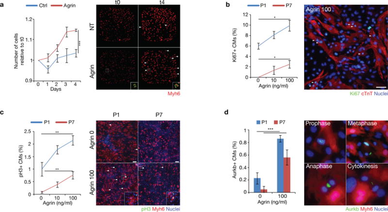
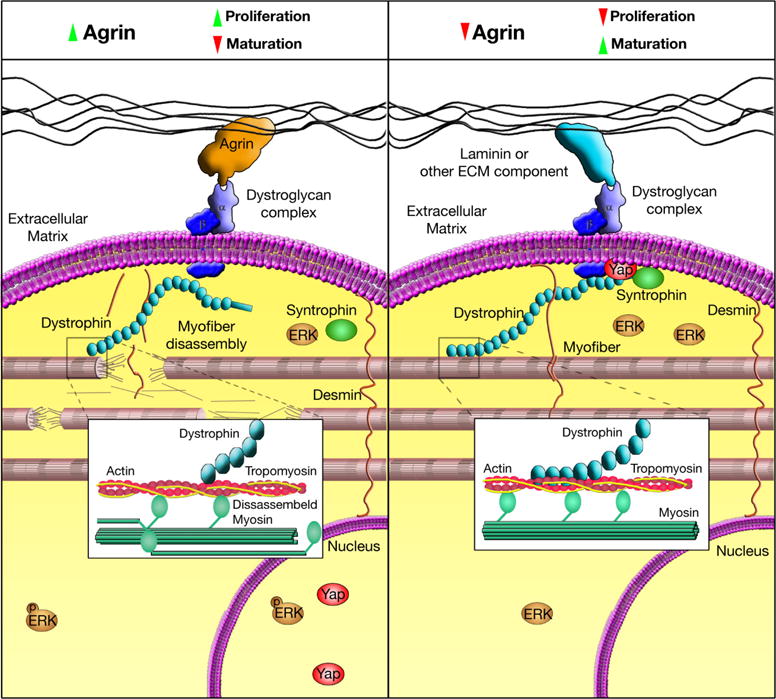
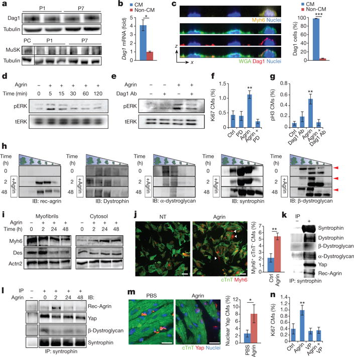
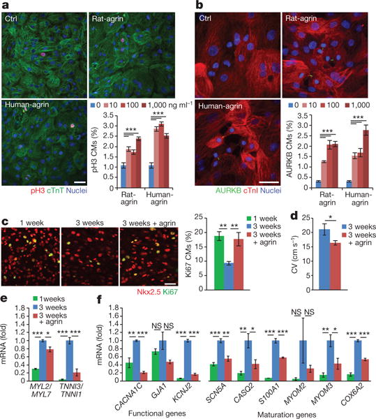
The adult mammalian heart is non-regenerative owing to the post-mitotic nature of cardiomyocytes. The neonatal mouse heart can regenerate, but only during the first week of life. Here we show that changes in the composition of the extracellular matrix during this week can affect cardiomyocyte growth and differentiation in mice. We identify agrin, a component of neonatal extracellular matrix, as required for the full regenerative capacity of neonatal mouse hearts. In vitro, recombinant agrin promotes the division of cardiomyocytes that are derived from mouse and human induced pluripotent stem cells through a mechanism that involves the disassembly of the dystrophin-glycoprotein complex, and Yap- and ERK-mediated signalling. In vivo, a single administration of agrin promotes cardiac regeneration in adult mice after myocardial infarction, although the degree of cardiomyocyte proliferation observed in this model suggests that there are additional therapeutic mechanisms. Together, our results uncover a new inducer of mammalian heart regeneration and highlight fundamental roles of the extracellular matrix in cardiac repair.


Conflict of interest statement

The authors declare no competing financial interests.

Figures

Extended Data Figure 1
Extended Data Figure 1. P1 and P7 ECM explants contain increased gelatinase activity
a, b, Cell removal assessment of heart sections by DAPI or scanning electron microscopy. Scale bars, 50 μm (a) and 20 μm (b). c, A schematic diagram of the in situ zymography assay. d, Immunofluorescence evaluation of Col1, Col4 and gelatin degradation in response to P1 and P7 ECM. e, Quantification of the in situ zymography assay. n = 2 samples.
Extended Data Figure 2
Extended Data Figure 2. Mass spectrometry results and validation
a, Venn diagram representing the LC–MS results. b, qPCR analysis of genes obtained from the LC–MS in P1 and P7 whole-heart lysates. n = 3 P1 hearts and 3 P7 hearts.
Extended Data Figure 3
Extended Data Figure 3. P1 and P8 cardiac cell separation
qPCR of mRNA from P1 and P8 heart lysates. qPCR analysis of six cell populations (fibroblasts, non-fibroblasts, cardiomyocytes, non-cardiomyocytes, endothelial cells, non-endothelial cells) for CD90 (also known as Thy1, a fibroblast marker), Myh6 (a cardiomyocyte marker) and Pecam1 (Pecam, an endothelial cell marker) (for P1, n = 4 cardiomyocyte, 4 non-cardiomyocyte, 4 fibroblast, 4 non-fibroblasts, 7 endothelial cell, 7 non-endothelial cell samples and for P8 n = 2 cardiomyocyte and 2 non-cardiomyocyte samples). Data are presented as mean ± s.e.m. *P < 0.05, **P <0.01, ***P< 0.001; statistical significance was calculated using a one-tailed t-test.
Extended Data Figure 4
Extended Data Figure 4. Agrin induces cardiomyocyte proliferation in vitro
ad, Immunofluorescence evaluation of P1 and P7 cardiomyocytes (cTnT+ or Myh6CretdTomato+, see Methods) cell number (a), cell-cycle activity (Ki67; b), mitosis (pH3; c) or cytokinesis (Aurkb, also known as Aim1; d) in response to agrin dosage in vitro. White arrowheads indicate proliferating cells. Analysis of five individual wells per treatment (a); n = 1,855 P1 and 1,328 P7 cardiomyocytes pooled from the analysis of three P1 and four P7 samples (b); n = 11,450 P1 and 13,469 P7 cardiomyocytes pooled from the analysis of four P1 and six P7 samples (c); n = 7,971 P1 and 3,856 P7 cardiomyocytes pooled from the analysis of three P1 and three P7 samples (d). Scale bars, 30 μm (b) and 100 μm (c). Data are presented as mean ± s.e.m. *P <0.05, **P< 0.01, ***P< 0.001; statistical significance was calculated using an ANOVA followed by Dunnett’s post hoc test relative to the control group(ac) or using a one-tailed t-test (d).
Extended Data Figure 5
Extended Data Figure 5. Agrin-cKO cardiac characterization
a, Tnni3:Tnni1 protein ratio in P8 agrin-cKO and wild-type mice. n = 4 wild-type and 7 cKO mice. b, Immunofluorescence analysis and Pearsons’s correlation coefficent analysis of P14 T-tubules by Cav3 colocalizion with α-actinin labelled z-lines. n = 5 wild-type and 6 cKO samples. c, Immunofluorescence analysis of WGA membrane staining in P1 wild-type and agrin-cKO depicting changes in cell size. n = 2 wild-type and 3 cKO samples. d, qPCR analysis of pathological hypertrophic marker Acta1 in P1 wild-type and agrin-cKO heart lysates. n = 21 wild-type and 26 cKO samples. e, f, Serial echocardiographic measurements of ejection fraction (EF) and wall thickness of 1-, 3- and 14-month-old agrin cKO and wild-type mice. n = 8 mice per group at 1 month, 4 wild-type and 5 cKO at 3 months and 4 wild-type and 3 cKO at 14 months. g, Heart to body weight ratio of 1-, 3- and 14-month-old agrin-cKO and wild-type mice. n = 9 wild-type and 4 cKO at 1 month, 4 wild-type and 5 cKO at 3 months and 6 wild-type and 5 cKO at 14 months. h, Histological sections of 1- and 3-month-old wild-type and agrin-cKO stained with Masson’s trichrome. i, Scheme of P1 heart resection. j, Representative images of agrin-cKO and wild-type mice 28 days after resection at P1. k, Functional cardiac recovery measurement (cardiac output), 28 days after resection. n = 5 wild-type and 3 cKO mice (j, k). Scale bars, 20 μm (b), 10 μm (c) and 1 mm (h). Data are presented as mean ± s.e.m. *P< 0.05, **P< 0.01; Statistical significance was calculated using a one-tailed t-test.
Extended Data Figure 6
Extended Data Figure 6. Agrin induces cardiac regeneration in juvenile mice
a, A schematic diagram depicting LAD ligation in juvenile and adult mice. b, c, In vivo evaluation of cardiomyocytes cell-cycle re-entry by Ki67 (b) or Aurkb (c) in heart sections seven days after myocardial infarction. n = 1,934 cardiomyocytes pooled from the analysis of three PBS-treated mice and three agrin-treated mice (b); n = 1,450 cardiomyocytes pooled from the analysis of three PBS- and six agrin-treated samples (c). d, Scar quantification based on Masson’s trichrome staining of heart sections of PBS- and agrin-treated juvenile mice. Representative pictures are provided. n = 4 PBS- and 8 agrin-treated mice. eg, Serial echocardiographic measurements of ejection fraction (EF), fractional shortening (FS) and wall thickness of uninjured and injured PBS- and agrin-treated juvenile mice following myocardial infarction, according to the schema in a. n = 3 uninjured, 3 PBS- and 3 agrin-treated mice at day 13; and n = 4 uninjured, 5 PBS- and 9 agrin-treated mice at day 43. Scale bars, 10 μm (b), 20 μm (c) and 1 mm (d). Data are presented as mean ± s.e.m. *P <0.05, **P< 0.01, ***P< 0.001; statistical significance was calculated using a one-tailed t-test(bd, g) or ANOVA followed by Dunnett’s post hoc test relative to the control group (e, f).
Extended Data Figure 7
Extended Data Figure 7. Pharmacokinetics of agrin injection in injured adult hearts
Mice were subjected to LAD ligation (as shown in Fig. 4a), and received either PBS or agrin treatment. Hearts were collected at respective time points after treatment, and were subjected to protein extraction and anti-His-tag immunopreciptation using Ni-NTA resin. a, Representative western blot of agrin from relevant immunoprecipitation samples. Anti-tubulin western blot of respective total extracts, as immunoprecipitation loading control is shown at the bottom. b, The kinetics of recombinant agrin up to 96 h after treatment. The amount of agrin is presented as a percentage of the first time point (t = 0), and normalized according to tubulin. n = 3 repeats.
Extended Data Figure 8
Extended Data Figure 8. Dag1 expression in P1 and P7 whole hearts and cardiac cell separation
a, qPCR of Dag1 mRNA from P1 and P7 heart lysates. n = 5 P1 and 3 P7 samples. b, qPCR analysis of Dag1 mRNA in distinct cardiac populations from P1 mice (fibroblasts, non-fibroblasts, cardiomyocytes, non-cardiomyocytes, endothelial cells, non-endothelial cells). n = 4 cardiomyocyte, 4 non-cardiomyocyte, 4 fibroblast, 4 nonfibroblast and 7 endothelial and 7 non-endothelial cell samples. c, Serial immunofluorecence counting of Myh6-lineage-derived tdTomato-labelled cardiomyocytes treated with ouabain and digoxin which inhibit Na+K+ pumps. Quantification was made using acuman (see Methods). n = 31 control, 16 agrin-, 11 ouabain- and 11 digoxin-treated individual wells per treatment. d, quantification of cardiomyocyte sarcomeric status by cTnT and Myh6-linage immunofluorescence analysis of P7 agrin-treated cardiomyocytes cultured in vitro for three days. n = 2,222 cardiomyocytes pooled from the analysis of three samples per group. Representative images of cardiomyocytes with intact, partially disassembled and severely disassembled sarcomeres are provided. Scale bars, 40 μm. Data are presented as mean ± s.e.m. *P < 0.05, ***P < 0.001; statistical significance was calculated using a one-tailed t-test (a, b, d) or ANOVA followed by Dunnett’s post hoc test relative to control group (c).
Extended Data Figure 9
Extended Data Figure 9. A model for the agrin-DGC-Yap signalling axis in cardiomyocyte maturation and cardiac regeneration
Agrin triggers mild cardiomyocyte dedifferentiation and proliferation by modulation of DGC integrity and signalling. During neonatal stages agrin suppresses the maturation of the DGC. At P7, agrin levels are reduced and ECM–DGC interaction through Dag1 promotes cardiomyocyte differentiation and maturation. Yap is tethered to the DGC upon cardiomyocyte maturation while upon agrin treatment, it translocates to the nucleus to facilitate cardiomyocyte cell-cycle re-entry.
Figure 1
Figure 1. Identification of agrin in a screen for mouse cardiac ECM-mediated cardiomyocyte proliferation
a, b, Representative fields of heart cultures stained with DAPI (blue), cTnT (green) and Ki67 (red). White arrows indicate Ki67+cTnT+ cells. CMs, cardiomyocytes. c, d, Percentage of proliferating cardiomyocytes from P1 (c) or P7 (d) hearts in response to P1 and P7 ECM particles. n = 2,069 cardiomyocytes from three samples (c); n = 2,221 cardiomyocytes from four samples (d). e, f, Percentage of proliferating cardiomyocytes (Ki67+cTnT+) in response to P1 and P7 ECM in P1 (e) or P7 (f) cultures, with or without the broad MMP inhibitor (Marimastat). n = 3,480 cardiomyocytes from three samples (e); n = 23,445 cardiomyocytes from four samples (f). g, h, Percentage of P1 (g) or P7 (h) proliferating cardiomyocytes in response to MMP9- or MMP12-cleaved ECM fragments. n = 11,820 cardiomyocytes from four samples (g); n = 15,509 cardiomyocytes from four samples (h). i, qPCR of Agrn mRNA in P1 and P7 hearts. n = 8 P1 and 3 P7 hearts. j, Quantification of western blots for agrin from P1, P7 and 12-week-old (12W) adult heart lysates. A.U., arbitrary units. n = 3 samples per group. k, Images of P1 and P7 heart sections stained for agrin (green) and DAPI (blue). n = 3 samples per group. l, qPCR analysis of cardiac populations (FB, fibroblasts; CM, cardiomyocytes; EC, endothelial cells). n = 4 cardiomyocyte, 4 non-cardiomyocyte, 4 fibroblast, 4 non-fibroblast, 7 endothelial cells and 7 non-endothelial cell samples. Scale bars, 40 μm (a) and 10 μm (k). Data are presented as mean ±s.e.m. *P < 0.05, **P< 0.01, ***P< 0.001; statistical significance was calculated using ANOVA followed by a Dunnett’s post hoc test relative to the control group (ch) or a Tukey’s post hoc test (j), statistical significance was calculated using a one-tailed t-test (i, l).
Figure 2
Figure 2. Agrin delays neonatal cardiomyocyte maturation and is required for P1 cardiac regeneration following surgical resection
a, Diagram showing the mesoderm conditional knockout of agrin (agrin-cKO) in mice. b, qPCR of Agrn mRNA in P1 wild-type and agrin-cKO hearts. n = 8 wild-type (WT) and 7 cKO samples. c, Immunofluorescence images of agrin in P1 wild-type and agrin-cKO heart sections. n = 3 samples of each group. d, Quantification of western blots for agrin from wild-type and agrin-cKO mice heart lysates. n = 8 samples of each group. e, Immunofluorescence analysis and Pearson’s correlation coefficient analysis of T-tubules labelled with Cav3 in the z-lines (as indicated by Actn2). n = 4 wild-type and 3 cKO samples. White arrow heads indicate colocalization of T-tubules and z-lines. f, Myh6:Myh7 protein ratio from wild-type and agrin-cKO mice. n = 5 wild-type and 6 agrin-cKO samples. g, Staining and mean pixel intensity quantification of mitochondrial content in cardiomyocytes measured by Tom20 staining. n = 5 wild-type and 3 agrin-cKO samples. h, i, In vivo evaluation of P1 cardiomyocyte cell-cycle markers (Ki67; h) and (Aurkb; i) by immunofluorescence analysis in wild-type and agrin-cKO left ventricle heart sections. n = 41,695 cardiomyocytes from 11 wild-type and 4 agrin-cKO samples (h); n = 3,212 cardiomyocytes from 3 samples per group (i). j, Histological sections of P1 resected wild-type and agrin-cKO mice, 28 days after injury, stained with Masson’s trichrome. Bottom left corner of the cKO (left panel) was cropped to remove the adjacent section. k, l, Scar quantification of heart sections four weeks after resection of wild-type and agrin-cKO hearts. LV, left ventricle; none, 0% of the left ventricular wall; moderated ≤1% of the left ventricular wall; large ≥1% of the left ventricular wall. n = 12 wild-type and 8 cKO mice. m, n, Functional cardiac recovery measurements (ejection fraction (EF) and fractional shortening (FS)) of hearts from agrin-cKO and wild-type mice 28 days after resection. n = 5 wild-type and 3 cKO mice. o, p, In vivo evaluation of cardiomyocyte cell-cycle re-entry in the peri-infarct region by immunofluorescence analysis of Ki67 (o) or Aurkb (p) in heart sections 7 days after resection of wild-type and agrin-cKO hearts. n = 5,556 cardiomyocytes from seven wild-type and six cKO samples (o); n = 2,235 cardiomyocytes from three wild-type and four cKO samples (p). Scale bars, 10 μm (c, e, gi, p), 20 μm (o), 100 μm (j, right) and 1 mm (j, left). Data are presented as mean ± s.e.m. *P < 0.05, **P < 0.01, ***P < 0.001; statistical significance was calculated using a one-tailed t-test.
Figure 3
Figure 3. Agrin induces cardiac regeneration in adult mice
a–c, In vivo evaluation of cardiomyocyte cell-cycle re-entry in the peri-infarct region seven days after myocardial infarction by Ki67 (a), Aurkb (b) or 21 days after myocardial infarction BrdU (c) n = 1,842 cardiomyocytes from five PBS and six agrin samples; n = 2,259 cardiomyocytes from five samples per group (b); n = 9,307 cardiomyocytes from six samples per group (c). For BrdU pulse-chase experiment see Methods. d, e, Heart section scar assessment following PBS or agrin treatment at indicated days after myocardial infarction (MI). Representative images are shown in d and quantified in e. n = 4 mice per group for day 0, 5 PBS- and 4 agrin-treated mice for day 4, 4 PBS- and 5 agrin-treated mice for day 14, 7 PBS- and 8 agrin-treated mice for day 35. f–h, Serial echocardiographic measurements of ejection fraction, fractional shortening and wall thickness of uninjured and injured hearts treated with PBS or agrin. LVAW, left ventricle anterior wall; LVPW, left ventricle posterior wall. n = 8 baseline, 5 uninjured, 6 PBS- and 8 agrin-treated mice (f, g); n = 2 uninjured, 5 PBS- and 6 agrin-treated mice (h). Scale bars, 10 μm (a, c), 20 μm (b) and 1 mm (d). Data are presented as mean ± s.e.m. *P < 0.05, **P < 0.01; statistical significance was calculated using ANOVA followed by Dunnett’s post hoc test relative to control group (f–h) or using a one-tailed t-test relative to PBS (a–e).
Figure 4
Figure 4. Agrin promotes cardiomyocyte proliferation through Dag1, ERK and Yap signalling
a, Western blot of Dag1 and MuSK from P1 and P7 heart lysates. n = 3 samples per group for MuSK, n = 6 samples per group for Dag1. Skeletal muscle extract was used as positive control (PC) for MuSK expression (lower panel). b, c, P7 Dag1 expression by qPCR (b) or membrane staining (c) in cardiomyocytes relative to non-cardiomyocytes (z–x plane presented). WGA, wheat-germ agglutinin. n = 2 cardiomyocyte and 2 non-cardiomyocyte RNA samples; n = 17,012 cardiomyocytes from four samples per group (c). d, e, Western blots of phosphorylated ERK (pERK) and total ERK (tERK) in P7 cultures with the indicated treatments. Ab, antibody. n = 5 samples (d); n = 3 samples (e). f, g, P7 cardiomyocytes cell-cycle analysis by immunofluorescence staining following agrin treatment with a MEK inhibitor (f) or Dag1 inhibitory antibody (g). Ctrl, control; PD, PD0325901. n = 2,743 cardiomyocytes from six samples (f); n = 6,649 cardiomyocytes from four samples (g). h, Glycerol-gradient fractionation of whole-cell extracts from P7 cells with or without (time 0) agrin treatment for 2 and 48 h. Fractions were analysed by SDS–PAGE and immunoblotting (IB) with indicated antibodies. n = 3 samples. Rec-agrin, recombinant agrin. Arrowheads indicate glycosylated form of β-dystroglycan. i, Isolated myofibrillar pellets and cytosol from P7 cells treated with agrin for 48 h analysed by SDS–PAGE and immunoblotting. n = 3 samples. j, Quantification of cardiomyocyte dedifferentiation using Myh6-lineage-derived cardiomyocytes (red mostly nuclear staining) stained with cTnT (green). Arrows indicate cardiomyocytes that have lost cTnT expression. n = 2,869 cardiomyocytes from five samples per group. k, Co-immunoprecipitation (IP) assay of Yap, agrin and various DGC proteins immunoprecipitated with syntrophin. n = 3 samples.l, Co-immunoprecipitation assay from 0, 2, 24 and 48 h agrin-treated cell membranes immunoprecipitated by syntrophin and blotted for Yap, agrin and other DGC proteins. n = 3 samples. m, In vivo quantification of nuclear Yap of heart sections from 12-week-old mice with PBS or agrin treatments one day after myocardial infarction. Representative images are shown. n = 1,167 cardiomyocytes from four mice per group. n, Cardiomyocyte proliferation assay of P7 heart cultures treated with agrin and the Yap–TEAD inhibitor. VP, verteporfin. n = 13,680 cardiomyocytes from eight samples. Scale bars, 10 μm (j) and 20 μm (m). Data are presented as mean ± s.e.m. *P < 0.05, **P < 0.01, ***P < 0.001; statistical significance was calculated using a one-tailed t-test (b, c, j, m) or ANOVA with Dunnett’s post hoc test (f, g, n).
Figure 5
Figure 5. Agrin promotes proliferation and attenuates maturation of human iPSC–CMs
a, b, Immunofluorescence of iPSC–CM cell-cycle activity by pH3 (a) or AURKB (b) in response to rat and human agrin administration (10–1,000 ng ml−1). Representative images of cultures treated with 1,000 ng ml−1 are shown. pH3 quantification was performed automatically using imageXpress software (n = 174,850 cardiomyocytes from 35 images of control (Ctrl) and 37 images of rat-agrin-treated cultures and 32 images for all other samples (a); n = 38,542 cardiomyocytes from nine control samples and six samples in all other groups (b). c, Human iPSC–CM proliferation assay in the 3D patch culture system. n = 3 samples for one week, 4 samples for other treatments. Scale bars, 20 μm (a–c). d–f, Effects of human agrin on conduction velocity (d) and structural and functional maturation genes (e, f). n = 4 samples (d); n = 3 samples per group (e, f). mRNA expression ratios of MYL2/MYL7 and TNNI3/TNNI1 are shown in e. CV, conduction velocity. n = 4 samples (d); n = 3 samples per group (e, f). Data are presented as mean ± s.e.m. proliferation per field. *P<0.05, **P<0.01, ***P<0.001; statistical significance was calculated using a one-tailed t-test (d) or relative to untreated or 3-week control group using ANOVA followed by Dunnett’s post hoc test.

Comment in

Similar articles

Cited by

References

    1. Soonpaa MH, Kim KK, Pajak L, Franklin M, Field LJ. Cardiomyocyte DNA synthesis and binucleation during murine development. Am J Physiol Heart Circ Physiol. 1996;271:H2183–H2189. - PubMed
    1. Bergmann O, et al. Evidence for cardiomyocyte renewal in humans. Science. 2009;324:98–102. - PMC - PubMed
    1. Tzahor E, Poss KD. Cardiac regeneration strategies: staying young at heart. Science. 2017;356:1035–1039. - PMC - PubMed
    1. Porrello ER, et al. Transient regenerative potential of the neonatal mouse heart. Science. 2011;331:1078–1080. - PMC - PubMed
    1. Rupp F, Payan DG, Magill-Solc C, Cowan DM, Scheller RH. Structure and expression of a rat agrin. Neuron. 1991;6:811–823. - PubMed

Publication types

MeSH terms