. 2017 Jun 5;6(6):e344.
doi: 10.1038/oncsis.2017.47.

Regulation of cargo transfer between ESCRT-0 and ESCRT-I complexes by flotillin-1 during endosomal sorting of ubiquitinated cargo

Affiliations

Regulation of cargo transfer between ESCRT-0 and ESCRT-I complexes by flotillin-1 during endosomal sorting of ubiquitinated cargo

M Meister et al. Oncogenesis. .

Abstract

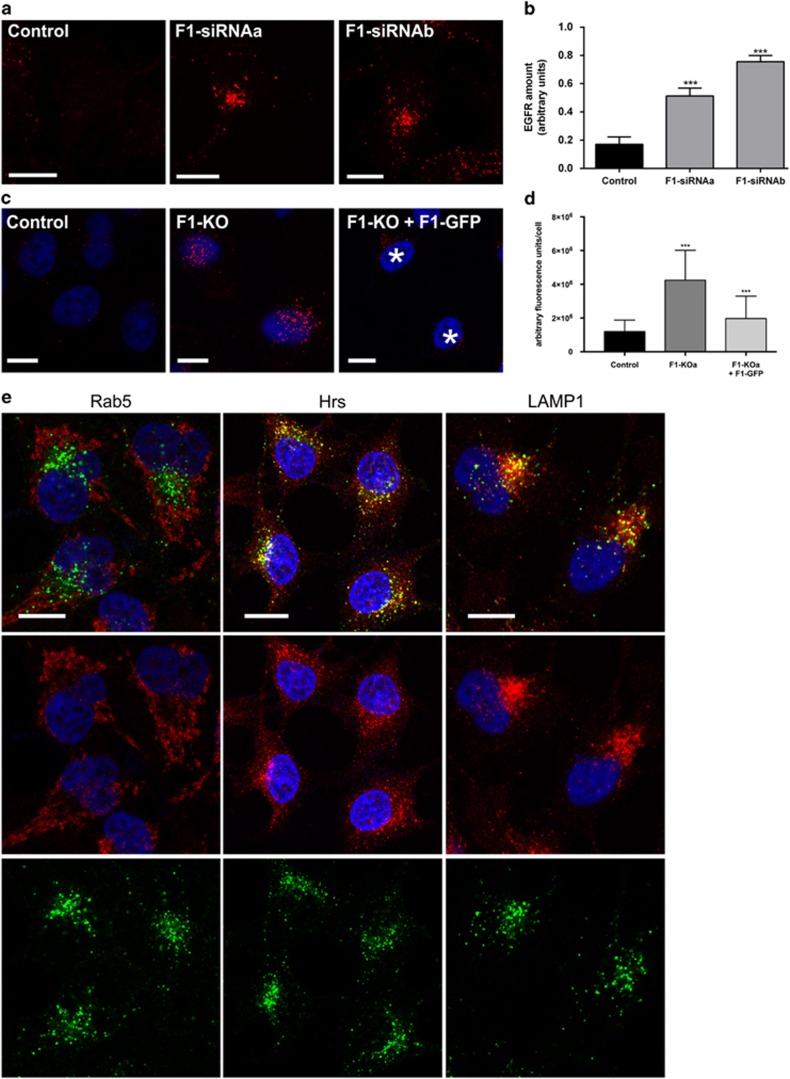
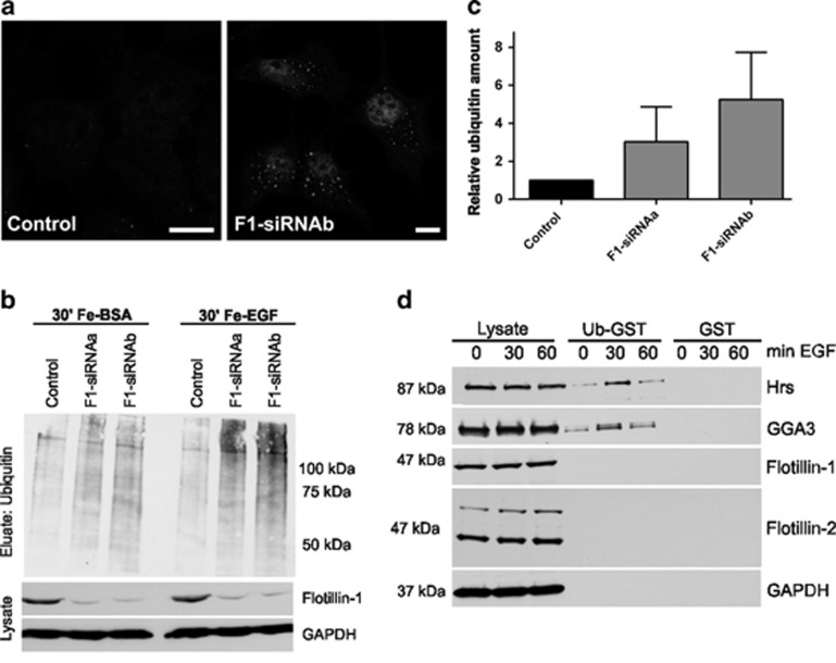
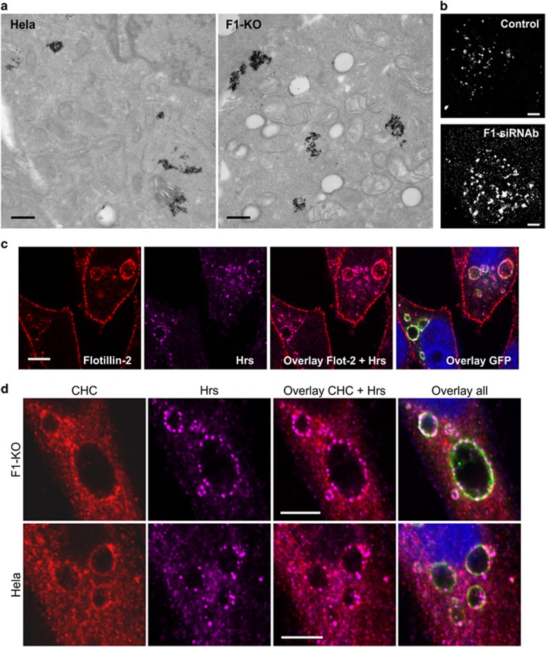
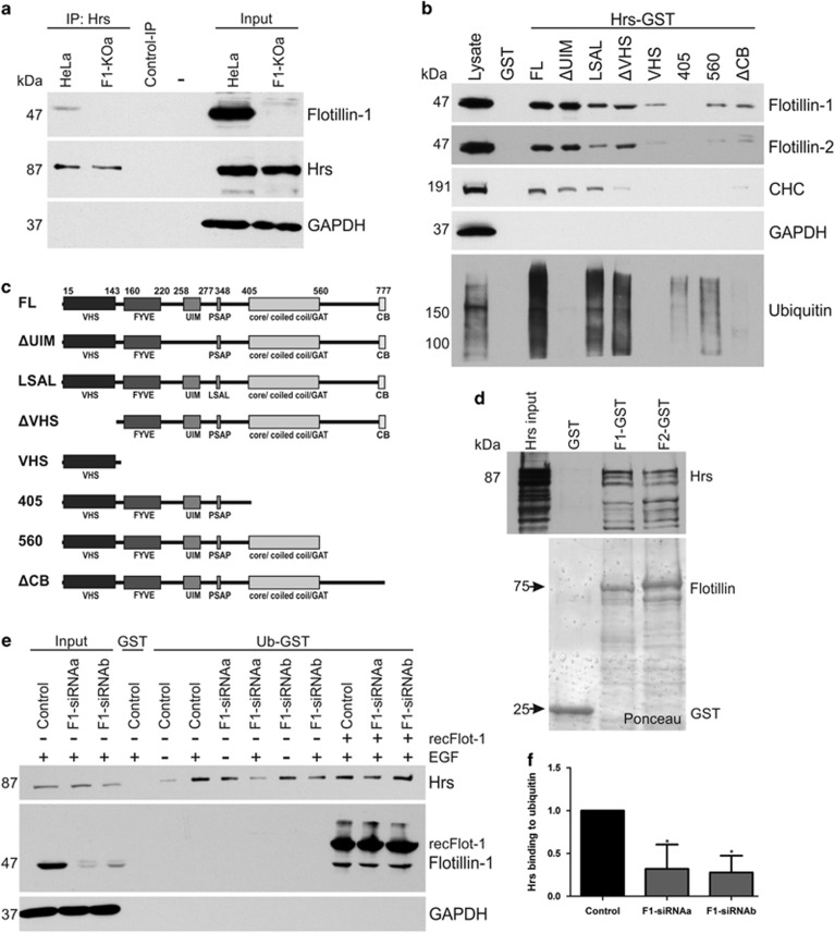
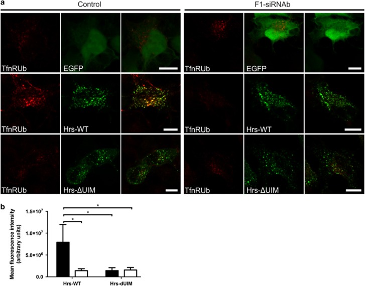
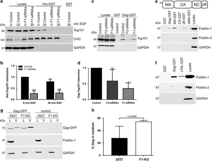
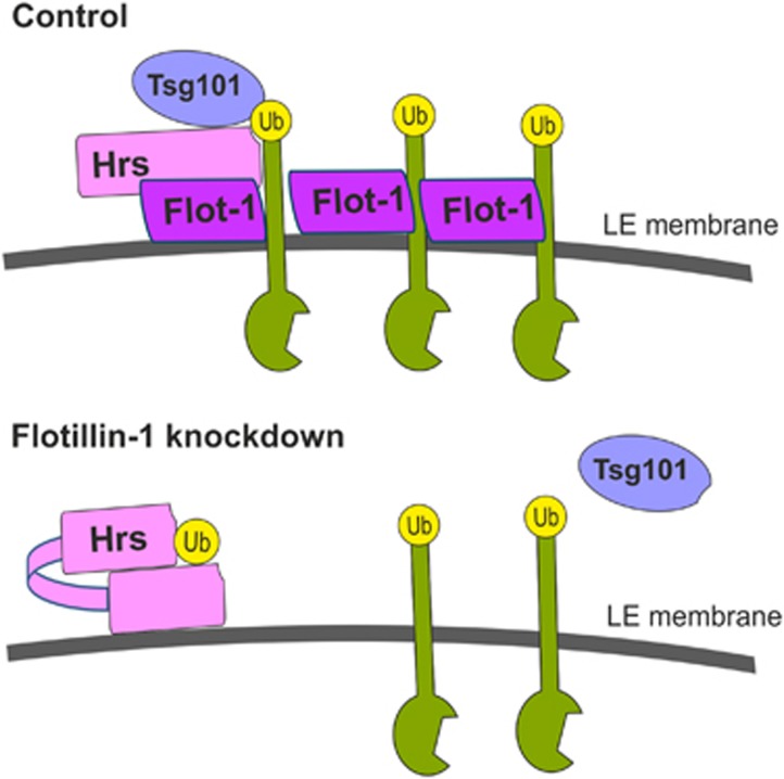
Ubiquitin-dependent sorting of membrane proteins in endosomes directs them to lysosomal degradation. In the case of receptors such as the epidermal growth factor receptor (EGFR), lysosomal degradation is important for the regulation of downstream signalling. Ubiquitinated proteins are recognised in endosomes by the endosomal sorting complexes required for transport (ESCRT) complexes, which sequentially interact with the ubiquitinated cargo. Although the role of each ESCRT complex in sorting is well established, it is not clear how the cargo is passed on from one ESCRT to the next. We here show that flotillin-1 is required for EGFR degradation, and that it interacts with the subunits of ESCRT-0 and -I complexes (hepatocyte growth factor-regulated tyrosine kinase substrate (Hrs) and Tsg101). Flotillin-1 is required for cargo recognition and sorting by ESCRT-0/Hrs and for its interaction with Tsg101. In addition, flotillin-1 is also required for the sorting of human immunodeficiency virus 1 Gag polyprotein, which mimics ESCRT-0 complex during viral assembly. We propose that flotillin-1 functions in cargo transfer between ESCRT-0 and -I complexes.


Conflict of interest statement

The authors declare no conflict of interest.

Figures

Figure 1
Figure 1
EGFR degradation is impaired in the absence of flotillin-1.(a, b) Control siRNA, flotillin-1 (F1-siRNAa and -b) and (c, d) control HeLa or flotillin-1-knockout cells (transfected with flotillin-1-EGFP or not) were starved overnight and stimulated with 100 ng/ml EGF for 60 min, fixed with methanol and immunostained for EGFR. (b) The amount of undegraded EGFR in flotillin-1-knockdown cells was quantified as fluorescence intensity in arbitrary units using the ImageJ Software. A significant increase of EGFR signal was observed in flotillin-knockdown cells as compared with the control. (d) EGFR amount after 60 min was quantified as in c, and the data show a rescue of EGFR degradation upon flotillin-1-EGFP expression (cells marked with *, images for GFP in Supplementary Figure S1d). The data in c, d are shown as mean±s.d. Cells from three independent experiments were evaluated (control siRNA cells: n=136, F1-siRNAa: n=135 and F1-siRNAb: n=149; control HeLa: n=358, flotillin-1 KO: n=181, rescue cells: n=252). Statistical analysis was performed with one-way analysis of variance (ANOVA) and Bonferroni post-test in comparison with the control. ***P<0.001. Scale bar in (a, c, e): 10 μm. (e) Flotillin-1-knockdown cells (F1-siRNAb) were treated as in a and immunostained for EGFR (green) and Rab5, Hrs or LAMP1 (red).
Figure 2
Figure 2
Increased amount of ubiquitinated cargo in endosomes of EGF-stimulated flotillin-1-knockdown cells.(a) Control siRNA and flotillin-1 siRNA-transfected cells were stimulated with 100 ng/ml EGF for 30 min, fixed and immunostained for ubiquitin. An enhanced ubiquitin staining was observed in punctate perinuclear structures in flotillin-1-knockdown cells. Scale bar: 10 μm. (b) Endosomes were isolated with magnetic columns from control siRNA and flotillin-1-depleted cells treated with Ferrofluid-coupled BSA or Ferro-EGF for 30 min. Samples from homogenate and eluate were analysed for ubiquitin by western blot. (c) Increased ubiquitin signal in endosomes of flotillin-1-knockdown cells. Densitometric analysis of ubiquitin signals shown in b from three independent experiments±s.d. Data were normalised to GAPDH in homogenates. (d) GST pulldown with ubiquitin-GST or GST from HeLa cells treated for 0, 30 or 60 min with EGF shows an increased binding of Hrs and GGA3 to ubiquitin upon EGF stimulation, whereas flotillins do not bind to ubiquitin-GST. Ubiquitin interactors were detected by western blot using specific antibodies, GAPDH was used as an input control.
Figure 3
Figure 3
Changes in endosomal and lysosomal morphology upon flotillin-1 knockdown. Immunofluorescent labelling for (a) LAMP3 or (b) LAMP1 in control siRNA and flotillin-1-knockdown HeLa cells. Quantification of the stainings for (c) LAMP3 and (d) LAMP1 show a significantly increased staining intensity for both endosomal markers in flotillin-1-knockdown cells. (e) Flotillin-1-knockout cells (F1-KO) show a similar phenotype with intense LAMP3 and LAMP1 staining that can be rescued upon expression of flotillin-1-EGFP (cells marked with *, images for GFP in Supplementary Figure S1g). (f) Quantification of the LAMP3 staining in e. The data in c, d, f are shown as mean±s.d. Cells from three independent experiments were evaluated (control siRNA cells: n=136, F1-siRNAa: n=135 and F1-siRNAb: n=149; control Hela: n=149, flotillin-1 KO: n=211, rescue cells: n=147). Statistical analysis in c, d was performed with Student’s t-test (two-tailed, unpaired), and in f with one-way analysis of variance (ANOVA) and Bonferroni post-test, in comparison with the control. ***P<0.001. Scale bar in a, b, e: 10 μm.
Figure 4
Figure 4
Endosomal morphology is altered in flotillin-1-knockdown/knockout cells, but endosomal domains persist.(a) Control and flotillin-1-knockout cells were chased for 2 h with Fe-EGF, fixed and analysed by transmission electron microscopy. Note the enlarged endosomes in flotillin-1-knockdown cells. Scale bar: 500 nm. (b) Control siRNA and flotillin-1-knockdown cells were stimulated with EGF for 30 min and labelled with an anti-mouse LAMP3 antibody detected with a secondary Alexa Fluor 647-coupled antibody. The specimens were analysed with Leica SR GSD 3D microscope. Scale bar: 2 μm. (c) Rab5-Q79L-GFP-expressing HeLa cells were immunostained for endogenous flotillin-2 and Hrs, which show a limited colocalization in the enlarged endosomes. Scale bar: 10 μm. (d) Rab5-Q79L-GFP-expressing HeLa and flotillin-1-knockout cells were immunostained for endogenous CHC and Hrs, which are localised in endosomal domains in both cell types and show some colocalization. Scale bar: 10 μm.
Figure 5
Figure 5
Flotillins directly interact with Hrs, and flotillin-1 is required for Hrs interaction with ubiquitin.(a) Co-immunoprecipitation of endogenous flotillin-1 with Hrs from HeLa cells stimulated with EGF for 30 min. Flotillin-1-knockout cells were used as a control to identify flotillin-1 bands. Control immunoprecipitation from HeLa cells with an isotype-matched antibody does not show any co-precipitation. (b) Purified Hrs-GST proteins were used to pull down flotillins from HeLa lysates for the mapping of the interaction sites of flotillins in Hrs. Flotillin interaction with Hrs is not dependent on the UIM domain in Hrs, whereas mutation of the PSAP motif into LSAL or deletion of the VHS domain partially abrogates flotillin interaction. VHS domain alone shows only a minor binding to flotillins. Deletion of the C-terminal region including the core/coiled-coil/GAT domains abolishes Hrs interaction with flotillins, and deletion of the C-terminal region beyond these domains results in decreased binding. Deletion of the clathrin-binding motif (CB) in Hrs C terminus also abrogates flotillin binding. (c) Structure of the constructs used in a. (d) Hrs and flotillins directly interact. GST pulldown was performed with recombinant, purified proteins. GST-tag in Hrs was removed before pulldown using thrombin digestion. (e) Interaction of endogenous Hrs with ubiquitin in an ubiquitin-GST-pulldown assay is reduced in EGF-stimulated (30 min) flotillin-1-knockdown cells, but can be rescued by the addition of purified recombinant flotillin-1. The pulldown samples were analysed by western blot using Hrs, flotillin-1 and GAPDH antibodies. (f) Quantification of Hrs binding to ubiquitin-GST upon 30 min EGF. Statistical analysis was performed using one-way analysis of variance (ANOVA) with Bonferroni post-test. The values shown represent mean±s.d. from three independent experiments. *P<0.05.
Figure 6
Figure 6
Flotillin-1 is necessary for the sorting of ubiquitinated cargo by ESCRT-0. (a) Control siRNA and flotillin-1-knockdown HeLa were transfected with TfnR-Ub chimera and Hrs-WT-GFP, ΔUIM-Hrs-GFP or EGFP and starved overnight. The cells were treated with Tfn-546 for 15 min, washed and chased for 60 min to allow for Tfn uptake. In control cells, Tfn accumulates in Hrs-WT-EGFP-positive structures, whereas in flotillin-1-knockdown cells, no accumulation is seen, indicating impaired Hrs–cargo binding. Expression of ΔUIM-Hrs does not result in Tfn accumulation, consistent with the inability of this mutant to bind ubiquitin. Scale bar: 10 μm. (b) Flotillin-1-knockdown cells accumulate significantly less Tfn after Hrs-WT-EGFP transfection. The mean fluorescence intensity of the Tfn-546 signal from control siRNA and flotillin-1-knockdown cells transfected with Hrs-WT-GFP, ΔUIM-Hrs-GFP and TfnR-Ub after 15 min uptake and 60 min chase was measured. Quantification of Tfn signals as arbitrary units was carried out with the ImageJ software. Bars show the mean±s.d. from three independent experiments (>90 cells per condition). Two-way analysis of variance (ANOVA) with Bonferroni post-test. *P<0.05.
Figure 7
Figure 7
Flotillin-1 is required for the binding of Hrs and HIV-1 Gag to Tsg101, and for endosomal localisation of Gag-EGFP.(a) Binding of Tsg101 and CHC from starved and EGF (100 ng/ml) stimulated cells transfected with control siRNA or flotillin-1 siRNA to Hrs-GST was tested by western blot. (b) Flotillin-1 depletion significantly impairs Hrs-GST binding to Tsg101. (c) Binding of endogenous Tsg101 to HIV-Gag-GST was determined in flotillin-1 and flotillin-2 knockdown HeLa cells. (d) Significantly less Tsg101 was bound to Gag-GST from flotillin-depleted cells. (e) Binding of endogenous flotillin-1 and flotillin-2 to full-length Gag and its subdomains (MA, CA, p6 and dMA-Gag) expressed as GST fusion proteins was analysed in HeLa cell lysates. Uppermost part shows the structural domain organisation of Gag. (f) Binding of flotillin-1 to Gag is reduced by ablation of the PTAP motif. (g) VLPs from Gag-EGFP-transfected 293T control and flotillin-1-knockout cells were prepared, and analysed by western blot. L: lysate; V: VLP. (h) The amount of Gag in the cell lysates and in the medium was determined by scanning densitometry. The percentage of released Gag was calculated taking into account the total volumes of each fraction, and the data are presented as % total Gag released in the medium. Densitometric quantification and statistical analysis in b, d, g were performed with Quantity One (Bio-Rad) or GraphPad Software. Statistical analysis was carried out with two-way analysis of variance (ANOVA) with Bonferroni post-test. Data in f were analysed with Student’s t-test (two-tailed, unpaired). Data are shown as the mean±s.d. of three to four independent experiments. *P<0.05; ***P>0.001.
Figure 8
Figure 8
Model for the function of flotillin-1 in endosomal sorting. The model shows the events that take place in the endosomal membrane after EGF stimulation. In control cells (upper part), flotillin-1 is required for Hrs to obtain a conformation that is compatible with the binding of cargo ubiquitin and Tsg101/ESCRT-I. Hereby, flotillin-1 interacts with both the cargo and Hrs. Upon flotillin-1 depletion (lower part), Hrs is capable of interacting with neither the cargo nor Tsg101, although it resides on the endosomal membrane. This may suggest that flotillin-1 is involved in the release of the autoinhibited conformation of Hrs that results from monoubiquitination upon EGF stimulation.

References

    1. Henne WM, Buchkovich NJ, Emr SD. The ESCRT pathway. Dev Cell 2011; 21: 77–91. - PubMed
    1. Henne WM, Stenmark H, Emr SD. Molecular mechanisms of the membrane sculpting ESCRT pathway. Cold Spring Harbor Perspect Biol 2013; 5: a016766. - PMC - PubMed
    1. Katzmann DJ, Babst M, Emr SD. Ubiquitin-dependent sorting into the multivesicular body pathway requires the function of a conserved endosomal protein sorting complex ESCRT-I. Cell 2001; 106: 145–155. - PubMed
    1. Komada M, Soriano P. Hrs, a FYVE finger protein localized to early endosomes, is implicated in vesicular traffic and required for ventral folding morphogenesis. Genes Dev 1999; 13: 1475–1485. - PMC - PubMed
    1. Raiborg C, Bremnes B, Mehlum A, Gillooly DJ, D'Arrigo A, Stang E et al. FYVE and coiled-coil domains determine the specific localisation of Hrs to early endosomes. J Cell Sci 2001; 114(Part 12): 2255–2263. - PubMed