

Angiopoietins and Tie2 in vascular inflammation
- PMID: 28582314
- PMCID: PMC5777595
- DOI: 10.1097/MOH.0000000000000361
Angiopoietins and Tie2 in vascular inflammation
Abstract
Purpose of review: As a subset of the organism-wide reaction to severe infection, the host vascular response has received increasing attention in recent years. The transformation that small blood vessels undergo to facilitate the clearance of pathogens may become harmful to the host if it occurs too broadly or if it is sustained too long. Adverse clinical manifestations of leaky and inflamed blood vessels include edema impairing the function of critical organs and circulatory shock.
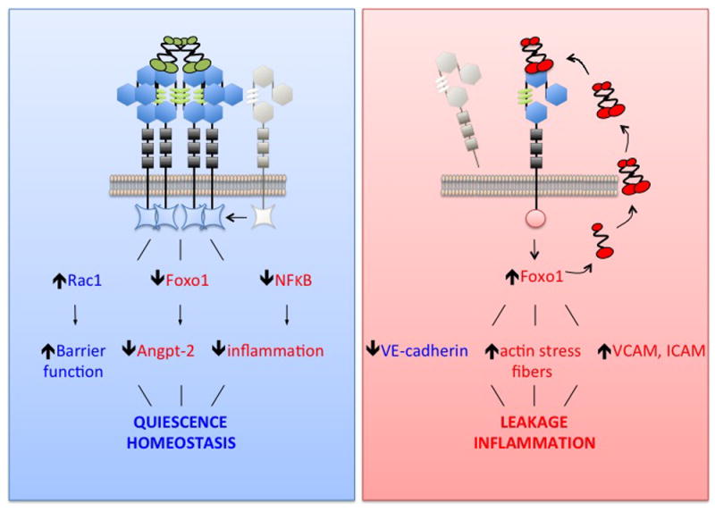
Recent findings: The study suggests that this host vascular response may be both measurable and potentially targetable. Tie2 is a receptor tyrosine kinase (RTK) heavily enriched in the vascular endothelium whose tonic signaling actively maintains vascular quiescence. When Tie2 becomes inactivated, important molecular brakes are released in the endothelium, which in turn potentiate inflammation and vascular leakage. The ligands of Tie2, Angiopoietin-1 and Angiopoietin-2, regulate its activation status. Genetic and molecular studies spanning thousands of humans link Tie2 and imbalance of the Angiopoietins to major adverse clinical events arising from bacterial sepsis, other severe infections, and even acute sterile inflammation.
Summary: The Tie2 signaling axis may constitute a molecular switch in systemic inflammation that can be measured and manipulated to target the host vascular response therapeutically.
Conflict of interest statement
Figures


Similar articles
-
Dysregulation of the angiopoietin-Tie-2 axis in sepsis and ARDS.Virulence. 2013 Aug 15;4(6):517-24. doi: 10.4161/viru.24906. Epub 2013 May 7. Virulence. 2013. PMID: 23652985 Free PMC article. Review.
-
Targeting Tie2 and the host vascular response in sepsis.Sci Transl Med. 2016 Apr 20;8(335):335fs9. doi: 10.1126/scitranslmed.aaf5537. Sci Transl Med. 2016. PMID: 27099172
-
Activation of Tie2 by angiopoietin-1 and angiopoietin-2 results in their release and receptor internalization.J Cell Sci. 2006 Sep 1;119(Pt 17):3551-60. doi: 10.1242/jcs.03077. Epub 2006 Aug 8. J Cell Sci. 2006. PMID: 16895971
-
The Angiopoietin-Tie2 Pathway in Critical Illness.Crit Care Clin. 2020 Apr;36(2):201-216. doi: 10.1016/j.ccc.2019.12.003. Epub 2020 Jan 31. Crit Care Clin. 2020. PMID: 32172809 Free PMC article. Review.
-
The angiopoietin:Tie 2 interaction: a potential target for future therapies in human vascular disease.Cytokine Growth Factor Rev. 2013 Dec;24(6):579-92. doi: 10.1016/j.cytogfr.2013.05.009. Epub 2013 Jul 6. Cytokine Growth Factor Rev. 2013. PMID: 23838360 Review.
Cited by
-
A dynamic nomogram for predicting diabetic macular edema in type 2 diabetes patients based on plasma cytokines.Aging (Albany NY). 2021 Mar 3;13(6):8369-8379. doi: 10.18632/aging.202647. Epub 2021 Mar 3. Aging (Albany NY). 2021. PMID: 33686950 Free PMC article.
-
Endothelial GNAQ p.R183Q Increases ANGPT2 (Angiopoietin-2) and Drives Formation of Enlarged Blood Vessels.Arterioscler Thromb Vasc Biol. 2022 Jan;42(1):e27-e43. doi: 10.1161/ATVBAHA.121.316651. Epub 2021 Oct 21. Arterioscler Thromb Vasc Biol. 2022. PMID: 34670408 Free PMC article.
-
Faricimab for the Treatment of Diabetic Macular Edema and Neovascular Age-Related Macular Degeneration.Pharmaceutics. 2023 May 5;15(5):1413. doi: 10.3390/pharmaceutics15051413. Pharmaceutics. 2023. PMID: 37242655 Free PMC article. Review.
-
Neutrophil Extracellular Traps, Angiogenesis and Cancer.Biomedicines. 2022 Feb 12;10(2):431. doi: 10.3390/biomedicines10020431. Biomedicines. 2022. PMID: 35203640 Free PMC article. Review.
-
Angiogenesis, Metabolism, Endothelial and Platelet Markers in Diabetes and Cardiovascular Disease.Br J Biomed Sci. 2022 Mar 22;79:10313. doi: 10.3389/bjbs.2022.10313. eCollection 2022. Br J Biomed Sci. 2022. PMID: 35996503 Free PMC article.
References
-
- Williams TW, Granger GA. Lymphocyte in vitro cytotoxicity: lymphotoxins of several mammalian species. Nature. 1968;219(5158):1076–7. Epub 1968/09/07. - PubMed
-
- Granger GA, Williams TW. Lymphocyte cytotoxicity in vitro: activation and release of a cytotoxic factor. Nature. 1968;218(5148):1253–4. Epub 1968/06/29. - PubMed
Publication types
MeSH terms
Substances
Grants and funding
LinkOut - more resources
Full Text Sources
Other Literature Sources
Medical
Research Materials
Miscellaneous
