. 2017 Oct 1;33(19):3051-3057.
doi: 10.1093/bioinformatics/btx358.

Glycan Reader is improved to recognize most sugar types and chemical modifications in the Protein Data Bank

Affiliations

Glycan Reader is improved to recognize most sugar types and chemical modifications in the Protein Data Bank

Sang-Jun Park et al. Bioinformatics. .

Abstract

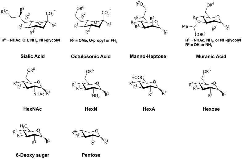
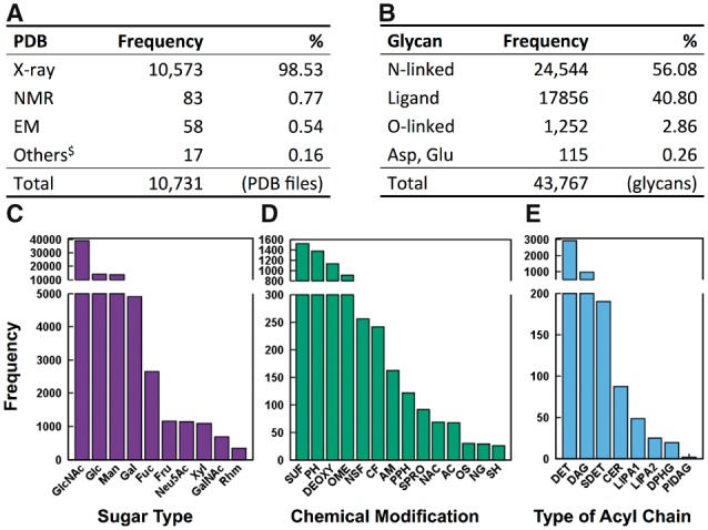
Motivation: Glycans play a central role in many essential biological processes. Glycan Reader was originally developed to simplify the reading of Protein Data Bank (PDB) files containing glycans through the automatic detection and annotation of sugars and glycosidic linkages between sugar units and to proteins, all based on atomic coordinates and connectivity information. Carbohydrates can have various chemical modifications at different positions, making their chemical space much diverse. Unfortunately, current PDB files do not provide exact annotations for most carbohydrate derivatives and more than 50% of PDB glycan chains have at least one carbohydrate derivative that could not be correctly recognized by the original Glycan Reader.

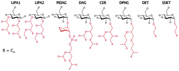
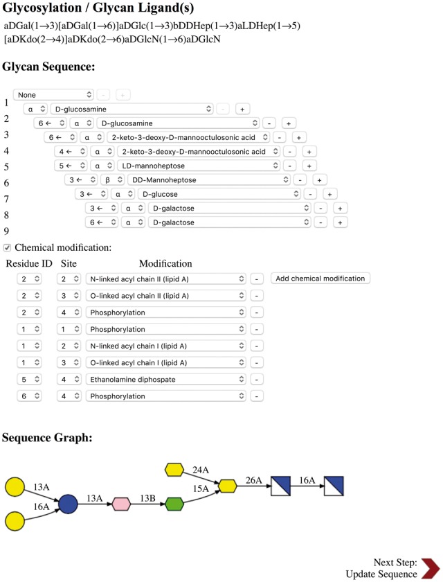
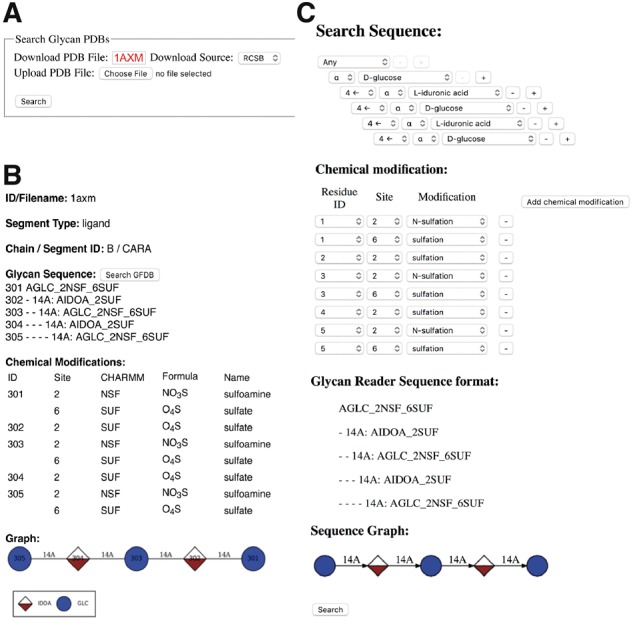
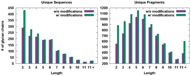
Results: Glycan Reader has been improved and now identifies most sugar types and chemical modifications (including various glycolipids) in the PDB, and both PDB and PDBx/mmCIF formats are supported. CHARMM-GUI Glycan Reader is updated to generate the simulation system and input of various glycoconjugates with most sugar types and chemical modifications. It also offers a new functionality to edit the glycan structures through addition/deletion/modification of glycosylation types, sugar types, chemical modifications, glycosidic linkages, and anomeric states. The simulation system and input files can be used for CHARMM, NAMD, GROMACS, AMBER, GENESIS, LAMMPS, Desmond, OpenMM, and CHARMM/OpenMM. Glycan Fragment Database in GlycanStructure.Org is also updated to provide an intuitive glycan sequence search tool for complex glycan structures with various chemical modifications in the PDB.

Availability and implementation: http://www.charmm-gui.org/input/glycan and http://www.glycanstructure.org.

Contact: wonpil@lehigh.edu.

Supplementary information: Supplementary data are available at Bioinformatics online.


Figures

Fig. 1.
Fig. 1.
Pyranose templates and available chemical modification sites. Six-membered rings that are composed of five carbon and one oxygen atoms are compared with the above templates through the VF2 isomorphism to identify the sugar types and assign the backbone atoms. R is the available position of chemical modifications
Fig. 2.
Fig. 2.
Glycolipid templates. The minimal substructures representing different acyl chain types are colored in red (Color version of this figure is available at Bioinformatics online.)
Fig. 3.
Fig. 3.
CHARMM-GUI Glycan Reader snapshot for a glycan chain in PDB:2FCP. When a PDB entry or uploaded PDB file contains glycan chains, their sequences are displayed in CASPER format under Glycosylation/Glycan Ligand(s) in the PDB manipulation section. When the edit button next to a specific CASPER sequence is clicked, Glycosylation/Glycan Ligand(s) is displayed in a new pop-up window, and one can view/edit glycosylation, sugar, linkage, and chemical modification types. All running steps of Glycan Reader are illustrated in Supplementary Figure S2
Fig. 4.
Fig. 4.
Snapshots from GlycanStructure.Org. (A) Glycan Reader in GlycanStructure.Org. (B) When a PDB entry is specified or a structure is uploaded in (A), detected glycan structure information is displayed. (C) Using the “Search GFDB” button in (B), the selected glycan chain information including the chemical modifications is transferred to GFDB (Glycan Fragment Database) to search the selected glycan structures in the PDB
Fig. 5.
Fig. 5.
Histograms for the numbers of unique and fragment glycan sequences in terms of glycan lengths in GFDB
Fig. 6.
Fig. 6.
RCSB glycan statistics as of December, 2016. (A) Numbers of PDB entries that have at least one glycan chain in terms of experimental methods. $Others are fiber diffraction, powder diffraction, neutron diffraction, solid-state NMR, and theoretical models. (B) Numbers of glycan chains in the PDB in terms of their types. (C) Histogram for the numbers of monosaccharides in terms of sugar types. (D) Histogram for the numbers of chemical modifications. (E) Histogram for the numbers of glycans in terms of lipid acyl chain types
Fig. 7.
Fig. 7.
MD simulations of FGF-1 monomer with a heparin analogue. (A) FGF-1 monomer in complex with a heparin analogue in PDB ID: 2ERM. (B) The hydrogen bonds between the FGF-1 monomer and heparin analogue are shown by dotted lines. (C) Symbolic representation of the heparin analogue. (D) Time series of ϕ and ψ glycosidic angles between Glc2NS (residue 6 in C) and IdoA2S (residue 5 in C) obtained from the 100-ns MD simulations starting from 20 different initial NMR models. (E) Frequency distribution curves of ϕ and ψ angles obtained from the MD simulations in comparison with those calculated from the 20 NMR models

References

    1. Abraham M.J. et al. (2015) GROMACS: high performance molecular simulations through multi-level parallelism from laptops to supercomputers. SoftwareX, 1–2, 19–25.
    1. Apweiler R. et al. (1999) On the frequency of protein glycosylation, as deduced from analysis of the SWISS-PROT database. Biochim. Biophys. Acta, 1473, 4–8. - PubMed
    1. Arthur E.J., Brooks C.L. 3rd (2016) Parallelization and improvements of the generalized born model with a simple sWitching function for modern graphics processors. J. Comput. Chem., 37, 927–939. - PMC - PubMed
    1. Berman H.M. et al. (2000) The Protein Data Bank. Nucleic Acids Res., 28, 235–242. - PMC - PubMed
    1. Bohne A. et al. (1999) SWEET - WWW-based rapid 3D construction of oligo- and polysaccharides. Bioinformatics, 15, 767–768. - PubMed