. 2017 Aug 21;45(14):8581-8595.
doi: 10.1093/nar/gkx499.

Unique localization of the plastid-specific ribosomal proteins in the chloroplast ribosome small subunit provides mechanistic insights into the chloroplastic translation

Affiliations

Unique localization of the plastid-specific ribosomal proteins in the chloroplast ribosome small subunit provides mechanistic insights into the chloroplastic translation

Tofayel Ahmed et al. Nucleic Acids Res. .

Abstract

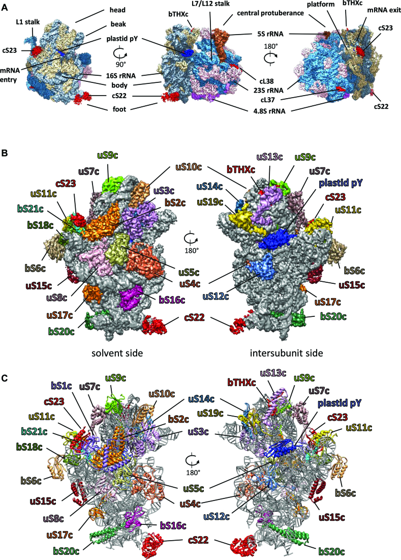
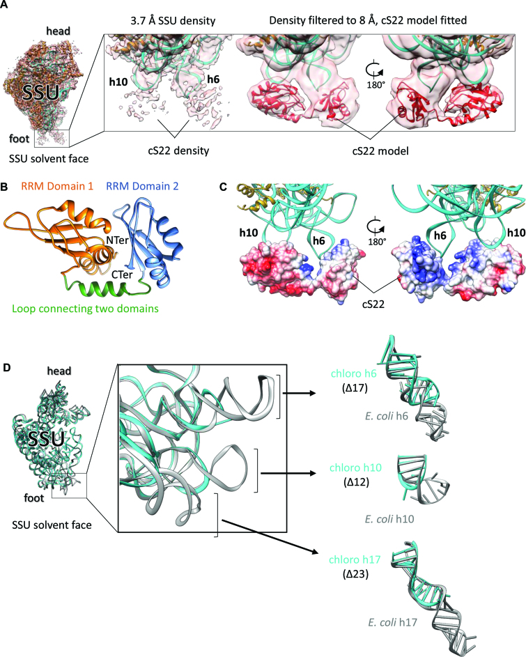
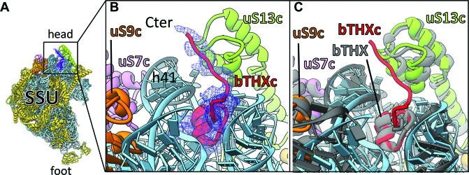
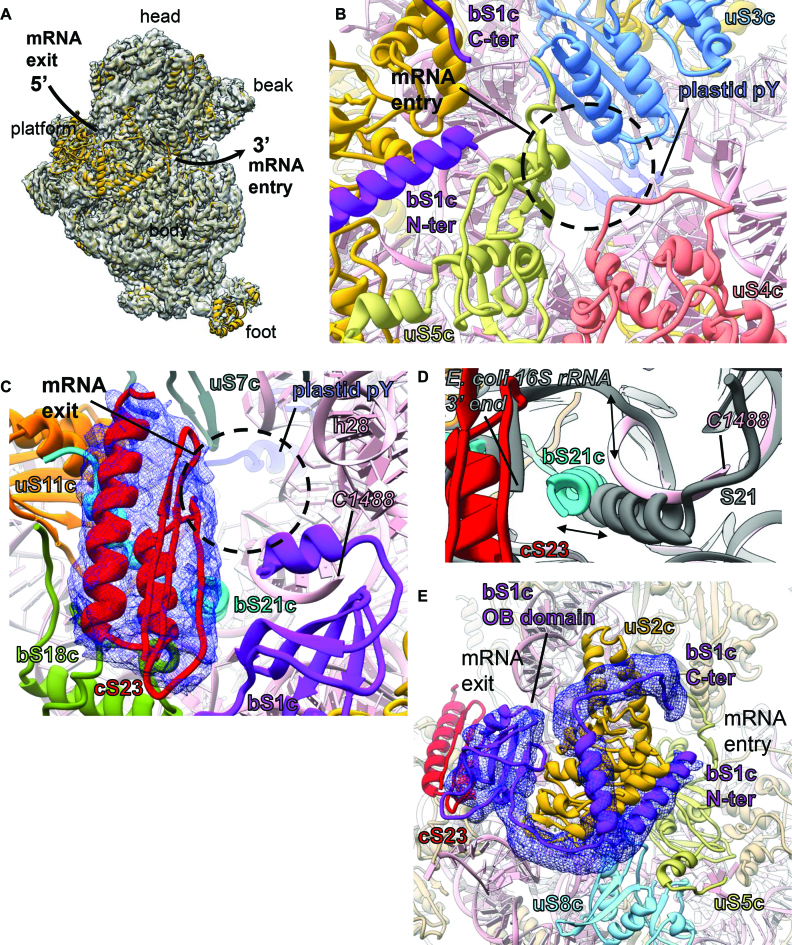
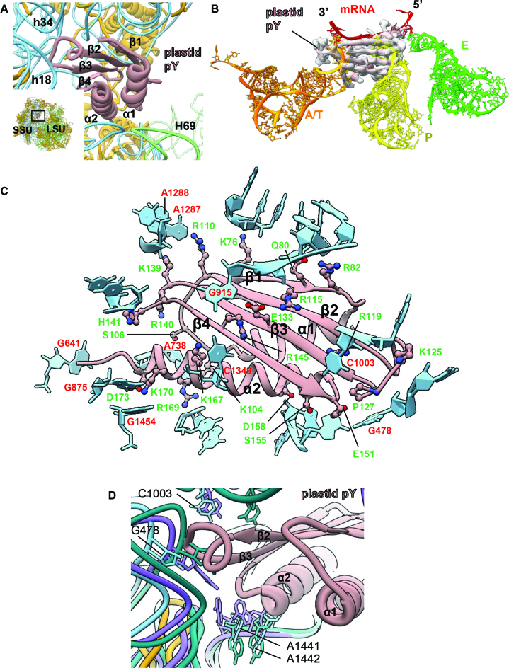
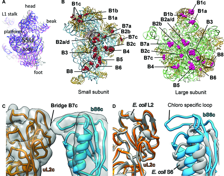
Chloroplastic translation is mediated by a bacterial-type 70S chloroplast ribosome. During the evolution, chloroplast ribosomes have acquired five plastid-specific ribosomal proteins or PSRPs (cS22, cS23, bTHXc, cL37 and cL38) which have been suggested to play important regulatory roles in translation. However, their exact locations on the chloroplast ribosome remain elusive due to lack of a high-resolution structure, hindering our progress to understand their possible roles. Here we present a cryo-EM structure of the 70S chloroplast ribosome from spinach resolved to 3.4 Å and focus our discussion mainly on the architecture of the 30S small subunit (SSU) which is resolved to 3.7 Å. cS22 localizes at the SSU foot where it seems to compensate for the deletions in 16S rRNA. The mRNA exit site is highly remodeled due to the presence of cS23 suggesting an alternative mode of translation initiation. bTHXc is positioned at the SSU head and appears to stabilize the intersubunit bridge B1b during thermal fluctuations. The translation factor plastid pY binds to the SSU on the intersubunit side and interacts with the conserved nucleotide bases involved in decoding. Most of the intersubunit bridges are conserved compared to the bacteria, except for a new bridge involving uL2c and bS6c.


Figures

Figure 1.
Figure 1.
Cryo-EM structure of the chloroplast 70S ribosome (A) Cryo-EM reconstruction (3.4 Å map) of the chloroplast 70S ribosome from spinach rendered at a density threshold value of 0.05 using UCSF Chimera (54). Density of 50S subunit proteins are shown in steel blue, 23S rRNA in pink, 5S rRNA in sienna, 4.8S rRNA in magenta, 30S subunit proteins in tan, 16S rRNA in slate gray, the translation factor pY in blue. PSRPs (cS22, cS23, bTHXc, cL37 and cL38) are shown in red. Structural landmarks of the ribosome are labeled. (B) Chloroplast ribosome SSU structure (3.7 Å map) from the solvent and the intersubunit side showing density for the PRPs and the PSRPs in differently colored surface at a density threshold value of 0.04 using UCSF Chimera (54). 16S rRNA is displayed in gray surface using the same threshold. Because of flexibility, density for bS1c does not appear at this threshold but can be observed in our 8-Å filtered map. (C) Models for the SSU proteins and 16S rRNA are displayed in the same colors as in (B).
Figure 2.
Figure 2.
Localization of cS22 at the chloroplast ribosome SSU foot (A) Fitting of cS22 at the SSU foot. SSU model fitted into high-resolution EM density showing extra density at the foot region near h6 and h10, which we assigned to cS22 (left). Proteins are in golden rod and 16S rRNA is in cyan. Zoomed-in view of the SSU foot showing high-resolution and 8 Å-filtered density map (right). Model of cS22 (in red) is fitted in the 8 Å-filtered map as indicated. (B) ITASSER (50) model of cS22 used for fitting the individual RRM domains in cS22 EM density. The two RRM domains of cS22 and connecting linker are labeled. (C) Coulombic surface coloring using UCSF Chimera (54) reveals that basic patches of cS22 interacts with negatively charged 16S rRNA (h6 and h10). (D) rRNA deletions in chloroplast ribosome 16S rRNA. Superimposition of 16S rRNA models from the chloroplast and the Escherichia coli ribosome (PDB ID: 4YBB) (51) (left). Zoomed view displays the three deletions in chloroplast ribosome 16S rRNA (center). Parts of h6, h10 and h17 are isolated to show comparison with E. coli counterparts (right). Chloroplast ribosome 16S rRNA is in cyan and E. coli 16S rRNA is in gray.
Figure 3.
Figure 3.
bTHXc localizes at the head of SSU (A) Position of bTHXc (red) at SSU head region. 16S rRNA is colored sky blue; ribosomal proteins except for those which surround bTHXc are colored golden rod. Isolated density of bTHXc is shown in blue mesh. (B) Zoomed-in view of (A) to show local environment of bTHXc. The core fold of bTHXc structure is buried in rRNA environment. (C) SSU structure of Thermus thermophilus ribosome (62) (gray) is overlaid with chloroplast ribosome SSU to show comparison between the structures of bTHXc and bTHX.
Figure 4.
Figure 4.
mRNA entry and exit sites, and localization of bS1c connecting both the sites (A) The mRNA entry and the exit sites are shown on the chloroplast ribosome SSU from the solvent exposed side. The arrow shows path of the mRNA. EM density for the SSU is shown in transparent surface (gray) with SSU model fitted. SSU proteins are colored golden rod and 16S rRNA is depicted in pink. (B) mRNA entry site. Three PRPs (uS3c, uS4c and uS5c) surround the mRNA entry site (black circle) in the chloroplast ribosome SSU. Fitted models of these PRPs are colored as: uS3c in sky blue, uS4c in salmon and uS5c in dark khaki. 16S rRNA is in pink. (C) mRNA exit site. Four PRPs (bS1c, uS7c, uS11c, bS21c) and one PSRP (cS23) along with h28 surround the mRNA exit site (black circle) in the chloroplast ribosome SSU and are colored as: bS1c in purple, uS7c in dim gray, uS11c in orange and bS21c in cyan. At the exit site, cS23 interacts with bS18c and bS21c. Isolated density of cS23 is displayed in blue mesh. 16S rRNA is colored pink. The nucleotide (C1488) at the 3′ end of our 16S rRNA model is indicated. (D) Shifted positions of bS21c (cyan) and the 3′ end of the chloroplast ribosome 16S rRNA (pink), compared to Escherichia coli (gray) ribosome SSU (PDB ID: 5AFI) (96). (E) Close association of bS1c (purple) with bS2c (golden rod). The N- and the C-terminals of bS1c extend toward the mRNA entry site while one of the three OB domains which is positioned closer to the C-terminal of bS1c reach out to the mRNA exit site. Proteins neighboring bS1c are colored as: cS23 in red, uS5c in dark khaki and uS8c in sky blue. 16S rRNA is in pink. Density for the N- and C-terminal of bS1c and the OB domain is displayed as blue mesh.
Figure 5.
Figure 5.
Binding of the plastid translation factor pY on SSU (A) Position of plastid pY at the intersubunit space is shown in the snippet at the bottom left. Both the ribosomal subunits are indicated. SSU proteins are colored golden rod, 16S rRNA is colored sky blue and LSU rRNAs (4.8S, 5S and 23S) are in green. The association of plastid pY exclusively to the SSU is displayed in the enlarged view. Plastid pY is colored rosy brown; orientation of the α helices and the beta strands of plastid pY in close association with 16S rRNA is indicated. (B) Plastid pY precludes binding of mRNA as well as A- and P-site tRNAs on the chloroplast ribosome. Overlaid tRNAs and mRNA from the Escherichia coli ribosome (PDB ID: 5AFI) (96) are colored as: A/T-site in orange, P-site in yellow, E-site in green and mRNA in red. EM density for plastid pY is depicted in transparent surface (gray) and fitted model of plastid pY is colored rosy brown. (C) Interactions between plastid pY and 16S rRNA are demonstrated. Plastid pY residues which contribute toward its binding to SSU are displayed in ball-and-stick and labeled in green. 16S rRNA is colored sky blue; only the nucleotide bases which directly interact with plastid pY are labeled (red). Interaction with plastid pY is also mediated by sugar-phosphate backbone of 16S rRNA but labels are hidden for clarity. (D) The conserved nucleotide bases (G478, C1003, A1441, A1442) involved in decoding are indicated. 16S rRNA from the apo- (PDB ID: 4YBB, in green) (51) and the translating states (PDB ID: 5AFI, in purple) (96) of the E. coli ribosome are overlaid with the chloroplast ribosome 16S rRNA to show relative positions of these conserved bases in our translationally inactive chloroplast 70S ribosome.
Figure 6.
Figure 6.
Intersubunit bridges of chloroplast 70S ribosome: (A) SSU models from PDB IDs: 4V6C (98) (gray) and 4V63 (97) (pink) overlaid with the chloroplast ribosome SSU displaying that plastid pY stabilizes a non-rotated state of the chloroplast ribosome. (B) The rRNA and protein residues of the SSU (left panel) and the LSU (right panel) which contribute in intersubunit bridge formation are represented as brown and pink spheres, respectively. 16S rRNA and the SSU proteins (left panel) are depicted in sky blue and golden rod, respectively. Plastid pY is displayed in rosy brown. 23S rRNA and LSU proteins (right panel) are depicted in green and golden rod, respectively. 5S rRNA and 4.8S rRNA (right panel) are colored in tan. In (B), both the ribosomal subunits are displayed from the subunit interface side. (C) Novel bridge in chloroplast 70S ribosome (B7c) is formed between proteins uL2c (golden rod) and bS6c (dodger blue). Density is 8 Å-filtered for clarity and shown in transparent surface (gray). Neighboring proteins and rRNAs are hidden for better visualization. (D) Models of the proteins S6 and L2 from Escherichia coli ribosome (PDB ID: 4YBB) (51), gray) overlaid with counterparts from chloroplast 70S ribosome showing absence of elongated loop in bacterial S6.

References

    1. Greber B.J., Ban N.. Structure and Function of the Mitochondrial Ribosome. Annu. Rev. Biochem. 2016; 85:103–132. - PubMed
    1. Hashem Y., des Georges A., Fu J., Buss S.N., Jossinet F., Jobe A., Zhang Q., Liao H.Y., Grassucci R.A., Bajaj C. et al. . High-resolution cryo-electron microscopy structure of the Trypanosoma brucei ribosome. Nature. 2013; 494:385–389. - PMC - PubMed
    1. Gupta A., Shah P., Haider A., Gupta K., Siddiqi M.I., Ralph S.A., Habib S.. Reduced ribosomes of the apicoplast and mitochondrion of Plasmodium spp. and predicted interactions with antibiotics. Open Biol. 2014; 4:140045. - PMC - PubMed
    1. Margulis L. Origin of Eukaryotic Cells: Evidence and Research Implications for a Theory of the Origin and Evolution of Microbial, Plant, and Animal Cells on the Precambrian Earth. 1970; New Have: Yale University Press.
    1. Jensen P.E., Leister D.. Chloroplast evolution, structure and functions. F1000Prime Reports. 2014; 6:40. - PMC - PubMed

MeSH terms

LinkOut - more resources