Review
. 2017 Jun 5;18(1):244.
doi: 10.1186/s12891-017-1608-z.

Magnetic resonance imaging of bacterial and tuberculous spondylodiscitis with associated complications and non-infectious spinal pathology mimicking infections: a pictorial review

Affiliations
Review

Magnetic resonance imaging of bacterial and tuberculous spondylodiscitis with associated complications and non-infectious spinal pathology mimicking infections: a pictorial review

Yogesh Kumar et al. BMC Musculoskelet Disord. .

Abstract

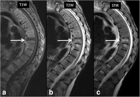
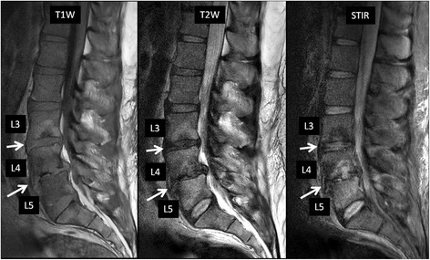
Magnetic resonance (MR) imaging plays an important role in the evaluation of bacterial and tuberculous spondylodiscitis and associated complications. Owing to its high sensitivity and specificity, it is a powerful diagnostic tool in the early diagnosis of ongoing infections, and thus provides help in prompt initiation of appropriate, therapy which may be medical or surgical, by defining the extent of involvement and detection of complications such as epidural and paraspinal abscesses. More specifically, MR imaging helps in differentiating bacterial from tuberculous infections and enables follow up of progression or resolution after appropriate treatment. However, other non-infectious pathology can demonstrate similar MR imaging appearances and one should be aware of these potential mimickers when interpreting MR images. Radiologists and other clinicians need to be aware of these potential mimics, which include such pathologies as Modic type I degenerative changes, trauma, metastatic disease and amyloidosis. In this pictorial review, we will describe and illustrate imaging findings of bacterial and tuberculous spondylodiscitis, their complications and non-infectious pathologies that mimic these spinal infections.

Keywords: Abscess; Discitis; Infection; MRI; Spine; Spondylitis.


Figures

Fig. 1
Fig. 1
42-year-old-diabetic-male patient presented with low back pain for 2 weeks. MR imaging showed L2–3 vertebral endplate marrow edema (red circle in a, b and c) and increased fluid signal in the L2–3 intervertebral disc on STIR sequence (blue arrow in image b). Post contrast images (c & d) show enhancing soft tissue in the epidural space (yellow arrows), consistent with phlegmon, which is a very early complication. Post contrast imaging can help in differentiating between phlegmon, which does not contain non-enhancing or liquefied components, from an abscess which does have those components. Streptococcus pneumoniae was the causative organism
Fig. 2
Fig. 2
MR imaging in a 36-year-old intravenous drug abuser female shows marrow edema involving L2–3 vertebra reaching up to the endplates (yellow arrows in a and b), with minimal enhancement on post contrast imaging consistent with osteomyelitis. Note increased amount of fluid signal in L2–3 intervertebral disc suggesting discitis-osteomyelitis complex. Post contrast imaging shows enhancing epidural soft tissue (white arrows in b, c and d) without areas of liquefaction, consistent with epidural phlegmon. Once again post contrast imaging can differentiate epidural abscess from phlegmon and thus alter management. Also note the presence of left psoas muscle involvement. Responsible bacteria in this case were Staphylococcus epidermidis
Fig. 3
Fig. 3
This is a companion case to the case shown in Fig. 2. 24-year-old male from India presented with low back pain. MR imaging showed involvement of the left diaphragmatic crura (white arrow in image a) and showing enhancement in post contrast imaging (yellow arrow in image b). Note the extensive prevertebral tubercular involvement (blue circles in c, d and e) on T1 W, STIR and post contrast imaging. Staphylococcus aureus was the causative organism
Fig. 4
Fig. 4
MR imaging in a 42-year-old female with paraparesis shows heterogeneous signal (red arrows) on T1 W image (a) and hyperintense epidural collection (yellow oval) on T2 W image (b) and show rim enhancement (yellow oval) on post contrast images (c), consistent with an epidural abscess. Axial T2 W (d) and post contrast images (e) distinctly show the epidural abscess (green arrows) causing indentation and posterior displacement of the thecal sac and cord (red asterisk). Escherichia coli was the causative organism. V- Vertebral body
Fig. 5
Fig. 5
64-year-old male with fever and low back pain. Sagittal T1 W (a) and STIR (b) MR images show extensive bone marrow edema involving entire L2 vertebral body and anterior portion of L1 vertebral body. High b-value (1000) axial diffusion weighted image (c) shows a right paraspinal soft tissue lesion with restricted diffusion (arrow) with associated decreased ADC values on ADC map (d, arrow). Biopsy of the lesion confirmed bacterial spondylodiscitis at L1/2 with a paraspinal abscess. Staphylococcus aureus was the causative organism
Fig. 6
Fig. 6
46-year-old-cashier presented with left lower back pain, especially with bending to the left for picking up shopping bags for customers. Physical exam showed left lower back point tenderness. T1 W images (a) showed a left sided hypointense posterior epidural lesion (white arrow in image a). STIR image (b) shows extensive edema in the region of left L2–3 lumbar facet joint (yellow arrows). T2 W image (c) shows fluid signal in left L2–3 facet joint with adjacent edema (white arrow in image c). Note the associated posterior epidural abscess (white arrow in image d) causing anterior displacement of the thecal sac. Staphylococcus aureus was the causative organism
Fig. 7
Fig. 7
MR imaging in a 24-year-old intravenous drug abuser male shows heterogeneous signal in the subdural space in the lower thoracic spine (yellow arrow in a) on T1 W images. There is peripheral rim enhancement on post contrast T1 W images (red arrows in b and c), consistent with subdural abscess causing anterior displacement of the cord (marked as S in c). Also note the preserved epidural fat suggesting subdural location of the abscess (blue arrows in a and b). Biopsy of the abscess confirmed Pseudomonas aeruginosa infection
Fig. 8
Fig. 8
27-year-old-male patient who was an immigrant from South America presented with back pain. Initial MRI was normal. Follow up MRI of the thoracolumbar spine for persistent back pain revealed marrow edema in the lower thoracic spine (yellow circle in a and b). Post contrast imaging showed discitis-osteomyelitis complex (red arrows in c). Note the enhancement of nerve roots suggesting extension of infectious process to the nerve roots causing arachnoiditis (green arrows in c and d)
Fig. 9
Fig. 9
An example of the Modic changes related to degenerative disease in a 68-year-old farmer. Note the Type I Modic changes at L4–5 endplates showing hyperintense signal on STIR image (a) and hypointense signal on T1-weighted image (b). Also note the absence of any surrounding paravertebral edema or psoas involvement (c), differentiating these Modic changes from vertebral osteomyelitis
Fig. 10
Fig. 10
74-year-old man presenting with acute low back pain following a fall. Sagittal STIR image at the time of presentation (a) shows end-plate deformities and bone marrow edema involving L2 and L4 vertebral bodies. When compared to prior study from 14 months ago (b), it became clear compression fracture of L2 is actually chronic. Change involving L4 is a new finding consistent with acute Schmorl’s node in this patient who had no clinical or laboratory evidence of active infection
Fig. 11
Fig. 11
52-year-old man with known history of ankylosing spondylitis presented with acute back pain following a fall. a T1-weighted, b T2-weighted and c STIR images in the sagittal plane shows a horizontal fracture line with associated bone marrow edema involving the superior endplate of T8 vertebral body (arrows). The patient showed no clinical evidence of active infection
Fig. 12
Fig. 12
64-year-old man with diabetes mellitus with low back and bilateral leg pain consistent with diabetic neuropathy. Sagittal T2-weighted image (a) shows end plate fragmentation at multiple lumbar levels (L2/3, L3/4, L4/5, arrows). MR neurography image (b) shows diffuse lumbosacral plexopathy with patchy thickening of all nerves. The patient showed no clinical evidence of active infection
Fig. 13
Fig. 13
39-year-old man with chronic renal failure on dialysis, with biopsy-proven amyloid spondyloarthropathy involving the lumbar spine. There are disc space narrowing and end-plate desctruction with associated bone marrow edema at L3/4 and L4/5. There is also grade 1 anterolisthesis of L4 over L5 as well as a disc herniation causing severe central canal stenosis at L4/5. From the imaging findings, initially infectious spondylodiscitis was suspected. However, failure to respond to antibiotic therapy prompted biopsy, which was negative for infectious organism and positive for amyloid stain

References

    1. Tali ET. Spinal infections. Eur J Radiol. 2004;50:120–133. doi: 10.1016/j.ejrad.2003.10.022. - DOI - PubMed
    1. Kourbeti IS, Tsiodras S, Boumpas DT. Spinal infections: evolving concepts. Curr Opin Rheumatol. 2008;20:471–479. doi: 10.1097/BOR.0b013e3282ff5e66. - DOI - PubMed
    1. Tins BJ, Cassar-Pullicino VN. MR imaging of spinal infection. Semin Musculoskelet Radiol. 2004;8:215–229. doi: 10.1055/s-2004-835362. - DOI - PubMed
    1. Galhotra RD, Jain T, Sandhu P, Galhotra V. Utility of magnetic resonance imaging in the differential diagnosis of tubercular and pyogenic spondylodiscitis. J Nat Sci Biol Med. 2015;6:388–393. doi: 10.4103/0976-9668.160016. - DOI - PMC - PubMed
    1. Ledbetter LN, Salzman KL, Shah LM. Imaging psoas sign in lumbar spinal infections: evaluation of diagnostic accuracy and comparison with established imaging characteristics. AJNR Am J Neuroradiol. 2016;37:736–741. doi: 10.3174/ajnr.A4571. - DOI - PMC - PubMed

MeSH terms