Randomized Controlled Trial
. 2017 Jun 5;8(1):129.
doi: 10.1186/s13287-017-0564-8.

Effect of single intralesional treatment of surgically induced equine superficial digital flexor tendon core lesions with adipose-derived mesenchymal stromal cells: a controlled experimental trial

Affiliations
Randomized Controlled Trial

Effect of single intralesional treatment of surgically induced equine superficial digital flexor tendon core lesions with adipose-derived mesenchymal stromal cells: a controlled experimental trial

Florian Geburek et al. Stem Cell Res Ther. .

Abstract

Background: Adipose tissue is a promising source of mesenchymal stromal cells (MSCs) for the treatment of tendon disease. The goal of this study was to assess the effect of a single intralesional implantation of adipose tissue-derived mesenchymal stromal cells (AT-MSCs) on artificial lesions in equine superficial digital flexor tendons (SDFTs).

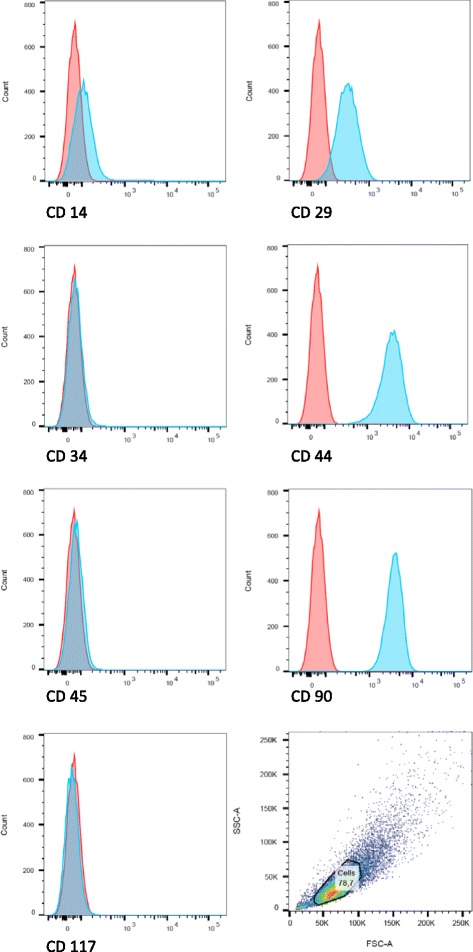
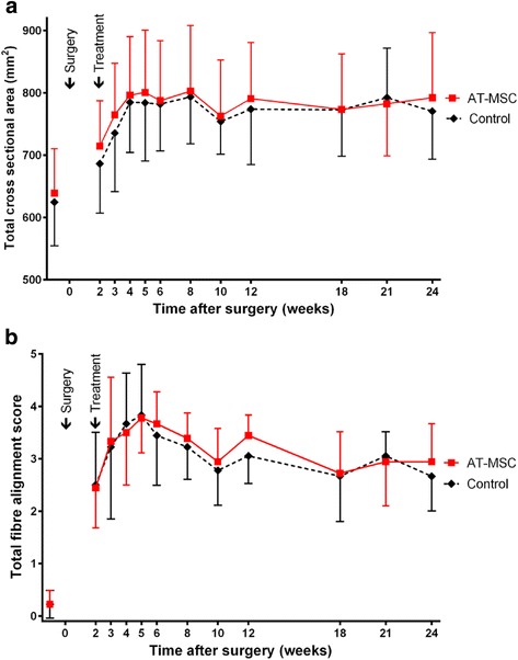
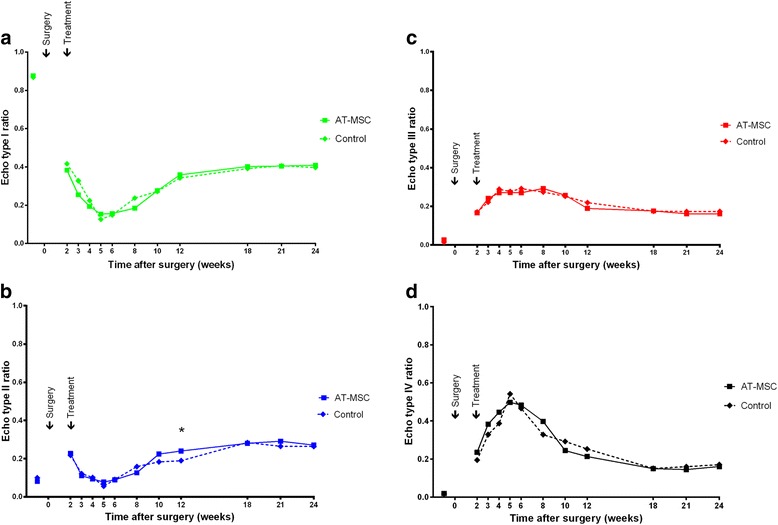
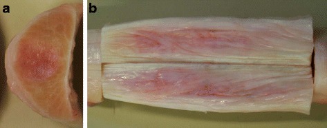
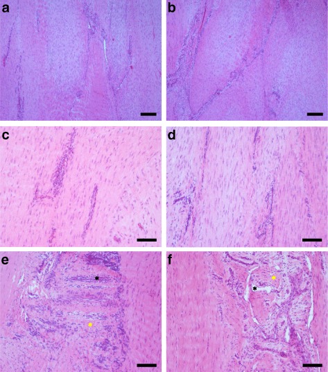
Methods: During this randomized, controlled, blinded experimental study, either autologous cultured AT-MSCs suspended in autologous inactivated serum (AT-MSC-serum) or autologous inactivated serum (serum) were injected intralesionally 2 weeks after surgical creation of centrally located SDFT lesions in both forelimbs of nine horses. Healing was assessed clinically and with ultrasound (standard B-mode and ultrasound tissue characterization) at regular intervals over 24 weeks. After euthanasia of the horses the SDFTs were examined histologically, biochemically and by means of biomechanical testing.

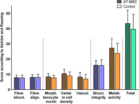
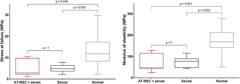
Results: AT-MSC implantation did not substantially influence clinical and ultrasonographic parameters. Histology, biochemical and biomechanical characteristics of the repair tissue did not differ significantly between treatment modalities after 24 weeks. Compared with macroscopically normal tendon tissue, the content of the mature collagen crosslink hydroxylysylpyridinoline did not differ after AT-MSC-serum treatment (p = 0.074) while it was significantly lower (p = 0.027) in lesions treated with serum alone. Stress at failure (p = 0.048) and the modulus of elasticity (p = 0.001) were significantly lower after AT-MSC-serum treatment than in normal tendon tissue.

Conclusions: The effect of a single intralesional injection of cultured AT-MSCs suspended in autologous inactivated serum was not superior to treatment of surgically created SDFT lesions with autologous inactivated serum alone in a surgical model of tendinopathy over an observation period of 22 weeks. AT-MSC treatment might have a positive influence on collagen crosslinking of remodelling scar tissue. Controlled long-term studies including naturally occurring tendinopathies are necessary to verify the effects of AT-MSCs on tendon disease.

Keywords: Biochemistry; Biomechanical testing; Histology; Horse; MSC, mesenchymal stem cells; Tendon; Ultrasonography; Ultrasound tissue characterization.


Figures

Fig. 1
Fig. 1
Flow cytometric analysis of cultured AT-MSCs from a representative study horse. Histograms indicate the immunophenotype of AT-MSCs for CD14, CD29, CD34, CD44, CD45, CD90 and CD117. Results are displayed for the distribution of immunostained (green) and unstained (red) AT-MSCs. All stained cells were positive for CD29, CD44 and CD90 while the signal for CD14 was weaker. No signal was detected for CD34, CD45 and CD117 (Colour figure online)
Fig. 2
Fig. 2
Adipogenic differentiation of equine AT-MSCs from a representative study horse. Photomicrographs of AT-MSCs (passage 2) taken 28 days after induction of adipogenic differentiation (a). After Oil Red staining a high number of intracellular lipid-containing vesicles was detected compared with the control without differentiation medium (b) (Colour figure online)
Fig. 3
Fig. 3
Osteogenic differentiation of equine AT-MSCs from a representative study horse. Photomicrographs of AT-MSCs (passage 2) taken on day 28 after induction of osteogenic differentiation (A1–A3; C1–C3). By contrast to controls without differentiation medium (B1–B3; D1–D3), deposition of extracellular calcium was detected by alkaline phosphatase (A1, C1), von Kossa staining (A2, C2) and Alizarin red staining (A3, C3) (Colour figure online)
Fig. 4
Fig. 4
Chondrogenic differentiation of equine AT-MSCs from a representative study horse. Photomicrographs of AT-MSCs (passage 2) taken on day 21 after induction of chondrogenic differentiation. The presence of glycosaminoglycans and collagen was detected by Toluidine Blue (a) and Safranin O (b) (Colour figure online)
Fig. 5
Fig. 5
B-mode ultrasonographic parameters. Development of ultrasonographic scores (mean ± SD) of SDFTs treated with AT-MSCs suspended in autologous inactivated serum (solid lines) and autologous inactivated serum alone (dotted lines) over 24 weeks after surgical induction of tendon injuries. a TCSA. b TFAS. AT-MSC adipose-derived mesenchymal stromal cell
Fig. 6
Fig. 6
Ultrasound tissue characterization. Development of echo type ratios for SDFTs treated with AT-MSCs suspended in autologous inactivated serum (solid lines) and autologous inactivated serum alone (dotted lines) over 24 weeks after surgical induction of tendon injuries. a Echo type I. b Echo type II. c Echo type III. d Echo type IV. *Significant difference between groups. AT-MSC adipose-derived mesenchymal stromal cell
Fig. 7
Fig. 7
Gross pathologic examination of SDFTs. a Transverse and b longitudinal section of a serum-treated tendon segment 24 weeks after surgical induction of tendinopathy, 2 and 2–5 cm proximal to the surgical entrance into the tendon, respectively. The centrally located scar tissue is pale to intensively pink, partly reddish and well demarcated from surrounding ivory-coloured tendon tissue. The scar is nearly circular in shape on the transverse and appears as an oblong area on the longitudinal section (Colour figure online)
Fig. 8
Fig. 8
Histology of surgically induced SDFT lesions 22 weeks after treatment. af Longitudinal specimens of tendon lesions treated with AT-MSCs suspended in autologous inactivated serum (a, c, e) and autologous inactivated serum alone (control; b, d, f) stained with H&E (a, b, scale bar = 200 μm; c, d, e, f, scale bar = 100 μm). Fibril arrangement was mostly unidirectional (ad), with some cases showing large regions without any regular fibre arrangement (e, f; yellow asterisk) in both groups. Specimens showed variations in cell density (ad) and regions with high cellularity and vascularization after both treatment modalities (e, f; black asterisks) (Colour figure online)
Fig. 9
Fig. 9
Histomorphological scores (Aström and Rausing [60], modified by Bosch et al. [45]). Mean histomorphological scores (± SD) for surgically induced SDFT lesions treated with AT-MSCs suspended in autologous inactivated serum (AT-MSC) and with autologous inactivated serum alone (control) 22 weeks after treatment. Total scores (green) include values from sub-scores for fibre structure (struct.), fibre alignment (align.), morphology (morph.) of tenocyte nuclei, variations (variat.) in cell density in cell density and vascularization (vascul.). Scores for structural (struct.) integrity and metabolic (metab.) activity summarize values from sub-scores displayed in blue and red, respectively. Scores and sub-scores did not differ between treatment modalities (p < 0.05). AT-MSC adipose-derived mesenchymal stromal cell (Colour figure online)
Fig. 10
Fig. 10
Biomechanical parameters. Stress at failure and modulus of elasticity (measure of tensile stiffness of materials) of surgically induced SDFT lesion tissue treated with AT-MSCs suspended in autologous inactivated serum (AT-MSC + serum) and autologous inactivated serum alone (serum) 24 weeks after creation of lesions and 22 weeks after treatment. Macroscopically normal tendon tissue (normal) was harvested from a proximal segment of the same tendons. AT-MSC adipose-derived mesenchymal stromal cell

References

    1. Cassel M, Baur H, Hirschmuller A, Carlsohn A, Frohlich K, Mayer F. Prevalence of Achilles and patellar tendinopathy and their association to intratendinous changes in adolescent athletes. Scand J Med Sci Sports. 2015;25(3):e310–8. doi: 10.1111/sms.12318. - DOI - PubMed
    1. Maffulli N, Wong J. Rupture of the Achilles and patellar tendons. Clin Sports Med. 2003;22(4):761–76. doi: 10.1016/S0278-5919(03)00009-7. - DOI - PubMed
    1. Ross MW. Movement. In: Ross MW, Dyson SJ, editors. Diagnosis and Management of Lameness in the Horse. 2. St. Louis: Elsevier Saunders; 2011. pp. 64–80.
    1. O’Meara B, Bladon B, Parkin TD, Fraser B, Lischer CJ. An investigation of the relationship between race performance and superficial digital flexor tendonitis in the Thoroughbred racehorse. Equine Vet J. 2010;42(4):322–6. doi: 10.1111/j.2042-3306.2009.00021.x. - DOI - PubMed
    1. Williams RB, Harkins LS, Hammond CJ, Wood JL. Racehorse injuries, clinical problems and fatalities recorded on British racecourses from flat racing and National Hunt racing during 1996, 1997 and 1998. Equine Vet J. 2001;33(5):478–86. doi: 10.2746/042516401776254808. - DOI - PubMed

Publication types