. 2017 Jun 5;36(1):75.
doi: 10.1186/s13046-017-0545-x.

ERβ1 inhibits metastasis of androgen receptor-positive triple-negative breast cancer by suppressing ZEB1

Affiliations

ERβ1 inhibits metastasis of androgen receptor-positive triple-negative breast cancer by suppressing ZEB1

Wei Song et al. J Exp Clin Cancer Res. .

Abstract

Background: Increasing evidence has indicated an important role for estrogen receptor beta 1 (ERβ1) in breast cancer. However, the role of ERβ1 in the metastasis of androgen receptor (AR)-positive triple-negative breast cancer (TNBC) and the underlying mechanisms are still unknown.

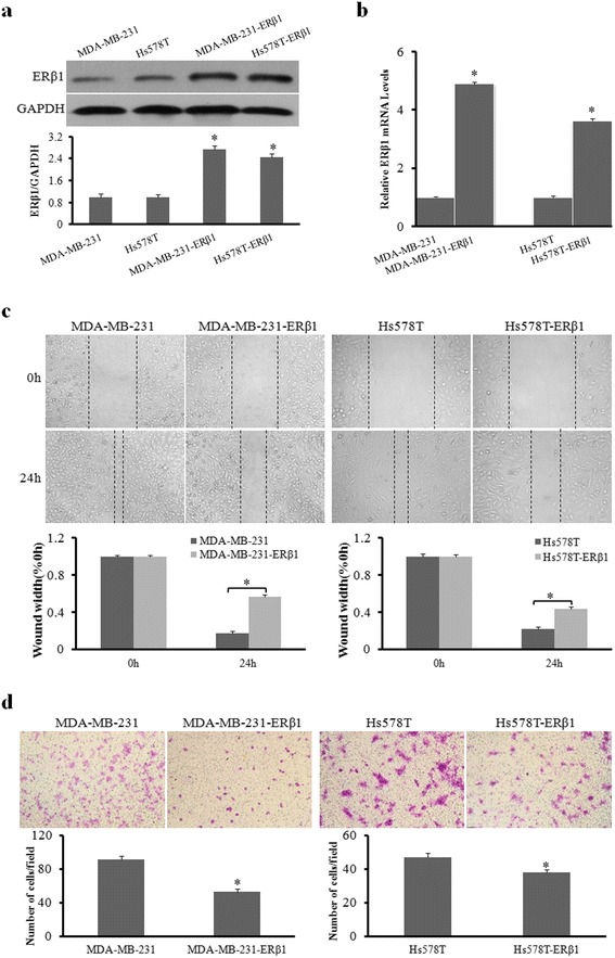
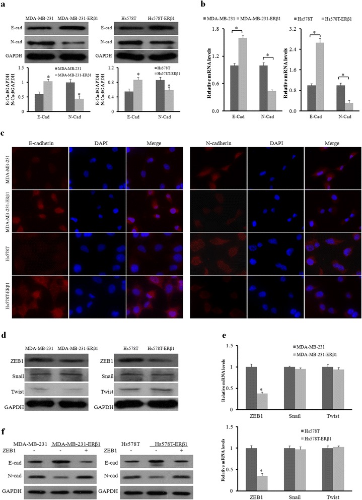
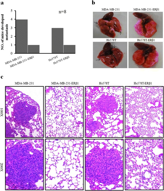
Methods: Stable ERβ1-expressing TNBC cell lines were generated for this study. We detected the abilities of cell migration and invasion by wound-healing and transwell assays and the expression of E-cadherin and N-cadherin by quantitative RT-PCR (qRT-PCR) and western blotting assays in TNBC cell lines. Chromatin immunoprecipitation (ChIP) analysis was performed to assess the effect of AR on ERβ1 promoter. Tumor metastasis was evaluated in vivo using a lung metastasis mouse model. Lastly, immunohistochemical expression of ERβ1 in TNBC tissues was analyzed and correlated with clinicopathological features.

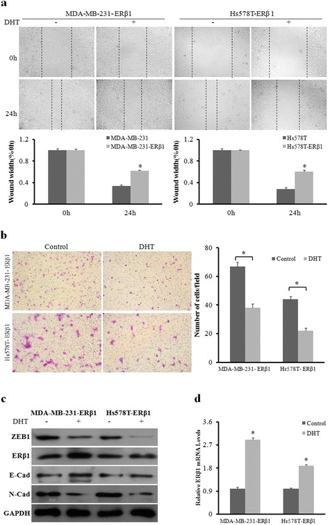
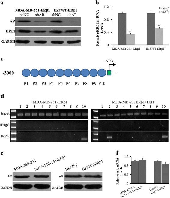
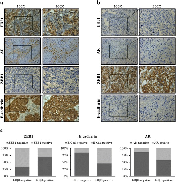
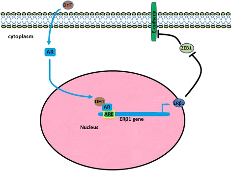
Results: ERβ1 suppressed the invasion and migration abilities of AR-positive TNBC cells and induced the downregulation of ZEB1. ZEB1 overexpression abrogated the increase in E-cadherin expression and the decrease in N-cadherin expression modulated by ERβ1. A lung metastasis mouse model showed that the incidence of metastasis was lower in ERβ1-expressing TNBC cells. Further, AR activation increased the anti-metastatic effect of ERβ1 in AR-positive TNBC cells, which accelerated ERβ1 transcription by functioning as a transcription factor that bound to the promoter of ERβ1. No significant change was observed in AR expression induced by ERβ1. Immunohistochemistry (IHC) analysis of TNBC clinical samples showed that ERβ1 and AR were positive in 31.7% and 23.2% of samples, respectively. ERβ1 expression was negatively correlated with ZEB1 expression and lymph node metastasis, and positively correlated with the expression of AR and E-cadherin.

Conclusion: Our findings suggested a potential role of ERβ1 in metastasis of AR-positive TNBC and provided novel insights into the mechanism of action of ERβ1 and the possible relationship between ERβ1 and AR.

Keywords: AR; ERβ1; Triple-negative breast cancer; ZEB1.


Figures

Fig. 1
Fig. 1
ERβ1 suppresses cell migration and invasion of AR-positive TNBC cells. a, b Western blot analysis and qRT-PCR of ERβ1 protein and mRNA expression in the control and stable ERβ1-transfected cell lines. c Representative images of wound-healing assays in the control and ERβ1-expressing cell lines. The widths of injury lines made in cells were examined at 0 and 24 h. Wound-healing migration is represented by the widths of injury lines. d Representative results of transwell assays showing the effect of ERβ1 overexpression on the invasion ability in control and ERβ1-expressing MDA-MB-231 and Hs578T cells. *p < 0.05
Fig. 2
Fig. 2
ERβ1 suppresses metastasis of AR-positive TNBC by inhibiting ZEB1. a Western blot analysis of E-cadherin and N-cadherin protein in control and ERβ1-expressing cells. GAPDH expression was used as the loading control. b qRT-PCR detection of E-cadherin and N-cadherin mRNA expression in control and ERβ1-expressing MDA-MB-231 and Hs578T cells. c Fluorescent microscopy analysis of the expression of E-cadherin and N-cadherin by immunofluorescence. The red signal represents E-cadherin or N-cadherin protein, and the blue signal represents the nuclear DNA staining by DAPI. d, e Western blot and qRT-PCR analyses of ZEB1, Snail and Twist in the control and ERβ1-expressing cells. f Western blot of E-cadherin and N-cadherin expression after transfection with ZEB1 or empty vector in the control and ERβ1-expressing MDA-MB-231 and Hs578T cells. *p < 0.05
Fig. 3
Fig. 3
ERβ1 inhibits metastasis of AR-positive TNBC cells in vivo. a Lung metastasis rate of mouse models in the control and ERβ1-expressing groups (n = 8). b Representative images of lung metastasis in mouse models injected with control and ERβ1-expressing MDA-MB-231 and Hs578T cells. c Representative HE staining of lung sections from the control or ERβ1-expressing groups
Fig. 4
Fig. 4
AR promotes the anti-metastatic effect of ERβ1. a Wound-healing assay for the cell migration of ERβ1-expressing MDA-MB-231 and Hs578T cells with or without DHT (10nM) treatment. The percentage of wound healing was calculated. b Representative images of transwell assays for the invasion of MDA-MB-231 and Hs578T cells treated with or without DHT. c Western blot analysis of ZEB1, ERβ1, E-cadherin and N-cadherin protein levels in ERβ1-expressing MDA-MB-231 and Hs578T cells after treatment with or without DHT. d qRT-PCR detection of ERβ1 mRNA in ERβ1-expressing MDA-MB-231 and Hs578T cells with or without DHT treatment. *p < 0.05
Fig. 5
Fig. 5
AR acts as a transcription factor that binds to the promoter of ERβ1. a, b Western blot and qRT-PCR analyses of ERβ1 protein and mRNA with or without AR knockdown in ERβ1-expressing MDA-MB-231 and Hs578T cells in the presence of DHT. c Primers used for PCR in the ChIP assay. d The gel electrophoresis of ChIP assay showed a binding site located in primer 10 without DHT treatment and two binding sites located in primers 2 and 10 with DHT treatment in ERβ1-expressing MDA-MB-231 cells. e, f Western blot and qRT-PCR analyses of AR protein and mRNA in the control and ERβ1-expressing cells. *p < 0.05
Fig. 6
Fig. 6
Relationships of ERβ1 with clinicopathological factors in TNBC. a, b Representative IHC staining of ERβ1 high-, AR high-, ZEB1 low-, and E-cadherin high-expression (a) or ERβ1 low-, AR low-, ZEB1 high-, and E-cadherin low-expression (b) in TNBC samples. c Expression ratio of AR, ZEB1 and E-cadherin in ERβ1-positive group or ERβ1-negative group, respectively
Fig. 7
Fig. 7
Proposed working model of activated AR regulation of the transcription of ERβ1, which subsequently suppresses ZEB1

References

    1. Jamdade VS, Sethi N, Mundhe NA, Kumar P, Lahkar M, Sinha N. Therapeutic targets of triple-negative breast cancer: a review. Br J Pharmacol. 2015;172(17):4228–4237. doi: 10.1111/bph.13211. - DOI - PMC - PubMed
    1. Bianchini G, Balko JM, Mayer IA, Sanders ME, Gianni L. Triple-negative breast cancer: challenges and opportunities of a heterogeneous disease. Nat Rev Clin Oncol. 2016;13(11):674–690. doi: 10.1038/nrclinonc.2016.66. - DOI - PMC - PubMed
    1. Jang G, Lee S, Ahn J, Jung K, Lee H, Gong G, Kim H, Ahn S, Ahn S, Kim S. Clinical features and course of brain metastases in triple-negative breast cancer: Comparison with HER2+ and other type. J Clin Oncol. 2009;27(15_suppl):1064.
    1. Collins LC, Cole KS, Marotti JD, Hu R, Schnitt SJ, Tamimi RM. Androgen receptor expression in breast cancer in relation to molecular phenotype: results from the Nurses’ Health Study. Mod Pathol. 2011;24(7):924–931. doi: 10.1038/modpathol.2011.54. - DOI - PMC - PubMed
    1. Mrklic I, Pogorelic Z, Capkun V, Tomic S. Expression of androgen receptors in triple negative breast carcinomas. Acta Histochem. 2013;115(4):344–348. doi: 10.1016/j.acthis.2012.09.006. - DOI - PubMed

Publication types

MeSH terms